25 Application of Isokinetics in Testing and Rehabilitation
Isokinetics plays a significant role in the evaluation and rehabilitation of injured athletes. The use of isokinetics has changed as interest in isokinetics has varied over the past 25 years. Isokinetics was developed in the 1960s and used increasingly during the 1970s. However, research on this subject was minimal, and the potential uses and applications of isokinetics were not clearly understood. In the 1980s the field of isokinetics came into its own, with increasing popularity and, most importantly, an increasing body of knowledge through numerous publications that supported the appropriate use of isokinetics in the testing and rehabilitation of athletes. During this period, isokinetics was used increasingly in many different areas and with many different applications. The first book dedicated solely to isokinetics was published in the early 1980 s1; it provided an overview of the testing and application of isokinetics through a combination of published research and empirically based clinical experience. However, in the 1990s there was a trend away from the use of isokinetics as part of the total evaluation and rehabilitation process. Despite extensive publications on isokinetics (more than 2000 published articles on the use and efficacy of isokinetics, an entire journal dedicated to the art and science of isokinetics [Isokinetics and Exercise Science], and four books dedicated exclusively to isokinetics1-4), many practicing clinicians have discontinued using isokinetics on the grounds that it is not functional. A PubMed search (performed on 12/16/2010) of the term isokinetics identified 4196 references. However, when the search was limited to higher levels of evidence, including randomized controlled trials, systematic reviews, and metaanalysis studies, the actual number was 513 high-quality studies. More than 220 of these articles have been published in the last decade. Interestingly, when a Google search was performed for the term isokinetics, 79,400 citations appeared. Although admittedly most athletes do not sit and flex and extend their knees as a functional activity, there is high correlation between isokinetic testing of the knee and functional testing. Unfortunately, many clinicians are disregarding the extensive documentation of isokinetics in the evaluation and treatment of athletes and are embracing closed kinetic chain (CKC) exercises as a panacea without significant documentation of efficacy. We do not advocate that only isokinetics should be used or that CKC exercises should not be used; we would, however, like to emphasize the need for an integrated approach that uses many modes of testing and rehabilitation.
Overview and terminology
Numerous modes of exercise can be used for the evaluation and rehabilitation of athletes, including isometrics, isotonics, plyometrics, isoacceleration, isodeceleration, and isokinetics.
The concept of isokinetic exercise was introduced by James Perrine in the late 1960s, and it proved to be a revolution in exercise training and rehabilitation. Instead of the traditional exercises that were performed at variable speeds against a constant weight or resistance, Perrine developed the concept of isokinetics, which involves a dynamic preset fixed speed with resistance that is totally accommodating throughout the range of motion (ROM). Since the inception of isokinetics, this form of testing and exercise has become increasingly popular in clinical, athletic, and research settings, with the first article describing isokinetic exercise being published in 1967.5 Since then, numerous articles and research presentations have documented the use of isokinetics for objective testing or for training.
Isokinetics means that exercise is performed at a fixed velocity (ranging from 1°/sec to approximately 1000°/sec) with an accommodating resistance. Accommodating resistance means that isokinetic exercise is the only way to dynamically load a muscle to its maximum capability at every point throughout ROM. Therefore, the resistance varies to exactly match the force applied by the athlete at every point in ROM. This is important because as the joint goes through ROM, the amount of torque that can be produced varies because of the Blix curve (musculotendinous length-to-tension ratio) and because of the physiologic changes in the length-to-tension ratio that occur in the muscle-tendon unit and in biomechanical skeletal leverage. The advantages and limitations of isokinetics are listed in Box 25-1.
Box 25-1 Advantages and Limitations of Isokinetics
Data from Davies, G.J. (1992): A Compendium of Isokinetics in Clinical Usage and Rehabilitation Techniques, 4th ed. Onalaska, WI, S & S Publishers.
Advantages
Open Kinetic Chain
An open kinetic chain (OKC) assessment or rehabilitation exercise is considered to be an activity in which the distal component of the extremity is not fixed but is free in space.6 It is questionable whether many exercises are pure OKC, CKC, or combinations of the two. Nevertheless, an operational definition of an OKC test or exercise, within the limitations of this chapter, is one in which the distal end of the extremity is free and not fixed to an object. One of the best examples of the OKC pattern is performance of a knee flexion-to-extension pattern while sitting. This OKC pattern will serve as the model to describe OKC exercises.
Closed Kinetic Chain
A CKC assessment or rehabilitation exercise is considered to be an activity in which the distal component of the extremity is fixed.6 The fixed end may be either stationary or movable.6 An example of a CKC exercise in which the distal end is stationary is a squat exercise in which the foot is fixed to the ground. An example of a CKC exercise in which the distal end is movable is an exercise on a leg press system in which the athlete’s body is stationary and there is a movable footplate.
The acronyms OKC and CKC will be used often throughout this chapter in describing both testing and rehabilitation applications of isokinetic exercise.
Isokinetic testing
In this section some general guidelines and principles of isokinetic testing are described briefly. For more detailed information the reader is referred to A Compendium of Isokinetics in Clinical Usage and Rehabilitation Techniques.7
The purposes of isokinetic testing are several: to obtain objective records, screen athletes, establish a database, quantify objective information, obtain objective serial reassessments, develop normative data, correlate isokinetic torque curves with pathologic conditions, and use the shape of the curve to individualize the rehabilitation program to a specific athlete’s needs.
Isokinetic assessment allows the clinician to objectively assess muscular performance in a way that is both safe and reliable.8 It produces objective criteria for the clinician and provides reproducible data for assessing and monitoring an athlete’s status. Isokinetic testing has been demonstrated to be reliable and valid.1,9-25
Absolute and relative contraindications to testing and using isokinetics in rehabilitation must be established, as with any methodology in medicine. Examples of such contraindications are soft tissue–healing constraints, pain, limited ROM, effusion, joint instability, acute strains and sprains, and occasionally, subacute conditions.
A standard test protocol should be established to enhance reliability of the testing. Numerous considerations should be taken into account when devising such a protocol, including the following: (1) educating the athlete regarding the particular requirements of the test, (2) testing the uninvolved side first to establish a baseline and to demonstrate the requirements so that the athlete’s apprehension is decreased, (3) providing appropriate warm-ups at each speed, (4) using consistent verbal commands for instructions to the athlete, (5) having a consistent protocol for testing different joints, (6) having properly calibrated equipment, and (7) providing proper stabilization. A standard orthopedic testing protocol should be followed during isokinetic testing.7Box 25-2 provides such an example.
Box 25-2 Orthopedic Testing Protocol
Isokinetic testing allows the use of a variety of testing protocols ranging from power to endurance tests (see Davies7 for a detailed description of the various isokinetic testing protocols). Our primary recommendation is to perform velocity spectrum testing so that the test will assess the muscle’s capabilities at different speeds, thus simulating various activities. Frequently, deficits in a muscle’s performance may be revealed at one speed and not at others. For example, athletes with a patellofemoral problem often have more deficits in power at slow speeds, whereas after various surgical procedures on the knee, athletes will have fast-velocity deficits.
Isokinetic data and analysis
One of the advantages of isokinetic testing is that it provides numerous objective parameters that can be used to evaluate and analyze an athlete’s performance. Various isokinetic testing data that are frequently used to analyze an athlete’s performance are peak torque, time rate of torque development, acceleration, deceleration, ROM, total work, average power, and shape of the torque curves.26 After these data are collected from the tests and analyzed to determine specific deficits and limitations of the athlete, the results need to be interpreted with use of the criteria presented in Box 25-3.7,27-29
Box 25-3 Criteria for Interpreting Isokinetic Tests Results
Recent research has demonstrated the possible relationship between the isokinetic torque curve and joint function. Bryant et al30 indicated that specific characteristics of the isokinetic torque curve of the knee extensor (extensor torque smoothness) may provide valuable clinical information regarding joint function. The morphology of knee extension torque-time curves demonstrated that following reconstruction of the anterior cruciate ligament (ACL), the involved knee had significant deficits. Eitzen et al31 evaluated isokinetic quadriceps strength profiles in ACL-deficient potential copers and noncopers. The results demonstrated that the peak torque did not identify the largest quadriceps muscle strength deficit; rather, it was established at knee flexion angles of less than 40°. This resulted in significant differences in angle-specific torque values between potential copers and noncopers. Furthermore, moderate to strong associations were disclosed between angle-specific torque values and single-legged hop performance, but only for the noncopers. Eitzen et al31 concluded that interpretation of isokinetic curve profiles seems to be of clinical importance for the evaluation of quadriceps muscle performance after ACL injury. Interestingly, more than a quarter of a century ago in the first book dedicated to isokinetics, entire chapters were dedicated to isokinetic analysis, including angle-specific torques and shapes of the torque curves.1
Although it is beyond the scope of this chapter to completely review all aspects of data interpretation, the reader is referred to several key texts1,3,4 and Ellenbecker and Davies32 for a more comprehensive review of the basic tenants of interpretation of data from isokinetic tests of the upper and lower extremity, as well as the trunk.
Rationale and need for isokinetic testing and rehabilitation
Even though the purpose of this chapter is to describe the rationale and need for isokinetic rehabilitation, a few comments about why CKC exercises should be used instead of just OKC exercises are necessary. Many articles have described the rationale for using CKC exercises,33-44 particularly in rehabilitating athletes after ACL reconstruction.42,45-67 However, Crandall et al68 performed a metaanalysis of 1167 articles published between 1966 and 1993 on the treatment of athletes with ACL injuries and found only 5 articles (and 3 of these articles included data on the same athletes) that met the criteria for metaanalysis of prospective, randomized, controlled, experimental clinical trials. Consequently, many of the articles that are commonly thought of as “definitive treatment articles” are simply descriptive studies. Therefore, although the benefits of using CKC exercises in rehabilitation have been described quite extensively, few scientifically based prospective, randomized, controlled, experimental clinical trials69-71 have documented the efficacy of CKC exercises. The reader is referred to a text that outlines these research studies and the application of CKC exercise as a complement to the material presented here on OKC isokinetic training and testing.6
The rationale for the use of CKC exercise only is thus founded not on scientific studies that have documented its efficacy but more on unverified empiric observations and descriptive studies.72
Rational for open kinetic chain isokinetic assessment in the lower extremity
Despite the many disadvantages described for OKC assessment, there are still several reasons why OKC exercises should be incorporated in both assessment and rehabilitation, as listed in Box 25-4.7,8,27,28,52,70,73-83
Box 25-4 Rationale for Incorporating Open Kinetic Chain Exercises into Assessment and Rehabilitation
CKC, Closed kinetic chain; OKC, open kinetic chain; ROM, range of motion.
The primary purpose for performing OKC isokinetic assessment is the need to test specific muscle groups of a pathologic joint in isolation. Although the muscles do not work in an isolated fashion, a deficit, or “weak link,” in a kinetic chain will never be identified unless specific isolated OKC isokinetic testing is performed. Furthermore, on serial retesting, one will not know how the athlete is progressing and whether and when the athlete meets the parameters for discharge. Examples of the importance of performing isolated testing of the kinetic chain to identify specific dysfunctions have been offered by several authors, including Nicholas et al27 and Gleim et al.28
Nicholas et al27 performed total leg strength isokinetic testing and developed a composite lower extremity score. They evaluated several groups of athletes with various pathologic conditions and determined that certain characteristic patterns of muscle weakness could be correlated with specific pathologic syndromes. Athletes with ankle and foot problems, knee ligamentous instability, intraarticular defects, and patellofemoral dysfunction had an irrefutable deficit in total leg strength (P < .01). For example, athletes with ankle and foot problems have statistically significant weakness of the ipsilateral hip abductors and adductors. Furthermore, there was a trend toward ipsilateral weakness of the quadriceps and hamstring muscles, although this trend was not statistically significant.
Gleim et al28 also determined that the total percent deficit in the injured leg was the one value that was most informative. Typically, when a single muscle group is compared bilaterally, values that fall within 10% are empirically determined to be normal. Because the total leg strength composite score is more sensitive and minimizes variability, Gleim et al28 suggested that even a 5% difference in bilateral comparison is significant.
It is important to note that the only way to document weakness in muscle groups distant to the site of injury is through performance of isolated OKC testing. Furthermore, specific muscle weakness at the site of injury can be identified only by isolated OKC testing.
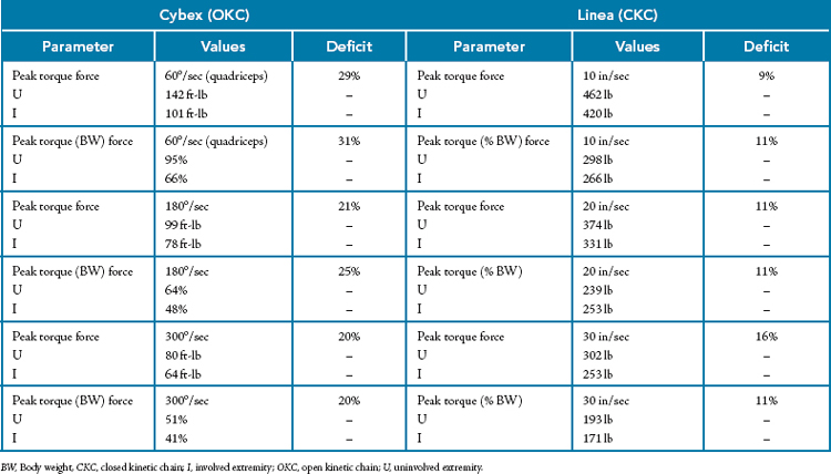
Another example of the need for isolated testing was illustrated in work by Davies,73 who performed CKC computerized isokinetic tests on athletes with various knee injuries and also analyzed bilateral comparison data. Dynamic CKC isokinetic testing, which required a linear motion with force production being measured in pounds at slow (10 in/sec), medium (20 in/sec), and fast (30 in/sec) velocities, was done on a Linea computerized CKC isokinetic dynamometer system.* The same athletes were also tested on a Cybex OKC computerized isokinetic dynamometer,† and bilateral analysis of the data was performed. Isolated joint testing was performed to provide rotational force and torque values, which were recorded at slow (60°/sec), medium (180°/sec), and fast (300°/sec) angular velocities. The results of the testing demonstrated that more significant deficits were shown to exist in athletes after OKC isolated joint and muscle testing than after CKC multiple joint and muscle testing (Table 25-1).
Table 25-1 Comparisons Between Open Kinetic Chain and Closed Kinetic Chain Computerized Isokinetic Dynamometer Testing of 300 Patients With Various Pathologic Knee Conditions or After Surgery
Similar results were reported by Feiring and Ellenbecker84 with isokinetic open and closed chain testing of 23 athletes 15 weeks after ACL reconstruction. Bilateral comparisons of OKC isokinetic knee extension muscle function ranged from 74% to 77% of the uninjured extremity, whereas the results of CKC isokinetic testing using a leg press extension-type movement pattern ranged between 91% and 93% of the uninjured extremity.
When testing multiple muscle groups and developing a composite score of their force, one sees that the proximal and distal muscles apparently compensate for the weak muscles and tend to demonstrate less of a deficit than actually exists in the area. We have made this empirically based observation for years, but now CKC isokinetic testing that objectively documents and quantifies performance has supported this observation. Again, if a muscle’s performance is not measured, a deficit cannot be identified. These research studies and examples provide justification of the need for OKC isokinetic testing.
Another major reason for performing OKC isokinetic testing is the clinical control that it provides. When testing, the clinician controls ROM, speed, translational stress (by shin pad placement), varus and valgus stress, and rotational force. However, when one begins CKC testing, control of these variables decreases, thereby increasing potential risk to the athlete.
An often-cited example is that performance of OKC isokinetic tests on an athlete who has undergone ACL reconstruction can stretch or injure the graft. This is a situation of good science being applied to an inappropriate clinical setting. If the graft were actually to be stretched during OKC testing, the problem is more one of the clinician performing an inappropriate test or testing at an inappropriate time rather than the OKC test itself.85Box 25-5 lists guidelines that should be followed when one is testing or rehabilitating an athlete after ACL reconstruction.86-88
Box 25-5 Guidelines for Testing or Rehabilitation of an Athlete After Anterior Cruciate Ligament Reconstruction
Correlation of Open Kinetic Chain with Closed Kinetic Chain Functional Performance
In addition to obtaining clinical control, another reason to perform OKC isokinetic testing is because of its correlation with CKC functional performance. Although athletes do not regularly function by sitting in a chair and flexing and extending their knees and even though some research indicates that there is no functional correlation,75,89 numerous studies do demonstrate a positive correlation between OKC testing and functional performance.49,64,76-80,90,91
Patel et al90 tested 44 normal subjects and 44 subjects with unilateral ACL deficiency isokinetically to assess knee extension and flexion strength. The group with ACL deficiency had significantly less isokinetic quadriceps strength than the control group did, and this difference in strength was related to a significant decrease in the peak external quadriceps moment during jogging, jog-stop, and jog-cut activities, as well as during stair ascent.90 Isokinetic quadriceps strength was significantly correlated with the external quadriceps moment for these functional activities in both the ACL-deficient and control groups. This study supports the use of isokinetic muscle testing because of the correlation with basic lower extremity functional measures.
Petschnig et al91 demonstrated the relationship between isokinetic strength testing and several lower extremity functional tests. A limb symmetry index of 95% or higher was regularly demonstrated in normal subjects and patients after ACL reconstruction via isokinetic testing, hop tests for distance, and one-legged vertical jump tests. Additionally, Jones et al92 compared isokinetic dynamometry at 60°/sec with functional field tests (seated unilateral leg press, horizontal hop, single-leg vertical, and drop jumps). No significant relationships were identified between the isokinetic variables and the field tests. However, it has previously been demonstrated by Wilk et al8 that testing at slower speeds does not correlate with functional tests whereas faster speeds (> 180°/sec) do in fact correlate with functional hop tests. Moreover, because of the specificity of the angular velocities involved in functional activities, it relates empirically to faster isokinetic testing velocities. Admittedly, isolated joint testing is performed at velocities slower than functional velocities, but most functional movements are really a summation of velocities through the kinematic chain. Therefore, if each link in the kinematic chain were evaluated independently, the velocities would be much slower than the summated force of the entire kinematic chain—hence the reason to perform faster isolated joint testing.
Sbriccoli et al93 investigated the neuromuscular response of the knee extensor and flexor muscles in elite and amateur karateka. Elite karateka had higher lower extremity isokinetic torques than amateurs did. Elite karateka demonstrated a typical neuromuscular activation strategy that seems to be dependent on task and skill level. Furthermore, the results in elite karateka suggested an improved ability to recruit fast motor units as a part of training-induced neuromuscular adaptations.
Specific applications of isokinetic testing in lower extremity rehabilitation
A plethora of research exists that provides both the rationale and objective guidance for the use of isokinetics in the rehabilitation of individuals with specific lower extremity conditions, including ACL reconstruction, patellofemoral pain, hip injury, and knee osteoarthritis (OA). A summary of pertinent research in these areas will provide additional framework for the application of isokinetic testing and training in these patient populations.
Use of Isokinetics to Assist in Prognosis Following Anterior Cruciate Ligament Reconstruction and Injury
Karanikas et al94 investigated the adaptations in walking, running, and muscle strength after ACL reconstruction and examined the interactions between muscle strength and walking and running kinematics. Isokinetics was used for dynamic muscle assessment, and the results demonstrated that adaptation of the motor tasks and muscle strength follows different time patterns. They showed that patients can function normally at submaximal levels; however, with a decrease in muscle strength after ACL reconstruction, documented isokinetically as significant weakness that exceeds a certain threshold in comparison to the uninvolved side, the kinematics of these patients’ locomotion strategies was abnormal.
Oiestad et al95 identified risk factors for knee OA 10 to 15 years after ACL reconstruction. Individuals with low self-reported knee function 2 years postoperatively and loss of quadriceps strength as measured with isokinetics between the 2-year and the 10- to 15-year follow-up had significantly higher odds for symptomatic, radiographically detected knee OA. However, quadriceps muscle weakness, by itself, after ACL reconstruction was not significantly associated with knee OA.
Keays et al96 used isokinetic testing to evaluate 10 factors involved in the development of OA after ACL reconstruction. The incidence of OA after ACL reconstruction is disturbingly high, with reports of mild to moderate OA developing in nearly 50% of patients 6 years after surgery. The five factors found to be predictive of tibiofemoral OA were meniscectomy, chondral damage, patellar tendon grafting, weak quadriceps, and low quadriceps-to-hamstring strength ratios. The quadriceps deficits and unilateral quadriceps-to-hamstring ratios were evaluated by isokinetic testing. Use of hamstring/gracilis grafts and restoration of quadriceps/hamstring strength balance were associated with less OA of the knee.
Segal et al97 used isokinetics to evaluate the effect of quadriceps strength and proprioception on risk for knee OA. The finding that quadriceps strength protected against incident symptomatic but not radiographic knee OA regardless of joint position sense (JPS) suggests that strength may be more important than JPS in mediating risk for knee OA. The clinical implications of these findings are interesting because quadriceps strength is often influenced by rehabilitation interventions. Because of the accommodating resistance afforded by isokinetics, it provides an excellent intervention technique that can be used in patients with OA of the knee.
Hiemstra et al98 used isokinetic testing to demonstrate specific muscle imbalances in a group after ACL reconstruction versus a control group. Angle- and velocity-matched hamstring-quadriceps ratio maps demonstrated systematic variation based on joint angle, velocity, and contraction type for both the control and ACL-reconstructed groups.
Ageberg et al99 used isokinetics as an outcome measure to determine muscle strength in patients with ACL injuries treated by training only or by ACL reconstruction. No differences were observed between the surgical and nonsurgical treatment groups in muscle strength or functional performance. The lack of difference in patients treated by training and surgical reconstruction or by training only indicates that reconstructive surgery is not a prerequisite for restoring muscle function.
Sekir et al100 investigated an early versus late start of isokinetic hamstring-strengthening exercise after ACL reconstruction with a patellar tendon graft. The results of this study demonstrated that hamstring and quadriceps strength can be increased by early hamstring strengthening after ACL reconstruction with no negative impact on knee function.
Stefanska et al101 demonstrated that 13 weeks following ACL reconstruction, patients had significant deficits in peak torque, maximum work, and average power in the injured limb. The deficit exceeded 30% for all measured values on isokinetic testing.
Eitzen et al102 used a variety of outcome measures, including isokinetics, to assess whether an early 5-week exercise therapy program following ACL injury improves function. A progressive 5-week exercise therapy program led to significant improvement in knee function from before to after the program in patients classified as both potential copers and noncopers. The authors concluded that short-term progressive exercise therapy programs are well tolerated and should be incorporated in early-stage ACL rehabilitation, either to improve knee function before ACL reconstruction or as a first step in further nonoperative management.
Isokinetic Testing Related to Patients With Osteoarthritis of the Knee
One of the most important reasons for locomotor dysfunction and disability in patients with knee OA is muscle weakness in the lower extremity. Isokinetics can be used in the treatment and assessment of functional outcomes in these patients very effectively. Important references in this section provide guidance and rationale for the application of isokinetic testing and training in patients with knee OA.
Diracoglu et al103 performed bilateral isokinetic testing for knee flexion/extension. Although manual muscle testing (MMT) produced normal or nearly normal results, significantly lower strength values were found in patients with knee OA with isokinetic testing than in healthy subjects. Muscle strength loss cannot be detected during clinical examination but can be identified during isokinetic measurements. This again demonstrates the accuracy of dynamic isokinetic muscle testing versus static muscle testing as an indicator of muscle performance.
Segal et al97 used isokinetics to evaluate the effect of quadriceps strength and proprioception on risk for knee OA. The finding that quadriceps strength protected against incident symptomatic but not radiographically detected knee OA, regardless of JPS, suggests that strength may be more important than JPS in mediating risk for knee OA. The clinical implications of these findings are interesting because the strength of the quadriceps is often influenced by rehabilitation interventions. Because of the accommodating resistance afforded by isokinetics, it provides an excellent intervention technique that can be used in patients with OA of the knee.
Segal et al104 also performed isokinetic testing of the quadriceps and hamstrings to examine the relationship between quadriceps strength and worsening of knee joint space narrowing over a 30-month period. It was demonstrated that women with the lowest quadriceps strength had increased risk for whole knee joint space narrowing. However, no associations were found between strength and joint space narrowing in men. Consequently, quadriceps weakness was associated with increased risk for tibiofemoral and whole knee joint space narrowing.
Kean et al105 examined test-retest reliability and quantified the minimal detectable change (MDC) in quadriceps strength (using isokinetics and isometrics) and voluntary activation in patients with knee OA. Intraclass correlation coefficients (ICCs) for all measures ranged from 0.93 to 0.98. Based on the standard error of measurement for the isokinetic tests, the MDC was 33.90 Nm for quadriceps strength. Therefore, based on maximal quadriceps isokinetic strength testing, the results demonstrated excellent test-retest reliability in patients with knee OA. The findings suggest that these measures are appropriate for use when evaluating change in neuromuscular function of the quadriceps in individual patients.
Rydevik et al106 compared functioning and disability in patients with hip OA. The patients with hip OA had mild to moderate pain and significantly lower knee extension strength based on isokinetic testing. Their conclusions recommended including quadriceps strengthening and hip ROM exercises when developing rehabilitation programs for patients with hip OA in the aim of improving function and reducing disability.
Isokinetic Testing of the Hip
Because of the positioning, stabilization required, muscle mass, free limb acceleration, and impact artifact caused by the size of the muscle mass, hip testing performed with isokinetics is limited. Julia et al107 demonstrated very good ICCs (values between 0.75 and 0.96) for concentric and eccentric peak torque values of the hip flexors and extensors. Additionally, they demonstrated no differences between the dominant and nondominant sides of the body, which enables use of the contralateral limb as a reference.
Boling et al108 compared the concentric and eccentric torque of the hip musculature in individuals with and without patellofemoral pain syndrome (PFPS). Patients with PFPS displayed weakness in eccentric hip abduction and hip external rotation (ER), which may allow increased hip adduction and internal rotation (IR) during functional movements.
With an increase in the past decade in both early recognition and diagnosis of hip injuries, such as acetabular labral tears and femoroacetabular impingement, it is expected that outcomes research profiling hip strength following important new procedures to treat these injuries in elite athletes and young active patients will result in greater application of both isokinetic testing and training of the hip. Further research and publication of normative data in this region will allow greater application of isokinetics to this important joint.
Specific application of isokinetic assessment in the upper extremity
Application of isokinetic exercise and testing for the upper extremity is imperative because of the demanding muscular work required in sport-specific activities. The large unrestricted ROM of the glenohumeral joint and its limited inherent bony stability necessitate dynamic muscular stabilization to ensure normal joint arthrokinematics.109 Objective information on the intricate balance of agonist and antagonist muscular strength at the glenohumeral joint is a vital resource for rehabilitation and preventive evaluation of the shoulder. Therapeutic exercise and isolated joint testing for the entire upper extremity kinetic chain, including the scapulothoracic joint, are indicated for overuse injury or postoperative rehabilitation of an isolated injury of the shoulder or elbow.110
Rationale for the Use of Isokinetics in Assessing Upper Extremity Strength
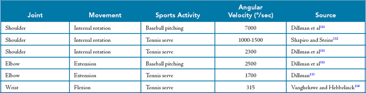
Unlike the lower extremities, in which most functional and sport-specific movements occur in a CKC environment, the upper extremities function almost exclusively in an OKC format.43 The throwing motion, tennis serve, and ground stroke are all examples of OKC activities for the upper extremity. OKC muscular strength assessment methodology allows the isolation of particular muscle groups, as opposed to CKC methods, which use multiple joint axes, planes, and joint and muscle segments. Traditional isokinetic upper extremity test patterns are open chain with respect to the shoulder, elbow, and wrist. The velocity spectrum (1°/sec to approximately 1000°/sec) currently available on commercial isokinetic dynamometers provides specificity for testing the upper extremity by allowing the clinician to assess muscular strength at faster, more functional speeds. Table 25-2 lists the angular velocities of sport-specific upper extremity movements.111-114
The dynamic nature of upper extremity movements is a critical factor in directing the clinician to the optimal testing methodology for the upper extremity. MMT provides a static alternative for assessment of muscular strength that uses well-developed patient positions and stabilization.68,115 Despite detailed descriptions of manual assessment techniques, the reliability of MMT is compromised because of differences in the size and strength of clinicians and patients and the subjective nature of the grading system.116
Ellenbecker117 compared isokinetic testing of the shoulder internal and external rotators with MMT in 54 subjects who exhibited symmetric normal grade (5/5) strength by manual assessment. With isokinetic testing, 13% to 15% bilateral differences in ER and 15% to 28% bilateral differences in IR were found. Of particular significance was the large variability in the size of this mean difference between extremities despite the presence of bilateral symmetry on MMT. The use of MMT is an integral part of a musculoskeletal evaluation. MMT provides a time-efficient, gross screening of the strength of multiple muscles by using a static, isometric muscular contraction, particularly in patients with neuromuscular disease or in athletes with large deficits in muscular strength.116,118 The limitations of MMT appear to be most evident in instances in which only minor impairment of strength is present, as well as in the identification of subtle isolated deficits in strength. Differentiation of agonist and antagonist muscular strength balance is also complicated when manual techniques are used rather than an isokinetic apparatus.117
Glenohumeral Joint Testing
Dynamic assessment of the strength of the rotator cuff musculature is of primary importance in rehabilitation and preventive screening of the glenohumeral joint. The rotator cuff forms an integral component of the force couple in the shoulder, as described by Inman et al.119 The approximating role of the supraspinatus muscle for the glenohumeral joint, as well as the inferior (caudal) glide component action provided by the infraspinatus, teres minor, and subscapularis muscles, must stabilize the humeral head within the glenoid cavity against the superiorly directed force exerted by the deltoid muscle with humeral elevation.120 Muscular imbalances, primarily in the posterior rotator cuff, have been objectively documented in athletes with glenohumeral joint instability and impingement.121
Shoulder Internal Rotation and External Rotation Strength Testing
Initial testing and training using isokinetics for rehabilitation of the shoulder typically involve the modified base position. The modified base position is obtained by tilting the dynamometer approximately 30° from the horizontal base position.1,7 This causes the shoulder to be placed in approximately 30° of abduction (Fig. 25-1). The modified base position places the shoulder in the scapular plane 30° anterior to the coronal plane.122 The scapular plane is characterized by enhanced bony congruity and a neutral glenohumeral position that results in a midrange position for the capsular ligaments and scapulohumeral musculature.122 This position does not place the suprahumeral structures in an impingement situation and is well tolerated by athletes.1
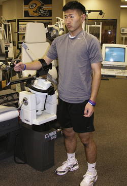
Figure 25-1 Modified neutral position for isokinetic testing of shoulder internal or external rotation.
Isokinetic testing using the modified base position requires consistent testing of the athlete on the dynamometer. Studies have demonstrated significant differences in IR and ER strength with varying degrees of abduction, flexion, and horizontal abduction and adduction of the glenohumeral joint.123-125 The modified base position requires the athlete to be standing, which compromises both isolation and test-retest reliability. Despite these limitations, valuable data can be obtained early in the rehabilitative process with this neutral, modified base position.7,126,127
Isokinetic assessment of IR and ER strength is also done with 90° of glenohumeral joint abduction. Specific advantages of this test position are greater stabilization in either a seated or supine test position on most dynamometers and placement of the shoulder in an abduction angle corresponding to the overhead throwing position used in sports activities.111,128 As a precursor to using the 90° abducted position, we require initial tolerance of the athlete to the modified base position; 90° abducted isokinetic testing can be performed in either the coronal or the scapular plane. The benefits of use of the scapular plane are similar to those discussed for the modified position and include protection of the anterior capsular glenohumeral ligaments and theoretic enhancement of the length-tension relationship of the posterior rotator cuff.123,129,130 Changes in the length-tension relationship and in the line of action of the scapulohumeral and axiohumeral musculature are reported with 90° of glenohumeral joint abduction instead of a more neutral adducted glenohumeral joint position.1 The 90° abducted position for isokinetic strength assessment is more specific for assessing the muscular functions required for overhead activities.131
Heavy emphasis is placed on assessing the IR and ER strength of the shoulder during rehabilitation. The rationale for this apparently narrow focus is provided by an isokinetic training study by Quincy et al.81 Six weeks of isokinetic training of the internal and external rotators produced statistically significant improvements not only in IR and ER strength but also in flexion-extension and abduction-adduction strength. Isokinetic training for flexion-extension and for abduction-adduction produced improvements only in the position of training. The physiologic overflow of strength caused by training the internal and external rotators provides a rationale for the heavy emphasis on strength development and assessment in rehabilitation. Additional research has identified the IR and ER movement pattern as the preferred testing pattern in athletes with rotator cuff tendinopathy.132
Interpretation of Shoulder Internal and External Rotation Testing
Bilateral differences
As with isokinetic testing of the lower extremities, assessment of the strength of an extremity relative to the contralateral side forms the basis for standard data interpretation. This practice is more complicated in the upper extremities because of limb dominance, particularly in athletes in unilaterally dominant sports. In addition to the complexity caused by limb dominance, isokinetic descriptive studies demonstrate disparities in the degree of limb dominance, as well as in strength dominance, only in specified muscle groups.26,133-139
In general, maximum limb dominance of the internal and external rotators of 5% to 10% is assumed in nonathletic persons and athletes engaging in recreational upper extremity sports.140 Significantly greater IR strength has been identified in the dominant arm in professional,137,141 collegiate,26 and high school139 baseball players, as well as in elite junior136 and adult135 tennis players. No difference between extremities has been demonstrated in concentric ER in professional129,142 and collegiate26 baseball pitchers or in elite junior134,136 and adult135 tennis players. This selective strength development in the internal rotators produces significant changes in agonist-antagonist muscular balance. Identification of such selectivity with isokinetic testing has implications for rehabilitation and prevention of injuries.
Use of normative data
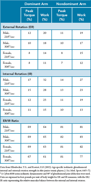
Normative or descriptive data can assist clinicians in further analyzing isokinetic test data. Care must be taken to use normative data that are both population and apparatus specific.11Tables 25-3 to 25-5 present data using two dynamometer systems from large samples of specific athletic populations. Data are presented with body weight used as the normalizing factor.
Table 25-3 Isokinetic Peak Torque–to–Body Weight Ratios in 150 Professional Baseball Pitchers*
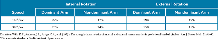
Table 25-4 Isokinetic Peak Torque–to–Body Weight and Work–to–Body Weight Ratios in 147 Professional Baseball Pitchers*
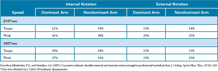
Table 25-5 Isokinetic Peak Torque–to–Body Weight Ratios, Single-Repetition Work-to–Body Weight Ratios, and External Rotation–to–Internal Rotation Ratios in Elite Junior Tennis Players*
Unilateral strength ratios (agonist to antagonist)
Assessment of the balance in muscular strength of the internal and external rotators is of vital importance when one interprets upper extremity strength tests. Alteration of this ER-to-IR ratio has been reported in athletes with glenohumeral joint instability and impingement.109 The initial description of the ER-to-IR ratio for normal female subjects was published by Ivey et al143 and confirmed by Davies1 in both men and women. An ER-to-IR ratio of 66% is the target in normal subjects. Biasing this ratio in favor of the external rotators has been advocated by clinicians,68,127,144 both for preventing injury in throwing and racquet sport athletes and after injury to or surgery on the glenohumeral joint.
Reports of alteration in the ER-to-IR ratio as a result of selective muscular development of the internal rotators without concomitant ER strength are widespread in the literature.26,135-139 This alteration has provided clinicians with an objective rationale for the global recommendation of preventive posterior rotator cuff ER-strengthening programs in athletes performing high-level overhead activities.127,144 Examples of ER-to-IR ratios in specific athletic populations and with specific apparatus are presented in Tables 25-5 and 25-6.
Table 25-6 Unilateral External Rotation–Internal Rotation Ratios in Professional Baseball Pitchers
| Speed | Dominant Arm | Nondominant Arm |
|---|---|---|
| 180°/sec | ||
| Torque | 65 | 64 |
| 300°/sec | ||
| Torque | 61 | 70 |
| 210°/sec | ||
| Torque | 64 | 74 |
| Work | 61 | 66 |
| 300°/sec | ||
| Torque | 65 | 72 |
| Work | 62 | 70 |
Data from Wilk, K.E., Andrews, J.R., Arrigo, C.A., et al. (1993): The strength characteristics of internal and external rotator muscles in professional baseball pitchers. Am. J. Sports Med., 21:61–66; and Ellenbecker, T.S., and Mattalino, A.J. (1997): Concentric isokinetic shoulder internal and external rotation strength in professional baseball pitchers. J. Orthop. Sports Phys. Ther., 25:323–328.
Additional Glenohumeral Joint Testing Positions
Shoulder Abduction and Adduction
Shoulder abduction-adduction strength is an additional pattern frequently evaluated isokinetically because of the key role of the abductors in the Inman force couple119 and the functional relationship of the adductors to throwing velocity.29 Specific factors important in this testing pattern are the limitation of ROM to approximately 120° to avoid glenohumeral joint impingement and the consistent use of gravity correction.145
Interpretation of abduction-adduction isokinetic test results involves traditional bilateral comparison, normative data comparison, and unilateral strength ratios. Ivey et al,143 in testing normal adult women, reported ratios of 50% bilaterally. Similar findings were reported by Alderink and Kluck133 in high school and collegiate baseball pitchers. Wilk et al146,147 reported dominant arm abduction-to-adduction ratios of 85% to 95% with a Biodex dynamometer.* They used a windowing technique that removed impact artifacts from the data after free limb acceleration and end-stop impact. Upper extremity testing using long input adapters and fast isokinetic testing velocities can produce a torque artifact that significantly changes the isokinetic test result. Wilk et al147 recommended windowing the data by excluding all data obtained at velocities outside 95% of the present angular testing velocity. (Because of free limb acceleration and deceleration, only a portion of the entire ROM is truly isokinetic. If the velocities differ from the actual test speed by 5% or more, the data are not valid isokinetic data and should not be used.)
Shoulder Flexion-Extension and Horizontal Abduction-Adduction
Additional isokinetic patterns used to obtain a more detailed profile of shoulder function are flexion-extension and horizontal abduction-adduction. Both motions are generally tested in a less functional supine position to improve stabilization. Normative data for these testing positions are less prevalent in the literature. Flexion-to-extension ratios reported for normal subjects by Ivey et al143 are 80% (4:5). Ratios for athletes with shoulder extension–dominant activities are reported to be 50% for baseball pitchers123 and 75% to 80% for highly skilled adult tennis players.135 Normative data need to be developed further to define strength more clearly in these upper extremity patterns. Body position and gravity compensation are, again, key factors affecting proper interpretation of the data.
Scapulothoracic Testing (Protraction-Retraction)
In addition to the supraspinatus-deltoid force couple, the serratus anterior–trapezius force couple is of critical importance for thorough evaluation of upper extremity strength. Gross MMT and screening that attempt to identify scapular winging are commonly used in clinical evaluation of the shoulder complex. Davies and Hoffman74 published normative data on 250 shoulders for isokinetic protraction-retraction testing. An approximately 1:1 relationship of protraction-retraction strength was reported. Testing and training the serratus anterior, trapezius, and rhomboid musculature enhance scapular stabilization and strengthen the primary musculature involved in the scapulohumeral rhythm. Emphasis on promotion of proximal stability to enhance distal mobility is a concept used and recognized by nearly all disciplines of rehabilitative medicine.83
Concentric Versus Eccentric Considerations
The availability of eccentric dynamic strength assessment has made a significant impact, primarily in research investigations. Extrapolation of research-oriented isokinetic principles to patient populations has been a gradual process. Eccentric testing of the upper extremity is clearly indicated because of the prevalence of functionally specific eccentric work. Maximal eccentric functional contractions of the posterior rotator cuff during the follow-through phase of the throwing motion and tennis serve provide a rationale for eccentric testing and training in rehabilitation and preventive conditioning.148 Kennedy et al149 found mode-specific differences between the concentric and eccentric strength characteristics of the rotator cuff. Saccol et al150 evaluated shoulder ER and IR strength variables in concentric and eccentric modes in elite junior tennis players. To determine the peak torque–functional ratio, the eccentric strength of ER and the concentric strength of IR were calculated. Elite junior tennis players without shoulder injuries have imbalances in muscle strength during shoulder rotation that alter the normal functional ratio between rotator cuff muscles. This is probably a normal adaptive physiologic response caused by the specificity of training at a high level of performance. Further research on eccentric muscular training is necessary before widespread use of eccentric isokinetics can be applied to patient populations.
The basic characteristics of eccentric isokinetic testing, such as greater force production than with concentric contractions at the same velocity, have been reported for the internal and external rotators.151-153 This enhanced force generation is generally explained by the contribution of the series elastic (noncontractile) elements of the muscle-tendon unit in eccentric conditions. An increase in postexercise muscle soreness, particularly of latent onset, is a common occurrence after periods of eccentric work. Therefore, eccentric testing would not be the mode of choice during the early inflammatory stages of an overuse injury.110 Many clinicians recommend the use of dynamic concentric testing before they perform an eccentric test. Both concentric and eccentric isokinetic training of the rotator cuff has produced objective improvements in concentric and eccentric strength in elite tennis players.152,153
Relationship of isokinetic testing to functional performance
Dynamic muscular strength assessment is used to evaluate the underlying strength and balance of strength in specific muscle groups. This information is used to determine the specific anatomic structure that requires strengthening, as well as to demonstrate the efficacy of treatment procedures. Isokinetic testing of the shoulder internal and external rotators has been used as one parameter for demonstrating the functional outcome after rotator cuff repair in select patient populations.154-157
Bigoni et al158 used isokinetics as an outcome measure to determine recovery of strength after rotator cuff tears treated with two different arthroscopic repair techniques. Isokinetic strength testing demonstrated a difference between the two repairs and can therefore be used as a measure to assess the efficacy of surgical procedures. Oh et al159 evaluated patients with an isokinetic dynamometer following rotator cuff repair. Isokinetic muscle performance testing is a validated and objective method for evaluating muscle function, but it is presently unknown whether it correlates with the severity of rotator cuff tears. Oh et al159 demonstrated a correlation between isokinetic testing and preoperative isokinetic muscle performance parameters. The isokinetic muscle performance testing deficit was greater in shoulders with larger rotator cuff tears and greater degrees of fatty degeneration/infiltration. Isokinetic muscle performance testing provides objective and quantitative data for estimating the preoperative status of rotator cuff tears and can provide baseline data for postoperative anatomic assessment in patients with rotator cuff disorders.
An additional purpose for isokinetic testing is to determine the relationship of muscular strength to functional performance. Several investigators have tested upper extremity muscle groups and correlated their respective levels of strength with sport-specific functional tests. Pedegana et al160 found a statistical relationship between elbow extension, wrist flexion, shoulder extension-flexion, and ER strength measured isokinetically and throwing speed in professional pitchers. In a similar study, Bartlett et al161 found that shoulder adduction correlated with throwing speed. These studies are in contrast to those of Pawlowski and Perrin,162 who did not find a significant relationship in throwing velocity.
Andrade-Mdos et al163 established an isokinetic profile of shoulder rotator muscle strength in female handball players. Concentric balance and functional balance ratios did not differ between sides at slower angular velocities, but at faster angular velocities the functional balance ratio in the dominant limb was lower than that on the nondominant side. The results suggest that concentric strength exercises should be used for the internal and external rotators on the nondominant side and that functional exercise should be used to improve eccentric rotation strength for prevention programs.
Edouard et al164 did not find any significant postoperative correlations between shoulder function (as judged by the Rowe and Walch-Duplay scores) and IR or ER muscle strength. However, it is necessary to objectively measure recovery of rotator cuff strength to adequately strengthen the rotator cuff muscles before resumption of sports activities. Isokinetic strength assessment may thus be a valuable decision support tool for resumption of sports activities and would complement the functional scores studied in this study.
Additionally, Mandalidis et al56 evaluated the relationship between handgrip isometric strength and isokinetic strength of the shoulder musculature. A positive relationship was found between handgrip isometric strength and isokinetic strength of the shoulder stabilizers. The results of the present findings suggest that handgrip isometric strength can be used to monitor the isokinetic strength of certain muscle groups contributing to the stability of the shoulder joint. However, handgrip strength may account for only approximately 16% to 50% of the variability in isokinetic strength of these muscle groups.
Several studies have examined the relationship between isokinetic strength and the tennis serve in elite players. Ellenbecker et al152 determined that 6 weeks of concentric isokinetic training of the rotator cuff resulted in a statistically significant improvement in serving velocity in collegiate tennis players. In a similar study, Mont et al153 found improvements in serving velocity after both concentric and eccentric IR and ER training. A direct statistical relationship between isokinetically measured upper extremity strength and tennis serve velocity was not obtained by Ellenbecker135 despite earlier studies showing increases in serving velocity after isokinetic training. The complex biomechanical sequence of segmental velocities and the interrelationship of the kinetic chain link with the lower extremities and trunk make delineation and identification of a direct relationship between an isolated structure and a complex functional activity difficult.
Finally, from a distal upper extremity strength perspective, Lin et al165 used an isokinetic dynamometer to assess dominant arm (elbow) flexor and extensor concentric and eccentric strength. Based on the results of isokinetic tests, regression analysis revealed that a ratio of biceps concentric to triceps concentric strength greater than 0.76 significantly predicted elbow injury. No other ratios or variables were predictive of injury status. Assessment of this ratio may prove useful in a practical setting for training purposes and for both diagnosis and rehabilitation of injury.
Isokinetic testing can provide a reliable, dynamic measurement of isolated joint motions and muscular contributions to assist the clinician in the assessment of underlying muscular strength and strength balance. Integration of isokinetic testing with a thorough, objective clinical evaluation allows the clinician to provide optimal rehabilitation after both overuse injuries and surgery.
Application of isokinetics in designing rehabilitation programs
Many types of exercise programs are in widespread use for rehabilitating injured athletes. This section focuses on resistive rehabilitation programs, as well as on the specific progression of resistive exercise recommended during rehabilitation. Resistive rehabilitation programs vary from isometric, concentric, and eccentric isotonics to concentric and eccentric isokinetics to isoacceleration and isodeceleration programs. The scientific and clinical rationale for progression through a resistive exercise rehabilitation program is described, including the specific progression and inclusion of isokinetic exercise in the clinical rehabilitation of upper extremity overuse injuries.
Patient Progression Criteria
Progression through the resistive exercise program is predicated on several important concepts, including athlete status, signs and symptoms, time after surgery, and soft tissue–healing constraints. The athlete’s progression through the various levels of the resistive exercise program is determined by continual charting and assessment of subjective and objective evaluation criteria (Table 25-7). This resistive exercise progression continuum7 is based on the concept of a trial treatment. If any adverse changes occur, the rehabilitation program continues at the previous level of intensity of repetitions, sets, or duration without the athlete progressing to the next level of the exercise progression continuum.
Table 25-7 Commonly Used Subjective and Objective Criteria for Patient Progression in a Rehabilitation Program
| Subjective Criteria (Symptoms) | Objective Criteria (Signs) |
|---|---|
| Pain | Anthropometric measurements |
| Stiffness | Goniometric measurements |
| Changes in function | Palpable changes in cutaneous temperature Redness Manual muscle testing Isokinetic testing Kinesthetic testing Functional performance testing KT1000 testing |
From Davies, G.J. (1992): A Compendium of Isokinetics in Clinical Usage and Rehabilitation Techniques, 4th ed. Onalaska, WI, S & S Publishers.
If, however, an athlete performs the trial treatment without any negative effects, the athlete progresses gradually to the next higher level in the exercise continuum. An athlete may enter the exercise rehabilitation continuum at any stage, depending on the results of the initial evaluation. Furthermore, an athlete may also progress through several stages from one treatment session to the next, depending primarily on the response to treatment. Before the athlete begins the actual resistive exercise portion of the rehabilitation program, various warm-up exercises and mobilization stretching exercises are appropriate.
Resistive Exercise Progression Continuum
The rehabilitation program is designed along a progression continuum. The program begins with the safest exercises and progresses to more stressful exercises. These are illustrated in Figure 25-2 and Table 25-8.
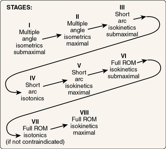
Figure 25-2 Stages of Davies’ resistive exercise progression continuum. ROM, Range of motion.
(From Davies, G.J. [1992]: A Compendium of Isokinetics in Clinical Usage and Rehabilitation Techniques, 4th ed. Onalaska, WI, S & S Publishers.)
Table 25-8 Davies’ Resistive Exercise Progression Continuum
| Exercise Effort (%) | Exercise Program |
|---|---|
| 100 | Submaximal multiple-angle isometrics SOAP TT of maximal multiple-angle isometrics |
| 50/50 | Submaximal multiple-angle isometrics + maximal multiple-angle isometrics SOAP |
| 100 | Maximal multiple-angle isometrics SOAP TT of submaximal short-arc isokinetics |
| 50/50 | Maximal multiple-angle isometrics + submaximal short-arc isokinetics SOAP |
| 100 | Submaximal short-arc isokinetics SOAP TT of maximal short-arc isokinetics or short-arc isotonics |
| 50/50 | Submaximal short-arc isokinetics + maximal short-arc isokinetics SOAP |
| 100 | Maximal short-arc isokinetics SOAP TT of submaximal full ROM isokinetics |
| 50/50 | Maximal short-arc isokinetics + submaximal full ROM isokinetics SOAP |
| 100 | Submaximal full ROM isokinetics SOAP TT of maximal full ROM isokinetics SOAP (full ROM isotonics here if not contraindicated) |
| 50/50 | Submaximal full ROM isokinetics + maximal full ROM isokinetics SOAP |
| 100 | Maximal full ROM isokinetics SOAP |
ROM, Range of motion; SOAP, subjective-objective assessment and plan; TT, trial treatment.
From Davies, G.J. (1992): A Compendium of Isokinetics in Clinical Usage and Rehabilitation Techniques, 4th ed. Onalaska, WI, S & S Publishers.
Multiple-Angle Isometrics
The exercise rehabilitation program typically begins with multiple-angle isometrics performed at a submaximal intensity level. The isometrics are performed approximately every 20° through the ROM that is indicated, based on the safe and comfortable ROM demonstrated during examination of the athlete. The rationale for using this particular exercise is the presence of a 20° physiologic overflow with the application of isometrics7 (Fig. 25-3). Therefore, as an example (Fig. 25-4), if the athlete has a painful arc syndrome, which is common in a shoulder with a pathologic rotator cuff condition, isometrics can be applied every 20° throughout the ROM, and the athlete will still obtain a concomitant strengthening effect throughout the entire ROM without increasing the symptomatic area. The painful arc that is typical in athletes with pathologic rotator cuff conditions occurs between 85° and 135° of elevation, at which point peak forces against the undersurface of the acromion occur.166 Performing isometric exercise around the painful arc during the rehabilitation process is a prime example of applying isometrics early in rehabilitation of the shoulder after an overuse injury or surgery.
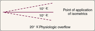
Figure 25-3 Isometric exercises and physiologic overflow through the range of motion.
(From Davies, G.J. [1992]: A Compendium of Isokinetics in Clinical Usage and Rehabilitation Techniques, 4th ed. Onalaska, WI, S & S Publishers.)
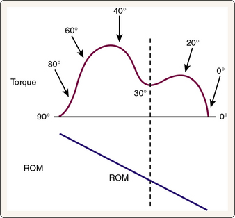
Figure 25-4 Application of isometric exercises through the range of motion (ROM) with a “painful” deformation. Isometrics are applied every 20° through the ROM. Note particularly the application of isometrics on each side of the “painful” deformation.
(From Davies, G.J. [1992]: A Compendium of Isokinetics in Clinical Usage and Rehabilitation Techniques, 4th ed. Onalaska, WI, S & S Publishers.)
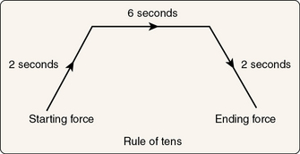
The next consideration with isometric exercise is that the athlete use the rule of 10s: 10-second contractions, 10-second rest, 10 repetitions, and so on. The athlete is usually taught to perform the isometrics in the following sequence: (1) take 2 seconds to gradually build up the desired tension, whether working at a submaximal or maximal intensity level; (2) hold the desired tension of the isometric contraction for 6 seconds, which is the optimal duration for an isometric contraction167; and (3) gradually relax and release tension in the muscle over the last 2 seconds (Fig. 25-5). This sequence allows controlled buildup and easing of the contraction with an optimal 6-second isometric contraction.
Gradient Increase and Decrease in Force Production
Gradient increase and decrease in muscle force production are concepts that athletes have taught us over the years. As an example, if an athlete has effusion or pain in a joint and performs a muscle contraction, pain is often induced. It is usually the result of capsular distention from the internal pressure of the effusion. The submaximal muscle contraction places external pressure on the capsule, which is highly innervated,168 and subsequently increases the pain. However, with a gradient increase in muscle contraction to the desired intensity (submaximal or maximal), an accommodation is often created that either eliminates or minimizes the pain. At completion of the 6-second isometric contraction, a gradient decrease in muscle contraction is performed. Again, when an effusion is present and the athlete suddenly releases the contraction, pain results. This is perhaps due to a rebound type of phenomenon in which effusion in the joint pushes the capsule out and the muscular contraction that was pushing in against the capsule and compressing it causes an “equalizing”; of the pressure. At release of the muscular contraction, the external pressure is relieved; therefore, the internal pressure causes a rebound phenomenon in which the capsule is stretched and discomfort results. If the athlete gradually releases the muscle contraction and some type of accommodation occurs, the pain is either eliminated or minimized.
Determining Submaximal Exercise Intensity
Submaximal exercise intensity can be distinguished from maximal exercise intensity in various ways. If a submaximal exercise is being performed, intensity can be determined by using symptom-limited submaximal exercises (exercises performed at less than maximal effort that do not cause pain) or a musculoskeletal rating of perceived exertion for submaximal effort. Furthermore, distinction must be made between good and bad pain after exercise. Good pain refers to the transient acute pain after an exercise bout that is due to accumulation of lactic acid, changes in pH in the muscle, and an ischemic response. However, bad pain is pain that occurs at the site of the actual injury or at the muscle-tendon unit of injury. An example of this pain classification used in shoulder rehabilitation would be posteriorly oriented discomfort or pain over the infraspinous fossa after an ER exercise (good pain) versus anteriorly directed pain over the greater tuberosity or tendon of the long head of the biceps (bad pain).
Guidelines for Pain During Exercise
The following are guidelines that we use during the rehabilitation program: (1) if no pain is present at the start of an exercise bout but develops after the exercise, that particular exercise is stopped, and modifications are made in the exercise; (2) if pain is present at the start of the exercise and the pain increases, that exercise is terminated; and (3) if pain is present at the start of an exercise and the pain plateaus, the athlete continues the exercise program.
Trial Treatment
When a rehabilitation program includes progression of the athlete through a resistive exercise continuum, a key element is how to determine the progression from one stage to the next in the continuum. One of the keys to this progression is the use of a trial treatment. A trial treatment essentially consists of the athlete performing one set from the next stage in the exercise progression continuum (see Fig. 25-2). After the athlete completes the exercise program at one level of the exercise progression continuum, a trial of the next stage of treatment is performed. The athlete’s signs and symptoms are then evaluated at the conclusion of that particular treatment session, as well as at the next scheduled visit, at which time the athlete’s condition is reevaluated and a decision made on the basis of the athlete’s signs and symptoms. If they have stayed the same or improved, the athlete can progress to the next level of exercise because the trial treatment has demonstrated that the athlete’s muscle-tendon unit or joint is ready for the higher exercise intensity. Any negative sequelae such as increased pain or effusion in the joint are an indication that the joint or muscle is not ready for progression, and consequently, the athlete continues to work at the same level of intensity. Further physical therapy is performed to decrease the irritability of the joint or muscle-tendon unit, and during the next visit, a trial treatment is once again attempted to determine whether the athlete’s injury can tolerate the progression.
Submaximal Exercise: Fiber Recruitment
Several exercise modes can be performed at a submaximal level to enhance selective fiber recruitment. Preferential muscle fiber recruitment is predicated on the intensity of the muscle contraction to recruit either slow-twitch or fast-twitch A or fast-twitch B fibers. It is generally accepted that during voluntary contractions of human muscle there is an orderly recruitment of motor units according to the size principle.169 In mixed muscle containing both slow-twitch and fast-twitch fibers, this implies that involvement of slow-twitch fibers is obligatory, regardless of the power and velocity being generated, with fast-twitch A and fast-twitch B muscle fibers being recruited once higher intensities are generated.170Figure 25-6 summarizes this preferential muscular recruitment. Slow-twitch motor units have relatively low contraction velocities and long contraction times that require only low levels of stimulus to contract. In contrast, fast-twitch motor units require a very high intensity stimulus to contract and have very short contraction times. The preferential recruitment of muscle fibers is an important concept for the clinician to understand with regard to the manipulation of submaximal and maximal exercise intensities in rehabilitative exercise. Submaximal exercise can stimulate the slow-twitch muscle fibers and allow athletes to exercise at lower, pain-free intensities early in the rehabilitation process, with progression to higher exercise intensities that preferentially stimulate the fast-twitch fibers occurring later in rehabilitation.
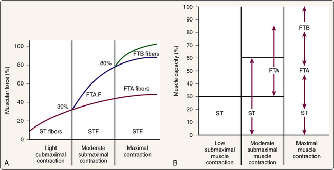
Figure 25-6 A and B, Preferential muscle fiber recruitment is predicated on the intensity of the muscle contraction. FTA, Fast-twitch A; FTB, fast-twitch B; ST, slow-twitch.
(From Davies, G.J. [1992]: A Compendium of Isokinetics in Clinical Usage and Rehabilitation Techniques, 4th ed. Onalaska, WI, S & S Publishers.)
Short-Arc Exercises
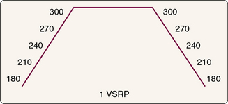
The athlete next progresses from static isometric exercises to more dynamic exercises. The dynamic exercises start with short-arc exercises and the ROM within symptom and soft tissue–healing constraints. Short-arc exercises are often started with submaximal isokinetics (Fig. 25-7) because the accommodating resistance inherent in submaximal isokinetic exercise makes it safe for the athlete’s healing tissues. With short-arc isokinetics, speeds ranging from 60 to 180°/sec are used (Fig. 25-8). The athlete works with what is called a velocity spectrum rehabilitation protocol (VSRP). When the athlete is performing short-arc isokinetics, slower contractile velocities (60 to 180°/sec) are chosen because of the acceleration and deceleration response (Fig. 25-9). Isokinetic exercise contains three major components, as identified in Figure 25-9: acceleration, deceleration, and load range. Acceleration is the portion of the ROM in which the athlete’s limb is accelerating to “catch” the preset angular velocity, deceleration is the portion of the ROM in which the athlete’s limb is slowing before cessation of that repetition, and the load range is the actual portion of the ROM in which the preset angular velocity is met by the athlete and a true isokinetic load is imparted to the athlete. Load range is inversely related to isokinetic speed. A larger load range is found at slower contractile velocities, and a statistically shorter load range occurs at faster contractile velocities.171
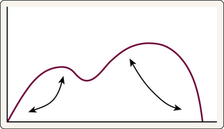
Figure 25-7 Short-arc isokinetic exercises being applied at different points in the range of motion. If an isokinetic torque curve has a deformity in the range of motion as illustrated, short-arc isokinetic exercises can be applied to each side of the deformity.
(From Davies, G.J. [1992]: A Compendium of Isokinetics in Clinical Usage and Rehabilitation Techniques, 4th ed. Onalaska, WI, S & S Publishers.)
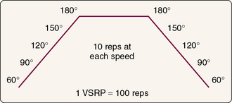
Figure 25-8 Short-arc isokinetic velocity spectrum rehabilitation protocol (VSRP) performed at intermediate contractile velocities. reps, Repetitions.
(From Davies, G.J. [1992]: A Compendium of Isokinetics in Clinical Usage and Rehabilitation Techniques, 4th ed. Onalaska, WI, S & S Publishers.)
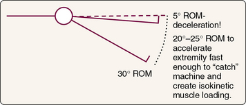
Figure 25-9 Acceleration and deceleration range of motion (ROM) with short-arc isokinetic exercise.
(From Davies, G.J. [1992]: A Compendium of Isokinetics in Clinical Usage and Rehabilitation Techniques, 4th ed. Onalaska, WI, S & S Publishers.)
Further support for short-arc or limited ROM exercise comes from research by Clark et al.172 These authors determined the influence of variable range of motion (VROM) training on neuromuscular performance and control of external loads. Subjects trained with either full ROM or partial ROM exercises. The partial ROM exercises demonstrated significant increases in several of the outcome measures, including isokinetic testing in terminal ROM. Analysis of the force-ROM relationship revealed that the VROM intervention enhanced performance at shorter muscle lengths. These findings suggest that VROM training improves gains in terminal and midrange performance, with the result that the athlete has improved ability to control external loading and produce dynamic force.
Consequently, the athlete’s available ROM must be evaluated to determine the optimal ROM for exercise. With short-arc isokinetic exercise, there is a physiologic overflow of approximately 30° throughout the ROM (Fig. 25-10). Therefore, when an athlete with a pathologic rotator cuff condition is exercising, an abbreviated ROM in IR-ER can be used in the pain-free range, with overflow into the painful ROM, without actually placing the injured structures into that movement range. Another example of isokinetic exercise for the upper extremities is limitation of external ROM to 90° during isokinetic training, even though the demands on the athletic shoulder in overhead activities exceed the 90° ER. Limiting ER to 90° protects the anterior capsular structures of the shoulder, with physiologic overflow improving strength at ranges of ER exceeded during training.
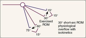
Figure 25-10 Thirty-degree short-arc range of motion (ROM) overflow with isokinetics.
(From Davies, G.J. [1992]: A Compendium of Isokinetics in Clinical Usage and Rehabilitation Techniques, 4th ed. Onalaska, WI, S & S Publishers.)
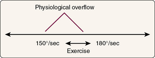
In addition to ROM, the speed selected with isokinetic exercise is also of vital importance in a VSRP. The speeds in the protocol are designed so that the athlete will exercise at 30°/sec through the velocity spectrum. The reason for using an interval of 30°/sec in the velocity spectrum is the physiologic overflow with respect to speed that has been identified in isokinetic research (Fig. 25-11).173-175
Rest Intervals
When the athlete is performing either submaximal or maximal short-arc isokinetics in a VSRP, the rest interval between each set of 10 training repetitions may be as long as 90 seconds.89 However, this is not a viable clinical rest time because it takes too much time to complete the exercise session. Consequently, rest intervals are often applied on a symptom-limited basis. If the athlete does complete a total VSRP, a rest period of 3 minutes after completion of the VSRP has been shown to be an effective rest interval176 (Fig. 25-12). Additional research has provided guidance for selection of rest intervals after isotonic and isokinetic exercise in rehabilitation. According to Fleck,177 50% of the adenosine triphosphate and creatine phosphate is restored in 20 seconds after an acute bout of muscular work. Seventy-five percent and 87% of intramuscular stores are replenished in 40 and 60 seconds, respectively. Knowledge of the phosphagen replenishment schedule allows clinicians to make scientifically based decisions on the amount of rest needed or desired after periods of muscular work. Another factor in determining optimal rest intervals with isotonic and isokinetic training is specificity. For example, during rehabilitation of the shoulder of a tennis player, a high-repetition format is used to improve local muscular endurance. Rest cycles are limited to 25 to 30 seconds because that is the time allotted during tennis play for rest between points. Applying activity or sport-specific muscular work rest cycles is an important consideration during rehabilitation.
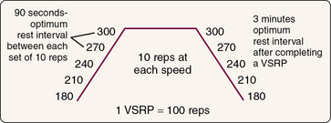
Figure 25-12 Optimum rest intervals. reps, Repetitions; VSRP, velocity spectrum rehabilitation protocol.
(From Davies, G.J. [1992]: A Compendium of Isokinetics in Clinical Usage and Rehabilitation Techniques, 4th ed. Onalaska, WI, S & S Publishers.)
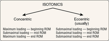
When isotonic exercises are performed, they are implemented between isokinetic submaximal and maximal exercises (see Fig. 25-2). The reason is that isotonic muscle loading loads a muscle only at its weakest point in the ROM. Figure 25-13 demonstrates the effects of isotonic muscle loading through the ROM. Consequently, when isotonic muscle exercise is performed through the ROM, a combination of maximal and submaximal loading occurs, whereas with isokinetics, submaximal exercises can be performed throughout the ROM, and loading of the muscle is maximal in intensity throughout the ROM because of the accommodating resistance phenomena inherent in isokinetic exercise.
Full Range-of-Motion Exercises
The athlete next progresses to full ROM isokinetic exercise beginning with submaximal exercise and then progressing to maximal intensity (Fig. 25-14). Straight planar movements are used initially to protect the injured plane of movement. Faster contractile velocities are also used from 180° up to the maximum capabilities of the isokinetic dynamometer. Numerous reasons have been proposed for using faster isokinetic speeds: physiologic overflow to slower speeds, specificity response, motor-learning response, and decreased joint compressive force.7 Joint compressive force is decreased based on the Bernoulli principle that at faster speeds, there is decreased pressure on the articular surface because of the synovial fluid interface.178 This is probably due to interfacing of the hydrodynamic pattern of the articular cartilage and movement of the synovial fluid. Another consideration is positioning of the athlete to use the length-tension curve of the muscle. With isokinetic exercise, the athlete’s position is often modified to bias the respective muscles, for example, to stretch them to facilitate contraction or to place them in a shortened position if that is the functional position. Obviously, of greatest importance is the attempt to replicate the ultimate functional performance position of the individual.
General isokinetic training issues
Remaud et al179 measured neuromuscular adaptations with isotonic versus isokinetic training. Significant increases in strength in both dynamic and static conditions were identified in both groups; however, no significant differences were noted between groups. Remaud et al179 recommended that either training method can be used. If isokinetic equipment is available and no contraindications are present, these authors recommend using isokinetics because of the accommodating resistance. Accommodating resistance allows maximal muscle loading throughout the entire ROM, thereby improving total work and not just peak torque in the middle of the ROM.
Oliveria et al180 evaluated the effectiveness of a single training session on power output in different contraction types; however, little is known about the neuromuscular adaptations to reach this enhancement. They demonstrated that a single training session improves neural strategies to contract muscles stronger and faster at the slowest velocity whereas higher velocities present different adaptations and might need more practice to further adaptations.
Isoacceleration and Deceleration
Because functional activities are primarily accelerative and decelerative movement patterns, it is important to try to replicate these patterns when one performs different types of rehabilitation activities. In addition, because of the functional activities involved in various sports, such as the deceleration phase of tennis or baseball that is applied to the posterior rotator cuff or to the forearm or biceps muscles, the potential use of eccentric exercise may also be important. Few studies have demonstrated the efficacy of performing eccentric exercise or eccentric isokinetic rehabilitation programs at this time.38 Ellenbecker et al152 reported improvement in IR and ER concentric strength after 6 weeks of eccentric isokinetic training of the internal and external rotators in elite tennis players. Mont et al153 found improvements in both concentric and eccentric strength with eccentric isokinetic training of the rotator cuff in elite tennis players. Despite the lack of research on eccentric exercise training, particularly in athletes, specific application of eccentric exercise programs to the posterior rotator cuff, quadriceps, and other important muscle-tendon units that must perform extensive eccentric work may be indicated. Empirically, we support the integration and application of eccentric isokinetics as part of the whole rehabilitation program.
Outcomes research
The evolution of rehabilitation modes over the past few decades can best be described as follows:
Bynum et al69 published the results of the first prospective, randomized study comparing OKC and CKC exercises. With respect to the parameters listed, their conclusions indicate the following about CKC exercises:
The authors stated: “As a result of this study, we now use the CKC protocol exclusively after anterior cruciate ligament reconstruction.”69 Surprisingly, Bynum et al69 came to several conclusions on data that were not statistically significant and probably not clinically significant either. Yet they based their entire protocol exclusively on these findings.
CKC exercises have almost replaced OKC exercises in the rehabilitation of athletes after ACL reconstruction. As indicated earlier, this change is not founded on solid experimental or clinical studies, with limited published prospective, randomized, experimental studies to prove the efficacy of CKC exercises.10 In contrast, the literature on OKC isokinetics and OKC isotonics is extensive, but most clinicians have ignored past successes with OKC exercises and have chosen to use CKC exercises without documentation.50
Snyder-Mackler et al71 described prospective, randomized clinical trials and the effects of intensive CKC rehabilitation programs and different types of electrical stimulation on athletes after ACL reconstruction. These researchers had previously demonstrated that the strength of the quadriceps femoris muscle correlates well with the function of the knee during the stance phase of gait. In their later study,71 after an intensive CKC rehabilitation program, they reported residual weakness in the quadriceps that produced alterations in the normal gait function of these athletes. The authors concluded that CKC exercise alone does not provide an adequate stimulus to the quadriceps femoris to permit more normal knee function in the stance phase of gait in most athletes soon after ACL reconstruction. They suggested that the judicious application of OKC exercises for the quadriceps femoris muscle (with the knee in a position that does not stress the graft) improves the strength of this muscle and the functional outcome after reconstruction of the ACL.
Isokinetic assessment and treatment techniques are only one part of the evaluation and rehabilitation process. The diversity in assessment and rehabilitation is tremendous, as illustrated by the fact that after ACL reconstruction, some athletes return to sport after 12 weeks and others return after 12 months. Therefore, we strongly encourage clinicians to use an integrated approach to assessment and rehabilitation, to review the literature critically, and to contribute to the advancement of the art and science of sports medicine by performing research and sharing results through peer-reviewed publications.
Conclusion
Overview and Terminology
Isokinetic Testing
Closed Kinetic Chain Versus Open Kinetic Chain Isokinetic Assessment and Rehabilitation
Use of Isokinetics in Upper Extremity Testing and Rehabilitation
Use of Isokinetics in Rehabilitation Programs
1 Davies G.J. A Compendium of Isokinetics in Clinical Usage and Rehabilitation Techniques. In La Crosse. WI: S & S Publishers; 1984.
2 Chan K.M., Maffulli N. Principles and Practice of Isokinetics in Sports Medicine and Rehabilitation. Hong Kong: Williams & Wilkins; 1966.
3 Dvir Z. Isokinetic Exercise and Assessment. In Champaign. IL: Human Kinetics; 1993.
4 Perrin D.H. Isokinetic Exercise and Assessment. In Champaign. IL: Human Kinetics; 1993.
5 Hislop H.J., Perrine J.J. The isokinetic concept of exercise. Phys. Ther.. 1967;47:114-117.
6 Ellenbecker T.S., Davies G.J. Closed Kinetic Chain Exercise. In Champaign. IL: Human Kinetics; 2000.
7 Davies G.J. A Compendium of Isokinetics in Clinical Usage and Rehabilitation Techniques 4th ed 1992 S & S Publishers Onalaska, WI
8 Wilk K.E., Romaniello W.T., Soscia S.M., et al. The relationship between subjective knee scores, isokinetic (OKC) testing and functional testing in the ACL reconstructed knee. J. Orthop. Sports Phys. Ther.. 1994;20:60-73.
9 Barbee J., Landis D. Reliability of Cybex computer measures. Phys. Ther.. 1984;68:737.
10 Farrell M., Richards J.G. Analysis of the reliability and validity of the kinetic communicator exercise device. Med. Sci. Sports Exerc.. 1986;18:44.
11 Francis K., Hoobler T. Comparison of peak torque values of the knee flexor and extensor muscle groups using the Cybex II and Lido 2.0 isokinetic dynamometers. J. Orthop. Sports Phys. Ther.. 1987;8:480-483.
12 Griffin J.W. Differences in elbow flexion torque measure concentrically, eccentrically, and isometrically. Phys. Ther.. 1985;67:1205.
13 Jackson A.L., Highgenboten C., Meske N., et al. Univariate and multivariate analysis of the reliability of the kinetic communicator. Med. Sci. Sports Exerc.. 1987;19(Suppl.):23.
14 Johnson J., Siegel D. Reliability of an isokinetic movement of the knee extensors. Res. Q.. 1978;49:88.
15 Mawdsley R.H., Knapik J.J. Comparison of isokinetic measurements with test repetitions. Phys. Ther.. 1982;62:169.
16 Moffroid M., Whipple R., Hofkosh J., et al. A study of isokinetic exercise. Phys. Ther.. 1969;49:735.
17 Molnar G.E., Alexander J. Objective, quantitative muscle testing in children: A pilot study. Arch. Phys. Med. Rehabil.. 1973;54:225-228.
18 Molnar G.E., Alexander J., Gudfeld N. Reliability of quantitative strength measurements in children. Arch. Phys. Med. Rehabil.. 1979;60:218.
19 Perrin D.H. Reliability of isokinetic measures. Athl. Train.. 1986;23:319.
20 Reitz C.L., Rowinski M.J., Davies G.J. Comparison of Cybex II and Kin-Com reliability of the measures of peak torque, work and power at three speeds. Phys. Ther.. 1988;69:782.
21 Snow D.J., Johnson K. Reliability of two velocity controlled tests for the measurement of peak torque of the knee flexors during resisted muscle shortening and resisted muscle lengthening. Phys. Ther.. 1988;68:781.
22 Thorstensson A., Grimby G., Karlsson J. Force-velocity relations and fiber composition in human knee extensor muscles. J. Appl. Physiol.. 1976;40:12.
23 Timm K.E. Reliability of Cybex 340 and MERAC isokinetic measures of peak torque, total work, and average power at five test speeds. Phys. Ther.. 1988;69:782.
24 Wessel J., Mattison G., Luongo F., et al. Reliability of eccentric and concentric measurements. Phys. Ther.. 1988;68:782.
25 Wilk K.E., Johnson R.E. The reliability of the Biodex B-200. Phys. Ther.. 1988;68:792.
26 Cook E.E., Gray V.L., Savinor-Nogue E., et al. Shoulder antagonistic strength ratios: A comparison between college-level baseball pitchers. J. Orthop. Sports Phys. Ther.. 1987;8:451-461.
27 Nicholas J.A., Strizak A.M., Veras G. A study of thigh muscle weakness in different pathological states of the lower extremity. Am. J. Sports Med.. 1976;4:241-248.
28 Gleim G.W., Nicholas J.A., Webb J.N. Isokinetic evaluation following leg injuries. Physician Sportsmed.. 1978;6:74-82.
29 Boltz S., Davies G.J. Leg length differences and correlation with total leg strength. J. Orthop. Sports Phys. Ther.. 1984;6:23-29.
30 Bryant A.L., Pua Y.H., Clark R.A. Morphology of knee extension torque-time curves following anterior cruciate ligament injury and reconstruction. J. Bone Joint Surg. Am.. 2009;91:1424-1431.
31 Eitzen I., Eitzen T.J., Holm I., et al. Anterior cruciate ligament–deficient potential copers and noncopers reveal different isokinetic quadriceps strength profiles in the early stage after injury. Am. J. Sports Med.. 2010;38:586-593.
32 Ellenbecker T.S., Davies G.J. The application of isokinetics in testing and rehabilitation of the shoulder complex. J. Athl. Train.. 2000;35:338-350.
33 Arms S.W., Pope M.H., Johnson R.J., et al. The biomechanics of anterior cruciate ligament rehabilitation and reconstruction. Am. J. Sports Med.. 1984;12:8-18.
34 Brask B., Lueke R.H., Soderberg G.L. Electromyographical analysis of selected muscles during the lateral step-up exercise. Phys. Ther.. 1984;64:324-329.
35 Chandler T.J., Wilson G.D., Store M.H. The effects of the squat exercise on knee stability. Med. Sci. Sports Exerc.. 1989;21:299-303.
36 Cook T.M., Zimmerman C.L., Lux K.M., et al. EMG comparison of lateral step-up and stepping machine exercise. J. Orthop. Sports Phys. Ther.. 1992;16:108-113.
37 DeCarlo M., Porter D.A., Gehlsen G., et al. Electromyographic and cinematographic analysis of the lower extremity during closed and open kinetic chain exercise. Isokin. Exerc. Sci.. 1992;2:24-29.
38 Draganich L.F., Vahey J.W. An in vitro study of anterior cruciate ligament strain induced by quadriceps and hamstring forces. J. Orthop. Res.. 1990;8:57-63.
39 Graham V.L., Gehlsen G.M., Edwards J.A. Electromyographic evaluation of closed and open kinetic chain knee rehabilitation exercises. J. Athl. Train.. 1993;28:23-30.
40 Gryzlo S.M., Pateck R.M., Pink M., et al. Effects of position and speed on eccentric and concentric isokinetic testing of the shoulder rotators. J. Orthop. Sports Phys. Ther.. 1989;11:64-69.
41 Hsieh H., Walker P.S. Stabilizing mechanisms of the loaded and unloaded knee joint. J. Bone Joint Surg. Am.. 1976;58:87-93.
42 Ohkoski Y., Yasada K. Biomechanical analysis of shear force exerted to anterior cruciate ligament during half squat exercise. Orthop. Trans.. 1989;13:310.
43 Palmitier R.A., An K.N., Scott S.G., et al. Kinetic chain exercise in knee rehabilitation. Sports Med.. 1991;11:402-413.
44 Reynolds N.L., Worrell T.W., Perrin D.H. Effects of a lateral step-up exercise protocol on quadriceps isokinetic peak torque values and thigh girth. J. Orthop. Sports Phys. Ther.. 1992;15:151-155.
45 Anderson A.F., Lipscomb A.B. Analysis of rehabilitation techniques after anterior cruciate reconstruction. Am. J. Sports Med.. 1989;17:154-160.
46 DeCarlo M., Shelbourne K.D., McCarroll J.R., et al. Traditional versus accelerated rehabilitation following ACL reconstruction: A one-year follow-up. J. Orthop. Sports Phys. Ther.. 1992;15:309-316.
47 Draganich L.F., Jaeger R.J., Kralj A.R. Coactivation of the hamstrings and quadriceps during extension of the knee. J. Bone Joint Surg. Am.. 1989;71:1075-1081.
48 Grana W.A., Muse G. The effect of exercise on laxity in the anterior cruciate ligament deficient knee. Am. J. Sports Med.. 1988;16:586-588.
49 Harter R.A., Osternig L.R., Singer K.M., et al. Long-term evaluation of knee stability and function following surgical reconstruction for anterior cruciate ligament insufficiency. Am. J. Sports Med.. 1988;16:434-443.
50 Hefzy M.S., Grood E.S., Noyes F.R. Factors affecting the region of most isometric femoral attachments. Part II. The anterior cruciate ligament. Am. J. Sports Med.. 1989;17:208-216.
51 Henning C.E., Lynch M.A., Glick K.R. An in-vivo strain gauge study of elongation of the anterior cruciate ligament. Am. J. Sports Med.. 1985;13:22-26.
52 Kannus P.M., Jarvinen M., Johnson R., et al. Function of the quadriceps and hamstring muscles in knees with chronic partial deficiency of the ACL. Am. J. Sports Med.. 1992;20:162-168.
53 Lutz G.E., Palmitier R.A., An K.N., et al. Closed kinetic chain exercises for athletes after reconstruction of the anterior cruciate ligament. Med. Sci. Sports Exerc.. 1991;23:413.
54 Lutz G.E., Palmitier R.A., An K.N., et al. Comparison of tibiofemoral joint forces during open kinetic chain and closed kinetic chain exercises. J. Bone Joint Surg. Am.. 1993;75:732-739.
55 Maltry J.A., Noble P.C., Woods G.W., et al. External stabilization of the anterior cruciate ligament deficient knee during rehabilitation. Am. J. Sports Med.. 1989;17:550-554.
56 Mandalidis D., O’Brien M. Relationship between hand-grip isometric strength and isokinetic moment data of the shoulder stabilisers. J. Bodyw. Mov. Ther.. 2010;14:19-26.
57 Markolf K.L., Barger W.L., Shoemaker S.C., et al. Role of joint load in knee stability. J. Bone Joint Surg. Am.. 1981;63:579-585.
58 Markolf K.L., Gorek J.F., Kabo J.M., et al. Direct measurement of resultant forces in the anterior cruciate ligament. J. Bone Joint Surg. Am.. 1990;72:557-567.
59 Markolf K.L., Graff-Radford A., Amstutz H.C. In-vivo knee stability. J. Bone Joint Surg. Am.. 1978;60:664-674.
60 Markolf K.L., Kochan A., Amstutz H.C. Measurement of knee stiffness and laxity in patients with documented absence of anterior cruciate ligament. J. Bone Joint Surg. Am.. 1984;66:242-253.
61 Markolf K.L., Mensch J.S., Amstutz H.C. Stiffness and laxity of the knee. The contributions of the supporting structures. J. Bone Joint Surg. Am.. 1976;58:583-594.
62 More R.C., Karras B.T., Neiman R., et al. Hamstrings: An anterior cruciate ligament protagonist. An in vitro study. Am. J. Sports Med.. 1993;21:231-237.
63 Renstrom P., Arms S.W., Stanwyck T.S., et al. Strain within the anterior cruciate ligament during hamstring and quadriceps activity. Am. J. Sports Med.. 1986;14:83-87.
64 Sachs R.A., Daniel D.M., Stone M.L., et al. Patellofemoral problems after anterior cruciate ligament reconstruction. Am. J. Sports Med.. 1989;17:760-764.
65 Shelbourne K.D., Nitz P. Accelerated rehabilitation after anterior cruciate ligament rehabilitation. Am. J. Sports Med.. 1990;18:292-299.
66 Shoemaker S.C., Markolf K.L. In-vivo rotatory knee stability. J. Bone Joint Surg. Am.. 1982;64:208-216.
67 Shoemaker S.C., Markolf K.L. Effects of joint load on the stiffness and laxity of ligament-deficient knees. J. Bone Joint Surg. Am.. 1985;67:136-146.
68 Crandall D., Richmond J., Lau J., et al. A meta-analysis of the treatment of the anterior cruciate ligament. Palm Desert, CA: Presented at the American Orthopedic Society of Sports Medicine; 1994.
69 Bynum E.B., Barrack R.L., Alexander A.H. Open versus closed chain kinetic exercises after anterior cruciate ligament reconstruction: A prospective randomized study. Am. J. Sports Med.. 1995;23:401-406.
70 Davies G.J., Romeyn R.L. to present): Prospective, randomized single blind study comparing closed kinetic chain versus open and closed kinetic chain integrated rehabilitation programs of patients with ACL autograft infrapatellar tendon reconstructions. Research in progress; 1992.
71 Snyder-Mackler L., Delitto A., Bailey S.L., et al. Strength of the quadriceps femoris muscle and functional recovery after reconstruction of the anterior cruciate ligament. J. Bone Joint Surg. Am.. 1995;77:1166-1173.
72 Crowell J.R. College football: To brace or not to brace (Editorial). J. Bone Joint Surg. Am.. 1987;69:1.
73 Davies G.J. Descriptive study comparing OKC vs. CKC isokinetic testing of the lower extremity in 200 patients with selected knee pathologies Presented at the World Confederation of Physical Therapy. Washington, DC 1995
74 Davies G.J., Hoffman S.D. Neuromuscular testing and rehabilitation of the shoulder complex. J. Orthop. Sports Phys. Ther.. 1993;18:449-458.
75 Greenberger H.B., Paterno M.V. Comparison of an isokinetic strength test and functional performance test in the assessment of lower extremity function. J. Orthop. Sports Phys. Ther.. 1994;19:61.
76 Barber S.D., Noyes F.R., Mangine R.E., et al. Quantitative assessment of functional limitations in normal and anterior cruciate ligament deficient knees. Clin. Orthop. Relat. Res.. 1990;225:204-214.
77 Shaffer S.W., Payne E.D., Gabbard L.R., et al. Relationship between isokinetic and functional tests of the quadriceps. J. Orthop. Sports Phys. Ther.. 1994;19:55.
78 Tegner Y., Lysholm J., Lysholm M., et al. A performance test to monitor rehabilitation and evaluate anterior cruciate ligament injuries. Am. J. Sports Med.. 1986;14:156-159.
79 Timm K.E. Post-surgical knee rehabilitation: A five year study of four methods and 5,381 patients. Am. J. Sports Med.. 1988;16:463-468.
80 Wiklander J., Lysholm J. Simple tests for surveying muscle strength and muscle stiffness in sportsmen. Int. J. Sports Med.. 1987;8:50-54.
81 Quincy R., Davies G.J., Kolbeck K., et al. Isokinetic exercise: The effects of training specificity on shoulder power. J. Athletic Training. 2000;35:564.
82 Sullivan E.P., Markos P.D., Minor M.D. An Integrated Approach to Therapeutic Exercise: Theory and Clinical Application. Reston, VA: Reston Publishing; 1982.
83 Davies G.J. The need for critical thinking in rehabilitation. J. Sport Rehabil.. 1995;4:1-22.
84 Feiring D.C., Ellenbecker T.S. Open versus closed chain isokinetic testing with ACL reconstructed patients. Med. Sci. Sports Exerc.. 1995;17:S106.
85 Maitland M.E., Lowe R., Stewart S. Does Cybex testing increase knee laxity after anterior cruciate ligament reconstructions? Am. J. Sports Med.. 1993;21:690-695.
86 Jurist K.A., Otis J.C. Anteroposterior tibiofemoral displacements during isometric extension efforts. Am. J. Sports Med.. 1985;13:254-258.
87 Wilk K.E., Andrews J.R. The effects of pad placement and angular velocity on tibial displacement during isokinetic exercise. J. Orthop. Sports Phys. Ther.. 1993;17:23-30.
88 Grood E.S., Suntay W.J., Noyes F.R., et al. Biomechanics of the knee-extension exercise. J. Bone Joint Surg. Am.. 1984;66:725-734.
89 Anderson M.A., Gieck J.H., Perrin D., et al. The relationship among isometric, isotonic and isokinetic concentric and eccentric quadriceps and hamstring force and three components of athletic performance. J. Orthop. Sports Phys. Ther.. 1991;14:114-120.
90 Patel R.R., Hurwitz D.E., Bush-Joseph C.A., et al. Comparison of clinical and dynamic knee function in patients with anterior cruciate ligament deficiency. Am. J. Sports Med.. 2003;31:68-74.
91 Petschnig R., Baron R., Albrecht M. The relationship between isokinetic quadriceps strength test and hop tests for distance and one-legged vertical jump test following anterior cruciate ligament reconstruction. J. Orthop. Sports Phys. Ther.. 1998;28:23-31.
92 Jones P.A., Bampouras T.M. A comparison of isokinetic and functional methods of assessing bilateral strength imbalance. J. Strength Cond. Res.. 2010;24:1553-1558.
93 Sbriccoli P., Camomilla V., DiMario A., et al. Neuromuscular control adaptations in elite athletes: The case of top level karateka. Eur. J. Appl. Physiol.. 2010;108:1269-1280.
94 Karanikas K., Arampatzis A., Bruggemann G.P. Motor task and muscle strength followed different adaptation patterns after anterior cruciate ligament reconstruction. Eur. J. Phys. Rehabil. Med.. 2009;45:37-45.
95 Oiestad B.E., Holm I., Gunderson R., et al. Quadriceps muscle weakness after anterior cruciate ligament reconstruction: A risk factor for knee osteoarthritis? Arthritis Care Res. (Hoboken). 2010;62:1706-1714.
96 Keays S.L., Newcombe P.A., Bullock-Saxton J.E., et al. Factors involved in the development of osteoarthritis after anterior cruciate ligament surgery. Am. J. Sports Med.. 2010;38:455-463.
97 Segal N.A., Glass N.A., Felson D.T., et al. Effect of quadriceps strength and proprioception on risk for knee osteoarthritis. Med. Sci. Sports Exer.. 2010;42:2081-2088.
98 Hiemstra L.A., Webber S., MacDonald P.B., Kriellaars D.J. Hamstring and quadriceps strength balance in normal and hamstring anterior cruciate ligament–reconstructed subjects. Clin. J. Sport Med.. 2004;14:274-280.
99 Ageberg E., Thomee R., Neeter C., et al. Muscle strength and functional performance in patients with anterior cruciate ligament injury treated with training and surgical reconstruction or training only: A two to five-year followup. Arthritis Rheum.. 2008;59:1773-1779.
100 Sekir U., Gur H., Akova B. Early versus late start of isokinetic hamstring-strengthening exercise after anterior cruciate ligament reconstruction with patellar tendon graft. Am. J. Sports Med.. 2010;38:492-500.
101 Stefanska M., Rafalska M., Skrzek A. Functional assessment of knee muscles 13 weeks after anterior cruciate ligament reconstruction—pilot study. Orthop. Traumatol. Rehabil.. 2009;11:145-155.
102 Eitzen I., Moksnes H., Snyder-Mackler L., Risberg M.A. A progressive 5-week exercise therapy program leads to significant improvement in knee function early after anterior cruciate ligament injury. J. Orthop. Sports Phys Ther.. 2010;40:705-721.
103 Diracoglu D., Baskent A., Yagci I., et al. Isokinetic strength measurements in early knee osteoarthritis. Acta Reumatol. Port.. 2009;34:72-77.
104 Segal N.A., Glass N.A., Tomer J., et al. Quadriceps weakness predicts risk for knee joint space narrowing in women in the MOST cohort. Osteoarthritis Cartilage. 2010;18:769-775.
105 Kean C.O., Birmingham T.B., Garland S.J., et al. Minimal detectable change in quadriceps strength and voluntary muscle activation in patients with knee osteoarthritis. Arch. Phys. Med. Rehabil.. 2010;91:1447-1451.
106 Rydevik K., Fernandes L., Nordsletten L., Risberg M.A. Functioning and disability in patients with hip osteoarthritis with mild to moderate pain. J. Orthop. Sports Phys. Ther.. 2010;40:616-624.
107 Julia M., Dupeyron A., Laffont I., et al. Reproducibility of isokinetic peak torque assessments of the hip flexor and extensor muscles. Ann. Phys. Rehabil. Med.. 2010;53:293-305.
108 Boling M.C., Padua D.A., Alexander Creighton R. Concentric and eccentric torque of the hip musculature in individuals with and without patellofemoral pain. J. Athl. Train.. 2009;44:7-13.
109 Meister K., Andrews J.R. Classification and treatment of rotator cuff injuries in the overhand athlete. J. Orthop. Sports Phys. Ther.. 1993;18:413-421.
110 Davies G.J., Ellenbecker T.S. Total arm strength rehabilitation for shoulder and elbow overuse injuries. In: Timm K.E., editor. Orthopaedic Physical Therapy Home Study Course. Orthopaedic Section. La Crosse, WI: American Physical Therapy Association, 1993.
111 Dillman C.J., Fleisig G.S., Andrews J.R. Biomechanics of pitching with emphasis upon shoulder kinematics. J. Orthop. Sports Phys. Ther.. 1993;18:402-408.
112 Shapiro R., Steine R.L. Shoulder rotation velocities. Lexington, KY: Technical report submitted to the Lexington Clinic; 1992.
113 Dillman C.J. The upper extremity in tennis and throwing athletes. Tucson, AZ: Presented at a United States Tennis Association meeting; 1991.
114 Vangheluwe B., Hebbelinck K.M. Muscle actions and ground reaction forces in tennis. Int. J. Sport Biomech.. 1986;2:89-99.
115 Kendall F.D., McCreary E.K. Muscle Testing and Function 3 rd ed 1983 Lippincott Williams & WilkinsBaltimore
116 Wakin K.G., Clarke H.H., Elkins E.C., Martin G.M. Relationship between body position and application of muscle power to movements of joints. Arch. Phys. Med. Rehabil.. 1950;31:81-89.
117 Ellenbecker T.S. Muscular strength relationship between normal grade manual muscle testing and isokinetic measurement of the shoulder internal and external rotators. J. Orthop. Sports Phys. Ther.. 1994;1:72.
118 Nicholas J.A., Sapega A., Kraus H., Webb J.N. Factors influencing manual muscle tests in physical therapy. J. Bone Joint Surg. Am.. 1978;60:186.
119 Inman V.T., Saunders J.B., de C.M., Abbot L.C. Observations of the function of the shoulder joint. J. Bone Joint Surg.. 1944;26:1-30.
120 Kronberg M., Nemeth F., Brostrom L.A. Muscle activity and coordination in the normal shoulder: An electromyographic study. Clin. Orthop. Relat. Res.. 1990;257:76-85.
121 Warner J.P., Micheli L.J., Arslanian L.E., et al. Patterns of flexibility, laxity and strength in normal shoulders and shoulders with instability and impingement. Am. J. Sports Med.. 1990;18:366-375.
122 Saha A.K. Dynamic stability of the glenohumeral joint. Acta Orthop. Scand.. 1971;42:491-505.
123 Hageman P.A., Mason D.K., Rydlund K.W., et al. Effects of position and speed on eccentric and concentric isokinetic testing of the shoulder rotators. J. Orthop. Sports Phys. Ther.. 1989;11:64-69.
124 Soderberg G.J., Blaschak M.J. Shoulder internal and external rotation peak torque production through a velocity spectrum in differing positions. J. Orthop. Sports Phys. Ther.. 1987;8:518-524.
125 Walmsley R.P., Szybbo C. A comparative study of the torque generated by the shoulder internal and external rotator muscles in different positions and at varying speeds. J. Orthop. Sports Phys. Ther.. 1987;9:217-222.
126 Daniels L., Worthingham C. Muscle Testing: Techniques of Manual Examination, 5th ed. Philadelphia: Saunders; 1986.
127 Ellenbecker T.S., Derscheid G.L. Rehabilitation of overuse injuries in the shoulder. Clin. Sports Med.. 1988;8:583-604.
128 Elliot B., March T., Blanksby B. A three dimensional cinematographic analysis of the tennis serve. Int. J. Sport Biomech.. 1986;2:260-271.
129 Ellenbecker T.S., Feiring D.C., Dehart R.L., Rich M. Isokinetic shoulder strength: Coronal versus scapular plane testing in upper extremity unilateral dominant athletes. Phys. Ther.. 1992;72:S80-S81.
130 Greenfield B.H., Donatelli R., Wooden M.J., Wilkes J. Isokinetic evaluation of shoulder rotational strength between the plane of the scapula and the frontal plane. Am. J. Sports Med.. 1990;18:124-128.
131 Basset R.W., Browne A.O., Morrey B.F., An K.N. Glenohumeral muscle force and moment mechanics in a position of shoulder instability. J. Biomech.. 1994;23:405-415.
132 Holm I., Brox J.I., Ludvigsen P., Steen H. External rotation—best isokinetic movement pattern for evaluation of muscle function in rotator tendinosis. A prospective study with a 2-year follow up. Isokin. Exerc. Sci.. 1996;5:121-125.
133 Alderink G.J., Kluck D.J. Isokinetic shoulder strength in high school and college pitchers. J. Orthop. Sports Phys. Ther.. 1986;7:163-172.
134 Chandler T.J., Kibler W.B., Stracener E.C., et al. Shoulder strength, power, and endurance in college tennis players. Am. J. Sports Med.. 1992;20:455-458.
135 Ellenbecker T.S. A total arm strength isokinetic profile of highly skilled tennis players. Isokin. Exerc. Sci.. 1991;1:9-21.
136 Ellenbecker T.S. Shoulder internal and external rotation strength and range of motion of highly skilled junior tennis players. Isokin. Exerc. Sci.. 1992;2:1-8.
137 Ellenbecker T.S., Dehart R.L., Boeckmann R. Isokinetic shoulder strength of the rotator cuff in professional baseball pitchers. Phys. Ther.. 1992;72:S81.
138 Ellenbecker T.S., Mattalino A.J. Concentric isokinetic shoulder internal and external rotation strength in professional baseball pitchers. J. Orthop. Sports Phys. Ther.. 1997;25:323-328.
139 Hinton R.Y. Isokinetic evaluation of shoulder rotational strength in high school baseball pitchers. Am. J. Sports Med.. 1988;16:274-279.
140 Ohkoski Y., Yasuda K., Kaneda K., et al. Biomechanical analysis of rehabilitation in the standing position. Am. J. Sports Med.. 1991;19:605-610.
141 Brown L.P., Neihues S.L., Harrah A., et al. Upper extremity range of motion and isokinetic strength of the internal and external shoulder rotators in major league baseball players. Am. J. Sports Med.. 1988;16:577-585.
142 Wilk K.E., Andrews J.R., Arrigo C.A., et al. The strength characteristics of internal and external rotator muscles in professional baseball pitchers. Am. J. Sports Med.. 1993;21:61-66.
143 Ivey F.M., Calhoun J.H., Rusche K., et al. Normal values for isokinetic testing of shoulder strength. Med. Sci. Sports Exerc.. 1984;16:127.
144 Wilk K.E., Arrigo C.A. Current concepts in the rehabilitation of the athletic shoulder. J. Orthop. Sports Phys. Ther.. 1993;18:365-378.
145 Hellwig E.V., Perrin D.H., Tis L.L., Shenk B.S. Effect of gravity correction on shoulder external/internal rotator reciprocal muscle group ratios. J. Natl. Athl. Trainers Assoc.. 1991;26:154.
146 Wilk K.E., Arrigo C.A., Andrews J.R. Standardized isokinetic testing protocol for the throwing shoulder: The throwers series. Isokin. Exerc. Sci.. 1991;1:63-71.
147 Wilk K.E., Arrigo C.A., Andrews J.R. Isokinetic testing of the shoulder abductors and adductors: Windowed vs. nonwindowed data collection. J. Orthop. Sports Phys. Ther.. 1992;15:107-112.
148 Jobe F.W., Tibone J.E., Perry J., et al. An EMG analysis of the shoulder in throwing and pitching. A preliminary report. Am. J. Sports Med.. 1983;11:3-5.
149 Kennedy K., Altcheck D.W., Glick I.V. Concentric and eccentric isokinetic rotator cuff ratios in skilled tennis players. Isokin. Exerc. Sci.. 1993;3:155-159.
150 Saccol M.F., Gracitelli G.C., da Silva R.T., et al. Shoulder functional ratio in elite junior tennis players. Phys. Ther. Sport. 2010;11:8-11.
151 Davies G.J., Ellenbecker T.S. Eccentric isokinetics. Orthop. Phys. Ther. Clin. North Am.. 1992;1:297-336.
152 Ellenbecker T.S., Davies G.J., Rowinski M.J. Concentric versus eccentric isokinetic strengthening of the rotator cuff: Objective data versus functional test. Am. J. Sports Med.. 1988;16:64-69.
153 Mont M.A., Choen D.B., Campbell K.R., et al. Isokinetic concentric versus eccentric training of the shoulder rotators with functional evaluation of performance enhancement in elite tennis players. Am. J. Sports Med.. 1994;22:513-517.
154 Gore D.R., Murray M.P., Sepic S.B., Gardner G.M. Shoulder muscle strength and range of motion following surgical repair of full thickness rotator cuff tears. J. Bone Joint Surg. Am.. 1986;68:266-272.
155 Rabin S.J., Post M.P. A comparative study of clinical muscle testing and Cybex evaluation after shoulder operations. Clin. Orthop. Relat. Res.. 1990;258:147-156.
156 Walker S.W., Couch W.H., Boester G.A., Sprowl D.W. Isokinetic strength of the shoulder after repair of a torn rotator cuff. J. Bone Joint Surg. Am.. 1987;69:1041-1044.
157 Walmsley R.P., Hartsell H. Shoulder strength following surgical rotator cuff repair: A comparative analysis using isokinetic testing. J. Orthop. Sports Phys. Ther.. 1992;15:215-222.
158 Bigoni M., Gorla M., Guerrasio S., et al. Shoulder evaluation with isokinetic strength testing after arthroscopic rotator cuff repairs. J. Shoulder Elbow Surg.. 2009;18:178-183.
159 Oh J.H., Yoon J.P., Kim J.Y., Oh C.H. Isokinetic muscle performance test can predict the status of rotator cuff muscle. Clin. Orthop. Relat. Res.. 2010;468:1506-1513.
160 Pedegana L.R., Elsner R.C., Roberts D., et al. The relationship of upper extremity strength to throwing speed. Am. J. Sports Med.. 1982;10:352-354.
161 Bartlett L.R., Browne A.O., Morrey B.F., An K.N. Glenohumeral muscle force and moment mechanics in a position of shoulder instability. J. Biomech.. 1994;23:405-415.
162 Pawlowski D., Perrin D.H. Relationship between shoulder and elbow isokinetic peak torque, torque acceleration energy, average power, and total work and throwing velocity in intercollegiate pitchers. Athl. Train.. 1989;24:129-132.
163 Andrade Mdos S., Fleury A.M., de Lira C.A., et al. Profile of isokinetic eccentric-to-concentric strength ratios of shoulder rotator muscles in elite female team handball players. J. Sports Sci.. 2010;28:743-749.
164 Edouard P., Beguin L., Fayolle-Minon I., et al. Relationship between strength and functional indexes (Rowe and Walch-Duplay scores) after shoulder surgical stabilization by the Latarjet technique. Ann. Phys. Rehabil. Med.. 2010;53:499-510.
165 Lin Y.C., Thompson A., Kung J.T., et al. Functional isokinetic strength ratios in baseball players with injured elbows. J. Sport Rehabil.. 2010;19:21-29.
166 Lucas D.B. Biomechanics of the shoulder joint. Arch. Surg.. 1973;107:425.
167 Astrand P., Rodahl K. Textbook of Work Physiology. New York: McGraw-Hill; 1977.
168 Rowinski M.J. Afferent neurobiology of the joint. In: Davies G.J., Gould J.A., editors. Orthopaedic and Sports Physical Therapy. Mosby: St. Louis, 1985.
169 Henneman E., Somjen G., Carpenter D.O. Functional significance of cell size in spinal motorneurons. J. Neurophysiol.. 1965;28:560-580.
170 Green H.J. Muscle power: Fibre type, recruitment, metabolism and fatigue. In: Jones N.L., McCartney N., McCoas A.J., editors. Human Muscle Power. Champaign, IL: Human Kinetics, 1986.
171 Brown L.E., Whitehurse M., Findley B.W., et al. Isokinetic load range during shoulder rotation exercise in elite male junior tennis players. J. Strength Cond. Res.. 1995;9:160-164.
172 Clark R.A., Humphries B., Hohmann E., Bryant A.L. The influence of variable range of motion training on neuromuscular performance and control of external loads. J. Strength Cond. Res.. 2011;25:704-711.
173 Caizzo V.J., Perrine J.J., Edgerton V.R., et al. Alterations in the in-vivo force-velocity. Med. Sci. Sports Exerc.. 1980;12:134.
174 Lesmes G.R., Costill D.L., Coycle E.F., Fine W.J. Muscle strengthening and power changes during maximal isokinetic training. Med. Sci. Sports Exerc.. 1978;10:266-269.
175 Moffroid M., Whipple R.H. Specificity of speed of exercise. Phys. Ther.. 1970;50:1693-1699.
176 Ariki P., Davies G.J., Siweart M., et al. Rest interval between isokinetic velocity spectrum rehabilitation sets. Phys. Ther.. 1985;65:733-734.
177 Fleck S. Interval training: Physiological bases. J. Strength Cond. Res.. 1983;5:4-7.
178 Barnam J.N. Mechanical Kinesiology. Mosby: St Louis; 1978.
179 Remaud A., Comu C., Geuvel A. Neuromuscular adaptations to 8-week strength training: Isotonic versus isokinetic mode. Eur. J. Appl. Physiol.. 2010;108:59-69.
180 Oliveira A.S., Corvino R.B., Goncalves M., et al. Effects of a single habituation session on neuromuscular isokinetic profile at different movement velocities. Eur. J. Appl. Physiol.. 2010;110:1127-1133.