Streptococcus pyogenes
Amy E. Bryant, Dennis L. Stevens
Streptococcus pyogenes (group A streptococcus) is one of the most important bacterial pathogens of humans. This ubiquitous organism is the most frequent bacterial cause of acute pharyngitis, and it also gives rise to a variety of cutaneous and systemic infections. Its unique place in medical microbiology stems from its propensity to initiate two nonsuppurative sequelae, acute rheumatic fever and poststreptococcal acute glomerulonephritis. The former malady has been responsible for suffering, disability, and mortality in all parts of the world.
History
Streptococci were demonstrated in cases of erysipelas and wound infections by Billroth in 1874 and in the blood of a patient with puerperal sepsis by Pasteur in 1879. Fehleisen, in 1883, isolated chain-forming organisms in pure culture from erysipelas lesions and then demonstrated that these organisms could induce typical erysipelas in humans. Rosenbach applied the designation Streptococcus pyogenes to these organisms in 1884.
Initial progress toward a rational classification of streptococci dates from the description by Schötmuller in 1903 of the blood agar technique for differentiating hemolytic from nonhemolytic streptococci. In 1919, Brown1 made a systematic study of patterns of hemolysis and introduced the terms α-, β-, and γ-hemolysis (see Chapter 198).
Rebecca Lancefield's classification of β-hemolytic streptococci into distinct serogroups in 1933 was a major turning point in our understanding of the epidemiology of streptococcal infections.2 Most strains pathogenic for humans were found to belong to serogroup A (S. pyogenes). Systems of serotyping group A streptococci were developed on the basis of M protein precipitin reactions (Lancefield) or T protein agglutination reactions (Griffith). In addition, Lancefield established the critical role of M protein in streptococcal virulence and the type-specific nature of protective immunity to group A streptococcal infection. Studies by Dochez and collaborators and by George and Gladys Dick in the 1920s established the relationship of scarlet fever to hemolytic streptococcal infection. A few years later, Todd's description of the method for titration of anti–streptolysin O in serum added still another important tool to the armamentarium available for study of the immunology and epidemiology of streptococcal disease. Such tools were used by a number of investigators, including Coburn, Collis, Rammelkamp, Stollerman, and Wannamaker, to establish the relationship of group A streptococcal infection to acute rheumatic fever and acute glomerulonephritis. Much of our knowledge of the detailed epidemiology of streptococcal infections and of acute rheumatic fever derives from the pioneering studies performed at Warren Air Force Base, Wyoming, during 1949 to 1951 by Rammelkamp, Wannamaker, and Denny.3-5
Description of the Pathogen
Group A streptococci grow as spherical or ovoid cells 0.6 to 1.0 µm in diameter and occur as pairs or as short to moderate-length chains in clinical specimens. When growing in broth media enriched with serum or blood, long chains are frequently formed and many strains produce capsules of hyaluronic acid. The organisms are gram positive, nonmotile, non–spore forming, catalase negative, and facultatively anaerobic. Group A streptococci are nutritionally fastidious and are usually cultivated in complex media, often supplemented with blood or serum.
When cultured on blood agar plates, S. pyogenes appears as white-to-gray colonies 1 to 2 mm in diameter surrounded by zones of complete (β) hemolysis. (Strains that fail to produce such hemolysis occur but are rare.) Strains that produce copious amounts of the hyaluronate capsular material appear mucoid, at times resembling a water drop on the plate. Less mucoid strains assume a crinkled, so-called matte appearance. Small opaque colonies of organisms that lack capsules and detectable M protein are termed glossy.
The complete genome sequences from several S. pyogenes serotypes have been reported, and this information is providing insight into the subtle genetic differences among streptococcal types that arm them to produce specific syndromes. A large number of somatic constituents and extracellular products of group A streptococci have been identified. The most important of these are indicated in the following sections.
Somatic Constituents
The organism is enveloped in a hyaluronic acid capsule that serves as an accessory virulence factor in retarding phagocytosis by polymorphonuclear neutrophils (PMNs) and macrophages of the host.6,7 Streptococcal strains vary greatly in their degree of encapsulation, and those with the most exuberant capsule production have a mucoid appearance when cultivated on blood agar plates. In certain heavily encapsulated group A streptococcal strains, the capsule may take precedence over M protein in mediating resistance to phagocytosis.7 Group A streptococcal capsular hyaluronate is chemically similar to that found in human connective tissue. For this reason, it is a poor immunogen, and antibodies to group A streptococcal hyaluronic acid have not been demonstrated in humans.
The cell wall is a complex structure containing many different antigenic substances. The group-specific carbohydrate of group A strains is a dimer of rhamnose and N-acetylglucosamine in a ratio of approximately 2 : 1. The mucopeptide (peptidoglycan) layer provides rigidity to the cell wall; it is composed of polymers of repeating subunits of N-acetylglucosamine and N-acetylmuramic acid connected by amino acid side chains.
M protein is the major somatic virulence factor of group A streptococci. Strains rich in this protein are resistant to phagocytosis by PMNs, multiply rapidly in fresh human blood, and are capable of initiating disease. Strains that do not express M protein are avirulent.8 Group A streptococcus may be divided into serotypes on the basis of antigenic differences in M protein molecules and more recently into genotypes on the basis of nucleotide differences in the emm gene encoding the molecule. More than 150 such serotypes and/or genotypes are currently recognized.9 Acquired human immunity to streptococcal infection is based on the development of opsonic antibodies directed against the antiphagocytic moiety of M protein. Such immunity is type specific and quite durable, lasting for many years and perhaps indefinitely.
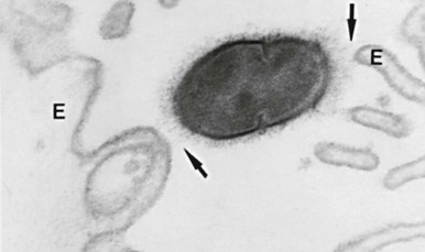
M protein is a filamentous macromolecule that exists as a stable dimer with an α-helical coiled coil structure.10 The molecule, which is anchored to the cell membrane, traverses and penetrates the cell wall. The more proximal portion of the molecule contains epitopes widely conserved among group A streptococci, whereas the more distal portion contains type-specific epitopes.11 This configuration localizes the type-specific moiety on the tips of fibrils protruding from the cell surface (Fig. 199-1). In the nonimmune host, M protein exerts its antiphagocytic effect by inhibiting activation of the alternate complement pathway on the cell surface.12,13 Such inhibition appears to be mediated by the binding to the M protein molecule of host proteins, among which are complement control proteins (factor H, a factor H–like protein, and human C4b-binding protein)14-16 and fibrinogen.17-19 The antiphagocytic effect is nullified in the presence of adequate concentrations of type-specific antibody. There is evidence that immunity caused by opsonic anti–M-type antibody may be strain and not type specific.20,21 M proteins analogous to those of group A streptococcus are present in many strains of groups C22 and G23 streptococci.
Additional surface proteins related to M protein have now been identified. Although their structure is overall similar to that of M protein, they differ in the types of repeats and in their ability to interact with different human proteins. Genes encoding these proteins (e.g., enn, mrp, fcrA, arp, protH) have been designated as members of the emm gene superfamily. A number of the M-like proteins bind IgG or IgA at the non–antigen-binding site and appear to be cooperative with M protein in antiphagocytic effect.24,25 Indeed, a notable function of the M protein family is its ability to bind to a wide range of host proteins, including, among others, albumin, fibrinogen, and plasminogen. Still other anti-opsonic surface proteins continue to be described.26 For example, Mac, a secreted group A streptococcal protein with homology to a human β2-integrin, binds to CD16 on the surface of human PMNs and inhibits phagocytosis and bacterial killing.27 An additional surface protein, streptococcal heme-associated protein (Shp), has been found in M1 strains of group A streptococci, which likely has a role in transport of iron intracellularly. Antibody against Shp has been found in convalescent sera and has opsonic capability.28 These observations underscore the extreme virtuosity with which the bacterium develops multiple mechanisms to evade phagocytic killing.
A protein antigen very closely associated with the M protein molecule of group A streptococci is the so-called serum opacity factor (OF). This factor is an α-lipoproteinase that is detected by its ability to opacify horse serum and that also has fibronectin-binding properties.29 Strains of a minority of the currently identified M types elaborate this antigen.30 OF itself is antigenic and type-specific—that is, its ability to opacify serum can be specifically inhibited by antiserum raised against homologous but not heterologous M types. Type-specific and non–type-specific immune responses to streptococcal M protein are generally weaker after pharyngeal infection with OF-positive than with OF-negative types.31 The former importance of this substance as an ancillary typing system for strains that could not be M serotyped has been obviated by the advent of emm genotyping. Interestingly, antibody against OF has opsonic activity and has been shown to synergize with anti–M protein antibody in protecting mice against challenge with OF-positive strains.32
A number of somatic streptococcal constituents play critical roles in the first step of colonization, namely, adherence to the surface of human epithelial cells. At least 17 adhesin candidates have been described,33 but the most extensively studied have been lipoteichoic acid (LTA), M protein, and fibronectin-binding proteins. Through hydrophobic interactions, LTA serves as a “first-step” adhesin, bringing the organisms into close contact with host cells and then allowing other adhesins to promote high-affinity binding.34 Although M protein does not appear to promote adhesion to human buccal or tonsillar epithelial cells,35,36 it does mediate adherence to skin keratinocytes via the attachment of the C repeat region to keratinocyte membrane cofactor CD46.37,38 Group A streptococcal surface proteins that bind fibronectin have been studied extensively and are important in adherence to both throat and skin. These include protein F1 (PrtF1),39 also known as SfbI (streptococcal fibronectin-binding protein I),40 and related proteins known as SbfII,41 FBP54,42 protein F2,43 and PFBB.44
Moreover, the expression of these adhesins has been reported to be environmentally regulated.45 Expression of protein F1 is enhanced in an oxygen-rich environment, whereas that of M protein is greater at higher partial pressures of carbon dioxide.46 Thus, teleologically, it might be postulated that the organism displays protein F1 on its surface when it seeks to adhere to the cutaneous surface but expresses M protein in the deeper tissues, where it is more likely to encounter phagocytic cells.
Extracellular Products
During the course of growth in vitro or in vivo, group A streptococcus elaborates numerous extracellular products, only a limited number of which have been well characterized. Two distinct hemolysins have been elaborated. Streptolysin O derives its name from its oxygen lability. It is reversibly inhibited by oxygen and irreversibly inhibited by cholesterol. In addition to its effect on erythrocytes, it is toxic to a variety of cells and cell fractions, including PMNs, platelets, tissue culture cells, lysosomes, and isolated mammalian and amphibian hearts. Streptolysin O is produced by almost all strains of S. pyogenes (as well as many group C and G organisms) and is antigenic. Measurement of anti–streptolysin O antibodies in human sera is useful as an indicator of recent streptococcal infection.
Streptolysin S is a hemolysin produced by streptococci growing in the presence of serum (hence the “S”) or in the presence of a variety of other substances such as serum albumin, α-lipoprotein, ribonucleic acid, or detergents such as Tween. Streptolysin S is nonantigenic, or at least no antibody to it has been detected that neutralizes its hemolytic activity. Streptolysin S shares with streptolysin O the capacity to damage the membranes of PMNs, platelets, and subcellular organelles. Unlike streptolysin O, it is not inactivated by oxygen, but it is thermolabile. Most strains of S. pyogenes produce both hemolysins. Hemolysis on the surface of blood agar plates is primarily caused by streptolysin S, whereas streptolysin O exerts its hemolytic effect best in subsurface colonies, in pour plates, or in anaerobic cultures. An occasional strain may produce only one of the two hemolysins. Rarely, strains are encountered that lack both hemolysins.
Several extracellular products may theoretically serve to facilitate the liquefaction of pus and the spreading of streptococci through tissue planes characteristic of streptococcal cellulitis and necrotizing fasciitis. These include the following: (1) four antigenically distinct enzymes that participate in the degradation of deoxyribonucleic acid (DNases A, B, C, and D); (2) hyaluronidase, which enzymatically degrades hyaluronic acid found in the ground substance of connective tissue; (3) streptokinase, which promotes the dissolution of clots by catalyzing the conversion of plasminogen to plasmin; (4) streptococcal pyrogenic exotoxin B (SpeB), which is a potent protease; and (5) C5a peptidase, which specifically cleaves the human chemotaxin C5a at the PMN binding site.47,48 SpeB also cleaves IgG bound to group A streptococcus, thus interfering with ingestion and killing by phagocytes.49
The streptococcal pyrogenic exotoxins are a family of bacterial superantigens believed to be associated with streptococcal toxic shock syndrome (strep TSS), necrotizing fasciitis, and other severe infections. This family includes the bacteriophage-encoded SpeA50 and SpeC, historically known as the scarlatinal toxins because of their association with scarlet fever, as well as the cysteine protease SpeB; a number of additional pyrogenic exotoxins (e.g., mitogenic factor [MF, SpeF], and streptococcal superantigen [SSA]) have more recently been identified.51
Superantigens are potent immunostimulators able to bind simultaneously to the major histocompatibility complex (MHC) class II molecule and specific V-β regions of the T-cell receptor.52 This binding results in clonal proliferation of these T cells. Superantigen activation of T cells leads to increased secretion of proinflammatory cytokines produced by both antigen presenting cells and T lymphocytes. This issue is discussed in more detail later in the section on pathogenesis of streptococcal toxic shock syndrome.
Emerging concepts regarding the molecular biology of streptococcal virulence, colonization, and tissue invasion have been reviewed.51,53 Control of the expression of the heretofore-mentioned virulence factors over time and under diverse environmental circumstances depends on a complex system of genetic modulation. Of the known transcriptional regulators in S. pyogenes, the two most intensively studied are Mga54 (multiple gene regulator) or Mry (regulator of M protein expression) and a two-component regulatory system known as CsrRS55 (capsule synthesis regulator) or, alternatively, CovRS56 (control of virulence genes), which represses the synthesis of the capsule and several exotoxins.57 The regulator of proteinase B (RopB) has been shown to have various polymorphisms that regulate the virulence of S. pyogenes.58
Streptococcal Pharyngitis
Epidemiology
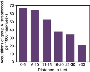
Streptococcal sore throat is among the most common bacterial infections of childhood. Group A streptococci are responsible for the great majority of such infections, but strains of other serogroups, especially groups C and G,59 are occasionally involved. The disease occurs primarily among children 5 to 15 years of age, with the peak incidence occurring during the first few years of school. All age groups are susceptible, however, and severe epidemics are common in military training facilities. There is no gender predilection. The disease is ordinarily spread by direct person-to-person contact, most likely via droplets of saliva or nasal secretions. Crowding such as occurs in schools or barracks favors interpersonal spread of the organism (Fig. 199-2) and may also enhance its virulence by processes of natural selection analogous to those that occur during mouse passage in the laboratory. The effect of crowding in facilitating transmission may account in part for the increased incidence of streptococcal pharyngitis in northern latitudes during the colder months of the year. Explosive foodborne or waterborne outbreaks are also well documented. Contamination of dust, clothing, blankets, or other fomites does not appear to play a significant role in contagion.
Group A streptococci frequently colonize the throats of asymptomatic persons. Pharyngeal carriage rates among normal schoolchildren vary with geographic location and season of the year. Carriage rates as high as 15% to 20% have been noted in several studies. The carriage rate among adults is considerably lower.
Studies of experimentally induced human infections and of transmission in military barracks have shed considerable light on the variables involved in interpersonal spread. During the acute phase of tonsillopharyngeal infection, M-typeable group A streptococci are frequently present in large numbers in both the nose and throat. In untreated infections, organisms may persist for many weeks, although the signs and symptoms of illness abate within a few days. During convalescence, the organisms decrease in numbers and tend to disappear from the anterior nares sooner than from the throat. In addition, the M protein content and virulence of persisting organisms gradually decline. The result of these qualitative and quantitative changes is that convalescent carriers are much less likely to transmit the organism to close contacts than acutely infected persons.
In patients who do not receive effective antibiotic therapy for acute streptococcal pharyngitis, type-specific antibodies are frequently detectable in the serum between 4 and 8 weeks after the infection. These opsonic antibodies protect against subsequent infection with organisms of the same M type, but the person remains susceptible to infection by heterologous types. Prompt and effective antibiotic therapy may ablate the type-specific immune response.
Clinical Manifestations
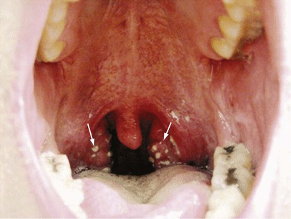
The usual incubation period of streptococcal pharyngitis is 2 to 4 days. The onset of illness is heralded by the rather abrupt onset of sore throat accompanied by malaise, feverishness, and headache. Nausea, vomiting, and abdominal pain are common in children. Prominent physical findings include redness, edema, and lymphoid hyperplasia of the posterior portion of the pharynx; enlarged, hyperemic tonsils; patchy discrete tonsillopharyngeal exudates (Fig. 199-3); enlarged, tender lymph nodes at the angles of the mandibles; and a temperature of 38.3° C (101° F) or higher. In the absence of these symptoms and signs, simple coryza, hoarseness, cough, or conjunctivitis does not suggest the presence of streptococcal infection. Laboratory findings include a positive throat culture for β-hemolytic streptococci and a total white blood cell count usually exceeding 12,000/mm3, with increased numbers of PMNs. The test for C-reactive protein is usually positive.60
Not all patients with streptococcal pharyngitis have the full-blown syndrome just described. Endemically occurring infections in open populations manifest a wide spectrum of clinical severity. For example, only approximately 50% of such patients with sore throats and positive throat cultures have tonsillar or pharyngeal exudates. Patients who have undergone tonsillectomy tend to experience a milder clinical syndrome. In infants, the response to streptococcal infection is much less sharply focalized to the lymphoid tissue of the faucial and posterior pharyngeal area. Rhinorrhea, suppurative complications, low-grade fever, and a more protracted course tend to characterize infections at this age. Exudative pharyngitis in children younger than 3 years is rarely streptococcal in cause.
In the absence of suppurative complications, the disease is self-limited. Fever abates within 3 to 5 days. Almost all acute signs and symptoms subside within 1 week, although several additional weeks may be required for tonsils and lymph nodes to return to their usual size. Penicillin shortens the period of fever, toxicity, and infectivity.61-63 Given the rather brief time course of untreated disease, however, such shortening of the clinical syndrome may not be striking unless therapy is initiated within the first 24 hours of illness. Antibiotic treatment shortens the clinical symptoms by 24 hours, and the main reason for treatment is the prevention of rheumatic fever.64
Scarlet Fever
Scarlet fever results from infection with a streptococcal strain that elaborates streptococcal pyrogenic exotoxins (erythrogenic toxins). Although this disease is usually associated with pharyngeal infections, it may follow streptococcal infections at other sites, such as wound infections or puerperal sepsis. The clinical syndrome is similar in most respects to that associated with nontoxigenic strains, save for the scarlatinal rash. The latter must be differentiated from those of viral exanthems, drug eruptions, staphylococcal toxic shock syndrome, and Kawasaki disease.
The rash usually appears on the second day of clinical illness as a diffuse red blush, with many points of deeper red that blanch on pressure. It is often first noted over the upper part of the chest and then spreads to the remainder of the trunk, neck, and extremities. The palms, soles, and usually the face are spared. Skin folds in the neck, axillae, groin, elbows, and knees appear as lines of deeper red (Pastia's lines). There are scattered petechiae, and the Rumpel-Leeds test of capillary fragility is positive. Occlusion of sweat glands imparts a sandpaper texture to the skin, which is a particularly helpful finding in dark-skinned patients.
The face appears flushed, except for marked circumoral pallor. In addition to findings of exudative pharyngitis and tonsillitis, patients display an enanthem characterized by small, red, hemorrhagic spots on the hard and soft palates. The tongue is initially covered with a yellowish white coat through which may be seen the red papillae (white strawberry tongue). Later, the coating disappears, and the tongue is beefy red in appearance (red strawberry tongue). The skin rash fades over the course of 1 week and is followed by extensive desquamation lasting for several weeks. A modest eosinophilia may be present early in the course of the illness.
Severe forms of scarlet fever, either associated with local and hematogenous spread of the organism (septic scarlet fever) or with profound toxemia (toxic scarlet fever), are characterized by high fever and marked systemic toxicity. The course may be complicated by arthritis, jaundice, and, very rarely, hydrops of the gallbladder. Such severe forms of the disease are infrequent in the antibiotic era. In the late 1800s, scarlet fever was associated with mortalities of 20% in Chicago, New York, and Scandinavia. Recently, an epidemic of 900 cases of scarlet fever occurred in 2011 in Hong Kong between January and July associated with emm12 strains of S. pyogenes.65
Suppurative Complications
Inflammation in the faucial area induced by acute streptococcal infection may affect structures that are directly contiguous to the pharynx or that drain that site. Such relatively rare complications include peritonsillar cellulitis, peritonsillar abscess, retropharyngeal abscess, suppurative cervical lymphadenitis, mastoiditis, acute sinusitis, and otitis media.66 Peritonsillar or retropharyngeal abscesses, however, frequently contain a variety of other oral flora, including anaerobes, with or without group A streptococci.67 Group A streptococci are responsible for only a small minority of cases of otitis media or sinusitis.
Extension up the cribriform plate of the ethmoid or via the mastoid bone may cause meningitis, brain abscess, or thrombosis of the intracranial venous sinuses. Streptococcal pneumonia, another potential suppurative complication, is discussed later. Finally, bacteremic spread of the streptococci may result in a variety of metastatic foci of infection, such as suppurative arthritis, endocarditis, meningitis, brain abscess, osteomyelitis, or liver abscess. Such complications of streptococcal pharyngitis are extremely rare since the advent of effective chemotherapy.
Nonsuppurative Complications
The nonsuppurative complications of streptococcal pharyngitis, acute rheumatic fever and acute poststreptococcal glomerulonephritis, are discussed in Chapter 200. The role of streptococci vis-à-vis other infectious and noninfectious agents in initiating certain other acute inflammatory disorders such as erythema nodosum and anaphylactoid purpura remains unresolved.
Diagnosis
Pharyngitis and tonsillitis may be caused by infectious agents other than S. pyogenes.59,68 Among these are streptococci of groups C69,70 and G.71-73 Corynebacterium diphtheriae, the other major bacterial pathogen associated with exudative pharyngitis, is now extremely rare in the United States74 and, when it occurs in the classic form, is differentiated by the appearance of the diphtheritic membrane, respiratory embarrassment, severe systemic toxicity, and myocardial and neurologic manifestations. Other bacterial agents such as Neisseria gonorrhoeae and perhaps Neisseria meningitidis occasionally cause pharyngitis, as does Mycoplasma pneumoniae.
Pharyngitis due to Arcanobacterium (formerly Corynebacterium) hemolyticum, although rare, may closely mimic that caused by S. pyogenes.75,76 Arcanobacterium hemolyticum affects primarily teenagers and young adults, and the patients may exhibit both an exudative pharyngitis and a scarlatiniform rash. The organism is more readily identified on rabbit or human blood agar than on sheep blood agar. Another rare cause of acute pharyngitis is Yersinia enterocolitica.77 Patients infected with this organism may appear quite ill and may or may not have associated enteric symptoms. When Y. enterocolitica pharyngitis is associated with disseminated yersiniosis, the mortality rate may be appreciable. Diagnosis depends on clinical clues because the organism is unlikely to be detected on routine throat cultures and antistreptococcal therapy is unavailing (see Chapter 231). The oropharyngeal form of tularemia is characterized by severe sore throat, exudative and ulcerative tonsillopharyngitis, and cervical adenopathy (see Chapter 229).
Acute pharyngitis is more frequently caused by viruses than by bacteria. Infectious mononucleosis and adenovirus infections frequently give rise to exudative pharyngitis and thus may closely mimic streptococcal sore throat. Herpes simplex viruses 1 and 2,78-80 influenza,81 and parainfluenza viruses may also simulate streptococcal pharyngitis, as may initially the acute retroviral syndrome in human immunodeficiency virus infection. Pharyngitis associated with the acute retroviral syndrome is, however, not exudative.82 Even when careful microbiologic techniques are used to detect bacteria, mycoplasma, and viruses, no causative agent can be detected in a substantial proportion of all cases of acute sore throat.83 A more complete discussion of the differential diagnosis of acute pharyngitis may be found in Chapter 59.
Approximately one fourth to one third of all children complaining of sore throat have a positive throat culture for group A streptococci. Of these, about half can be demonstrated to have immunologically significant infection, as judged by a significant rise in serum titer of one or more antistreptococcal antibodies. Many of the remainder are likely to be asymptomatic carriers, because the average carriage rate among school-aged children in temperate climates during the winter months may approximate 15% to 20%. Such asymptomatic carriers are at no risk for developing suppurative and nonsuppurative complications and do not require antibiotic therapy. Although acutely infected individuals tend to have more strongly positive throat cultures, this distinction cannot be made with confidence in patients whose signs and symptoms are compatible with those of streptococcal pharyngitis.
Numerous studies have tested the precision with which physicians may differentiate between streptococcal and nonstreptococcal sore throat by clinical criteria alone. In the presence of a classic scarlatinal rash or during a documented epidemic of streptococcal infections, such differentiation is usually easy. On the other hand, in the case of endemically occurring infections, the problem is much more complex. Certain clinical findings, particularly fever, sore throat, tonsillopharyngeal exudate, and tender, enlarged lymph nodes at the angles of the jaws, have a statistically significant correlation (approximately 85%) with the presence of positive throat cultures for group A streptococci.84 Such findings are not diagnostic, however. Although only approximately 50% of patients with immunologically proven streptococcal sore throat have tonsillar exudate, a substantial proportion of cases of exudative pharyngitis are nonstreptococcal in cause.
It is possible to identify individual patients in which “strep throat” can be effectively excluded on a combination of epidemiologic (see earlier) and clinical grounds. For example, symptoms of the common cold are not caused by S. pyogenes. Similarly, the presence of hoarseness and conjunctivitis and the absence of fever or pharyngeal erythema make streptococcal pharyngitis very unlikely. A number of investigators have developed clinical algorithms in children and adults to assist in determining the likelihood that a particular patient has group A streptococcal pharyngitis.84-90 These algorithms are useful and accurate in identifying patients whose risk for streptococcal infection is so low as to obviate the need for further microbiologic testing. Otherwise, such testing should be performed.
One published practice guideline91,92 has suggested that in adults with features strongly suggestive of streptococcal pharyngitis, empirical antimicrobial therapy without microbiologic confirmation is an acceptable alternative. That guideline uses an algorithm, developed by Centor and co-workers,90 using four clinical criteria—presence of tonsillar exudates, presence of swollen tender anterior cervical nodes (i.e., cervical lymphadenitis), lack of cough, and history of fever—that have been reported to be independently associated with the likelihood of a positive throat culture for group A streptococci.93 A subsequent cost-effectiveness analysis94 and two prospective clinical studies95,96 have concluded that such empirical therapy is neither the most effective nor least expensive strategy for diagnosis of “strep throat” in adults. Furthermore, empirical therapy in adults leads to considerable overuse of antibiotics.97 It is important to realize that the most common age group of streptococcal pharyngitis is in the 5- to 15-year age group. This is of particular concern in view of the fact that 73% of the 6.7 million adults visiting primary care providers annually in the United Sates with the complaint of sore throat receive a prescription for antibiotics.98
Thus, for adults and children, expert panels of the Infectious Diseases Society of America,64 American Heart Association,99 and American Academy of Pediatrics100 recommend that the presence of group A streptococci in the pharynx should be documented by a throat culture or rapid antigen detection test (RADT).63,99,100 It should be noted that a positive test does not discriminate between active streptococcal infection versus colonization and a concomitant viral infection. Clinical criteria for streptococcal pharyngitis should be present before antibiotic treatment is considered.
Throat Culture
Throat culture remains the gold standard for diagnosing streptococcal pharyngitis. Failure to isolate β-hemolytic streptococci in a carefully obtained and accurately interpreted throat culture rules out the diagnosis of streptococcal sore throat for practical purposes. In cases in which doubt exists as to the validity of a negative culture, it may be preferable to repeat the culture rather than to treat empirically with antimicrobial agents.
Although a negative culture eliminates the necessity for therapy, a positive culture does not differentiate between acute infection and asymptomatic carriage. Serum antibody titers do not rise until convalescence and thus are of no help in short-term management. Although the degree of positivity of the throat culture may assist in making this differentiation, it is best to assume that all positive cultures in patients with acute pharyngitis are significant and to treat accordingly while recognizing that, even with the use of the throat culture, some degree of overtreatment is inevitable.
Detailed instructions for obtaining and processing a throat culture have been published by the American Heart Association.101 Sheep blood agar is preferred because clear-cut patterns of hemolysis are obtained using this medium. In regard to isolation of group A streptococci, there is controversy in the literature as to the relative merits of plain sheep blood agar plates versus plates to which trimethoprim-sulfamethoxazole has been added to suppress competing normal pharyngeal flora. Similar controversy exists about the optimal atmosphere for incubation—aerobic, aerobic in the presence of 5% to 10% carbon dioxide, or anaerobic. Detailed analyses of these issues have been published.102,103 If blood agar plates are not immediately available, the swab may be placed in a dry sterile tube for transportation to the laboratory. After overnight incubation at 35° C to 37° C, culture plates from patients with streptococcal pharyngitis show colonies surrounded by clear zones of hemolysis as well as β-hemolysis around the agar stab. Plates that are negative on first reading should be reexamined after an additional 24 hours of incubation. Serologic grouping of β-hemolytic streptococcal isolates may now be readily performed by using commercially available kits. Fluorescent antibody techniques provide excellent results and specifically identify group A organisms. No quantitative information is gained about the degree of positivity of the culture. A less expensive screening procedure, the bacitracin sensitivity test, is best performed once the organism has been isolated in pure culture. This susceptibility procedure is based on the observation that more than 95% of all group A streptococcal strains are inhibited by low-potency (0.04 unit) bacitracin disks, whereas 80% to 90% of non–group A strains are resistant.
Because no group A streptococci resistant to penicillin or cephalosporins have yet been detected, antibiotic testing is unnecessary if these drugs are to be used. The same holds true in general for macrolides because group A streptococci resistant to this drug are rare in the United States at this time104 (see later section “Treatment” for caveats).
Rapid Antigen Detection Tests
RADTs allow detection of the presence of the group A carbohydrate antigen directly from throat swabs. Unlike the throat culture, which requires overnight or longer to yield a definitive result, RADTs can be completed in a matter of minutes. By facilitating early diagnosis and therapy, an RADT may possibly shorten the duration of illness, decrease secondary spread of the organism, and allow earlier return of patients and parents to school and work. Earlier tests based on latex agglutination methodology have been largely replaced by enzyme immunoassays that are easier to interpret and more sensitive. More recently, tests using optical immunoassay (OIA) and chemiluminescent DNA probes have become available.
Most currently commercially available RADTs are highly specific (95% or higher), so a positive result obviates the need for a throat culture. Unfortunately, the sensitivity of these tests is lower than that of the conventional throat culture, and therefore they may be negative in patients in whom conventional culture proves to be positive. Some investigators105,106 have found newer tests such as OIA to have a sensitivity equivalent to that of culture, but others have reached opposite conclusions.107-109 At present, the American Academy of Pediatrics recommends that a negative RADT be confirmed with a throat culture. In view of conflicting data about sensitivity of commercially available RADTs, as well as the paucity of studies directly comparing the various tests with each other, physicians who elect to use any RADT in children and adolescents without culture backup of negative results should do so only after confirming in their own practice that the rapid test is comparable in sensitivity to the throat culture.100
In considering appropriate laboratory diagnostic testing for adults, certain epidemiologic distinctions from pediatric disease deserve consideration. The group A streptococcus causes 15% to 30% of cases of acute pharyngitis in pediatric patients but only approximately 10% of such illnesses in adults.89,110,111 However, the risk for streptococcal pharyngitis may be higher in parents of school-aged children and adults whose occupation brings them into close association with children. Moreover, the risk for a first attack of acute rheumatic fever is extremely low in adults in the United States and most other developed countries, even if they experience an undiagnosed and untreated episode of streptococcal pharyngitis. These facts make performance of RADT without culture backup of negative results an acceptable alternative to throat culture.63,99 The generally high specificity of RADTs should minimize overprescribing of antimicrobial agents for adults. This latter point is of particular importance in view of national data indicating that antibiotics are prescribed for approximately 75% of adults consulting community primary care physicians for the complaint of sore throat and that the prescription of more expensive, broader-spectrum antibiotics is frequent.98 Physicians who wish to ensure they are achieving maximal sensitivity in diagnosis may continue to use the conventional throat culture or to back up a negative RADT with a culture. However, in one recent adult study,96 OIA without culture backup was performed in adults exhibiting two or more of Centor and co-workers' criteria.90 When compared with throat culture, this strategy led to nearly optimal treatment (94%) and antibiotic prescription (37%), with minimal antibiotic overuse (3%) and underuse (3%).
Therapy
Antimicrobial therapy is indicated for individuals with symptomatic pharyngitis after the presence of the organism in the throat is confirmed by culture or RADT. The goals of antimicrobial therapy are (1) prevention of acute rheumatic fever, (2) prevention of suppurative complications, (3) improvement in clinical symptoms and signs, and (4) rapid decrease in infectivity so as to reduce transmission of group A β-hemolytic streptococci to family members, classmates, and other close contacts and to allow the rapid resumption of usual activities. There is no firm evidence that poststreptococcal acute glomerulonephritis is preventable by treatment of the antecedent streptococcal infection.112
Treatment of group A streptococcal sore throat as long as 9 days after onset is still effective for the prevention of rheumatic fever.113 Thus, if the patient is seen early in the course of his illness, the delay in initiation of therapy occasioned by obtaining a positive throat culture is not ordinarily a matter of concern in this regard. As noted, patients with signs and symptoms of acute pharyngitis and a positive rapid test (properly performed and interpreted) for group A carbohydrate antigen should receive appropriate antimicrobial therapy.
In the minority of patients who are severely ill or toxic at presentation and in whom there is clinical and epidemiologic evidence resulting in a high index of suspicion, oral antimicrobial therapy can be initiated while awaiting the results of the throat culture (either as a primary diagnostic tool or in confirmation of a negative RADT). If oral therapy is prescribed, a positive throat culture serves as a guide to the necessity of completion of a full antimicrobial course or, alternatively, of recalling the patient for an injection of penicillin G benzathine. Early initiation of antimicrobial therapy results in faster resolution of the signs and symptoms,61-63 but group A streptococcal pharyngitis is usually a self-limited disease; fever and constitutional symptoms are markedly diminished within 3 or 4 days of onset, even without antimicrobial therapy.114 Thus, antimicrobial therapy initiated within the first 48 hours of onset hastens symptomatic improvement by only 1 to 2 days.
The drug of choice in the treatment of streptococcal infection is penicillin, because of its efficacy in the prevention of rheumatic fever, safety, narrow spectrum, and low cost (Table 199-1).63,99,100 Prevention of acute rheumatic fever is believed to require eradication of the infecting streptococcus from the pharynx, an effect that depends on prolonged rather than high-dose penicillin therapy. This objective may be accomplished by the administration of a single injection of 1.2 million units of penicillin G benzathine. For children weighing 60 pounds (27 kg) or less, the dose is reduced to 600,000 units. Most physicians in the United States, however, elect to administer oral therapy. In this case, penicillin V, in one of the regimens listed in Table 199-1, must be continued for a full 10 days, Amoxicillin is often prescribed in preference to penicillin V in children requiring liquid medication because of poor palatability of oral suspensions of penicillin V. Once-daily amoxicillin therapy is effective for the treatment of group A streptococcal pharyngitis115-117 in children. An oral time-release formulation of amoxicillin has recently been approved by the U.S. Food and Drug Administration (FDA) for once-daily treatment of group A streptococcal pharyngitis in adolescents and adults. Because of its convenience, low cost, and relatively narrow spectrum, once-daily amoxicillin is an acceptable alternative regimen for the treatment of group A β-hemolytic streptococcal pharyngitis.99
TABLE 199-1
| AGENT | DOSAGE | ROUTE | DURATION (DAYS) |
|---|---|---|---|
| Penicillins | |||
| Penicillin V (phenoxymethyl penicillin) | Children ≤27 kg (60 lb): 250 mg two to three times daily Children >27 kg, adolescents, and adults: 500 mg two to three times daily | Oral | 10 |
| or | |||
| Amoxicillin | 50 mg/kg once daily (maximum, 1 g) | Oral | 10 |
| or | |||
| Benzathine penicillin G | 600,000 U for patients ≤27 kg; 1.2 million U for patients >27 kg | Intramuscular | Once |
| For Individuals Allergic to Penicillin | |||
| Narrow-spectrum cephalosporin† (cephalexin, cefadroxil) | Variable | Oral | 10 |
| or | |||
| Clindamycin | 20 mg/kg/day divided in three doses (maximum, 1.8 g/day) | Oral | 10 |
| or | |||
| Azithromycin | 12 mg/kg once daily (maximum, 500 mg) | Oral | 5 |
| or | |||
| Clarithromycin | 15 mg/kg/day divided twice daily (maximum 250 mg twice daily) | Oral | 10 |
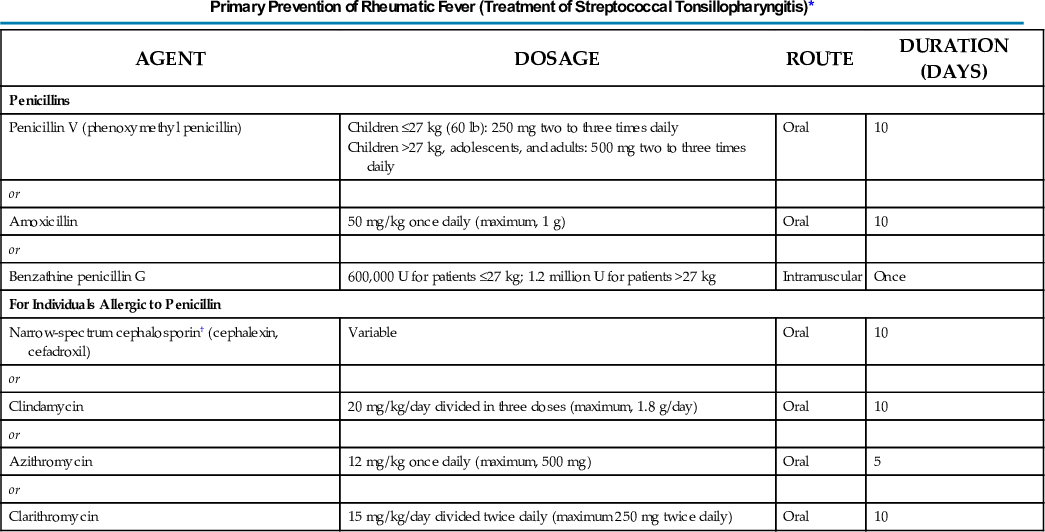
*The following are not acceptable: sulfonamides, trimethoprim, tetracyclines, and fluoroquinolones.
†To be avoided in those with immediate (type I) hypersensitivity to a penicillin.
From Gerber M, Baltimore R, Eaton C, et al. Prevention of Rheumatic Fever and Diagnosis of Acute Streptococcal Pharyngitis. A scientific statement from the American Heart Association Rheumatic Fever, Endocarditis, and Kawasaki Disease Committee, Council on Cardiovascular Disease in the Young, and Quality of Care and Outcomes Research Interdisciplinary Working Group. Circulation. 2009;119:1541-1551.
A variable percentage of patients continue to harbor group A streptococci of the originating serotype in their pharynx after completion of a course of oral penicillin.118 Such bacteriologic treatment failures are sometimes associated with symptomatic relapse. Because penicillin is ineffective in eradicating asymptomatic streptococcal pharyngeal carriage, apparent treatment failures may actually represent persistence of such carriage in patients with superimposed viral pharyngitis.119
Oral cephalosporins are highly effective in the treatment of streptococcal pharyngitis, and meta-analyses have suggested that streptococcal eradication rates and clinical cure rates attained with these agents are slightly higher in children and adults than those achieved with penicillin.118,120,121 These analyses have, however, been strongly challenged on methodologic grounds.122,123 It does appear that cephalosporins are more effective than penicillin in eradicating asymptomatic group A streptococcal carriage. Penicillin remains the recommended drug of choice.63,99,100 In penicillin-allergic patients, macrolide (erythromycin or clarithromycin) or azalide (azithromycin) antimicrobial agents, clindamycin, or first-generation cephalosporins are the agents of choice.63 The latter should be avoided in those with immediate (type I) penicillin hypersensitivity (see Table 199-1). Cephalosporins should not be administered to patients with a history of immediate (anaphylactic-type) hypersensitivity to penicillin. The physician should bear in mind the possibility of an increased risk for allergic reactions to cephalosporins when treating penicillin-allergic patients.
Erythromycin is less expensive than clarithromycin or azithromycin but may be associated with more gastrointestinal side effects. Although there have been reports of relatively high levels of resistance to macrolide antimicrobial agents from several countries,124-126 as well as isolated reports of increased rates of macrolide resistance in certain localized areas of the United States,127,128 such resistance does not appear to be widespread in this country at present. Three multistate surveillance studies conducted during 2002 and 2003 detected overall macrolide resistance rates of 3.8%,129 5.2%,130 and 6.8%.131 However, given the increasing use of azalides for upper and lower respiratory tract infections, the situation may change. Physicians should therefore be cognizant of local patterns of antimicrobial resistance. In areas in which macrolide resistance is known to be prevalent, antimicrobial susceptibility testing should be performed if these agents are used to treat group A streptococcal infections. Furthermore, continued surveillance of national trends in macrolide susceptibility is warranted.
There has been considerable recent interest in abbreviated courses of antimicrobial therapy. It has been reported that second- and third-generation cephalosporins such as cefuroxime,132,133 cefixime,134 ceftibuten,135 cefdinir,136 cefpodoxime,137 and cefditoren138 are effective in eradication of group A streptococci from the pharynx when administered for 5 days, although not all of these are approved for a 5-day course of therapy for acute streptococcal pharyngitis by the FDA at the time of this writing. Although such shortened courses might theoretically enhance patient compliance, the potential ecologic effects of using these broad-spectrum agents to treat such a common bacterial infection are of great concern. This is particularly true should these agents be used as first-line therapy for “strep throat.” Moreover, even when administered for short courses, they are considerably more expensive than penicillin. Clearly, penicillin treatment is also associated with potential adverse effects.
Similar favorable results of short-course therapy have also been reported for the newer macrolides or azalides, clarithromycin,139 and azithromycin.140-143 Because of its long intracellular half-life and slow release from tissue sites, a 5-day course of azithromycin is approved by the American Heart Association99 for use in penicillin-allergic patients. As noted, promiscuous use of macrolides has been associated with development of resistance by group A streptococci.124,125
Because tetracycline-resistant group A streptococci are prevalent in many areas, this drug is not recommended. Sulfonamides, which are effective for the secondary prophylaxis of rheumatic fever (see Chapter 200), are ineffective for the eradication of pharyngeal organisms or the prevention of rheumatic fever when used as therapy for acute pharyngeal infections.
Patients with more severe suppurative infections, such as those involving the mastoid or ethmoid, may require higher doses of penicillin or other β-lactam antibiotics administered parenterally. When streptococcal upper respiratory tract infection is complicated by the development of abscesses associated with suppurative cervical adenitis or in the peritonsillar or retropharyngeal soft tissues, aspiration or incision and drainage is usually required.
Because prevention of rheumatic fever appears to require eradication of the streptococcal organism from the pharynx, treatment failures are of concern. In addition to true treatment failure (i.e., re-isolation of the original infecting streptococcal serotype shortly after completion of a full course of antibiotic therapy), causes of post-treatment culture positivity include failure of compliance with oral medication schedules and reinfection with the same or different streptococcal types in the home or school environment. Apparent failure may also occur when the patient is in reality a streptococcal carrier suffering from an acute viral pharyngitis. In everyday practice, it is often impossible to differentiate among these alternatives.
Nevertheless, routine reculture of the throat after a course of antistreptococcal therapy in an asymptomatic patient is not advised63 because the cost-benefit ratio of such cultures continues to decline in parallel with the incidence of acute rheumatic fever in developed countries. Such cultures should be undertaken in high-risk circumstances (e.g., if the patient or a family member has a history of rheumatic fever) or when symptoms compatible with streptococcal infection persist or recur. When an increased incidence of acute rheumatic fever is detected in a community, as happened in a number of U.S. cities during the 1980s, the approach to streptococcal infection must be particularly rigorous, and serious consideration should be given to routine performance of post-treatment cultures. If reculture is undertaken, only a single re-treatment course is warranted for patients who still harbor group A streptococci. Re-treatment with an oral cephalosporin might be considered in view of the slightly increased eradication rates observed with these agents.
The presence of persistently but weakly positive throat cultures after repeated courses of antibiotic therapy in an otherwise asymptomatic patient is not a cause for alarm. Such persons are streptococcal carriers119 who are neither at risk for developing rheumatic fever nor likely to spread their infection to others. Their most frequent problem is anxiety produced by multiple medical consultations associated with the streptococcal colonization. In the event in which, for medical or psychological reasons, eradication of chronic streptococcal carriage becomes highly desirable, clindamycin,144 amoxicillin-clavulanate,145 or azithromycin may be efficacious.63,146 Any of these antibiotic treatments can have adverse events such as Clostridium difficile colitis.
Streptococcal acquisition rates of 25% or higher have been recorded in family contacts. Certainly, family contacts with symptoms of upper respiratory tract infection should be cultured and treated appropriately if positive. Asymptomatic family contacts should also be cultured in high-risk circumstances, such as a family member who has had rheumatic fever or known cases of rheumatic fever or poststreptococcal glomerulonephritis occurring in the general area. In situations of lesser risk, routine culture of asymptomatic family contacts is not recommended.63,99 The advisability of culture and/or prophylaxis of household contacts of patients with invasive group A streptococcal infection is discussed later.146
There is no firm evidence to suggest that tonsillectomy reduces the incidence of rheumatic fever, either in healthy persons or in persons who have had rheumatic fever and faithfully maintained continuous antibiotic prophylaxis. In certain patients with recurrent bouts of tonsillopharyngitis, however, tonsillectomy may decrease the frequency of incapacitating acute infections.147,148 Tonsillectomy should be considered only for the most severely affected patients.149
Streptococcal Pyoderma
Pyoderma, impetigo, and impetigo contagiosa are terms used synonymously to describe discrete purulent lesions that are primary infections of the skin and that are extremely prevalent in many parts of the world. In the great majority of cases, pyoderma is caused by β-hemolytic streptococci and/or Staphylococcus aureus.
Epidemiology
Pyoderma occurs most frequently in economically disadvantaged children dwelling in tropical or subtropical climates. It is also prevalent in northern climates during the summer.150 The peak incidence of impetigo is in children aged 2 to 5 years. This disorder also occurs among older children and adults whose recreational activities or occupation results in cutaneous cuts or abrasions.151-153 There is no gender predilection, and all races appear to be susceptible. The prevalence of streptococcal pyoderma is markedly influenced by several factors, the most important of which appear to be climate and level of hygiene.
Meticulous prospective studies of streptococcal impetigo have demonstrated that the responsible microorganisms initially colonize the unbroken skin,150 an observation that probably explains the influence of personal hygiene on disease incidence. Development of skin colonization with a given streptococcal strain precedes the development of impetiginous lesions by an average interval of 10 days. The mechanism of production of skin lesions is unproved, but it is most likely caused by intradermal inoculation of surface organisms by abrasions, minor trauma, or insect bites. Frequently, there is a transfer of the streptococcal strains from the skin and/or pyoderma lesions to the upper respiratory tract. The interval between colonization of the skin and colonization of nose or throat or both averages 2 to 3 weeks.
Bacteriology and Immunology
Streptococci isolated from pyodermal lesions are primarily group A, but occasionally representatives of other serogroups such as C and G are responsible. Group A streptococci that cause impetigo differ in several respects from those usually associated with tonsillitis and pharyngitis. Skin strains belong to different M serotypes or genotypes from the classic throat strains; because most have been identified more recently, they tend to comprise the higher numbered M types. Throat and skin strains can also be differentiated by genetic markers.154 A relatively small number of serotypes seem capable of regularly initiating both pharyngitis and pyoderma.155
Assays of streptococcal antibodies are of no value in the diagnosis and management of impetigo, but they provide helpful supporting evidence of recent streptococcal infection in patients suspected of having poststreptococcal glomerulonephritis. The anti–streptolysin O response is weak in patients with streptococcal impetigo,156,157 presumably because the activity of streptolysin O response is inhibited by skin lipids (cholesterol),158 whereas anti–DNase B levels are elevated.156,157
Clinical Manifestations
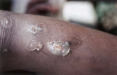
The lesion of streptococcal pyoderma begins as a papule that rapidly evolves into a vesicle surrounded by an area of erythema. The vesicular lesions are evanescent and rarely recognized clinically; they give rise to pustules that gradually enlarge and then break down over a period of 4 to 6 days to form characteristic thick crusts (Fig. 199-4). The lesions heal slowly and leave depigmented areas. A deeply ulcerated form of impetigo is known as ecthyma.
Streptococcal impetigo occurs on exposed areas of the body, most frequently on the lower extremities or face. The lesions remain well localized but are frequently multiple. Although regional lymphadenitis may occur, systemic symptoms are not ordinarily present.
In the past, the lesions just described could be rather confidently diagnosed as streptococcal. This was the predominant form of impetigo and could be distinguished from bullous impetigo caused by phage group II S. aureus. Although bullous impetigo remains almost exclusively staphylococcal in cause, the bacteriology of nonbullous impetigo has changed.159 S. aureus, either alone or in combination with S. pyogenes, is now the predominant causative agent.160-162 Almost all such staphylococci are penicillinase producers. Therefore, treatment with penicillin, which in the past had been highly effective against nonbullous impetigo, even when both streptococci and staphylococci were isolated from the lesions, now frequently fails.161
Therapy and Prevention
Because of the current frequency of isolation of S. aureus from nonbullous impetigo lesions and concomitant reports of penicillin failures,161,163,164 penicillinase-resistant penicillins or first-generation cephalosporins are preferred.161 Erythromycin has long been a mainstay of pyoderma therapy, but its use may be lessened in areas in which erythromycin-resistant strains of S. aureus or, more recently, S. pyogenes are prevalent. Topical therapy with mupirocin is equivalent to oral systemic antimicrobial therapy165,166 and may be used when lesions are limited in number. It is expensive, however, and some strains of staphylococci may be resistant.167 Retapamulin, a novel pleuromutilin antibacterial agent, has recently been approved by the FDA for treatment of bullous and nonbullous impetigo caused by group A streptococcus and methicillin-susceptible strains of S. aureus in children 9 months of age or older.168-170 In vitro data have suggested that it may be more effective than mupirocin against methicillin-resistant Staphylococcus aureus.
Adherence to good regimens of personal hygiene is the most effective measure currently available for prevention of impetigo.
Complications
Suppurative complications are uncommon. For as yet unexplained reasons, rheumatic fever has never been shown to occur after streptococcal pyoderma. On the other hand, cutaneous infections with nephritogenic strains of group A streptococci are the major antecedent of poststreptococcal glomerulonephritis in many areas of the world. There are as yet no conclusive data to indicate that treatment of an individual case of pyoderma prevents the subsequent occurrence of nephritis in these patients. Such therapy is nevertheless important as an epidemiologic measure in eradicating nephritogenic strains from the environment.
Invasive Streptococcal Infections of Skin and Soft Tissues
In the mid-1980s, outbreaks of acute rheumatic fever began to occur throughout the United States, concomitant with the reappearance of certain streptococcal strains exhibiting characteristics known to be associated with rheumatogenicity (see Chapter 200). Shortly thereafter, invasive streptococcal infections, of a frequency and severity not seen in the preceding decades, began to be reported both in the United States and abroad.171-174 Although strains of a number of group A streptococcal M types have been isolated from invasive infections, there has been a definite and consistent tendency for M types 1 and 3 to be associated with life-threatening infections.172-176 A high proportion of these cases has occurred in adults, and the portal of entry is frequently the skin or soft tissues. In some cases, the infections give rise to shock and multiorgan failure, features that simulate in certain respects the staphylococcal toxic shock syndrome.177 This entity has thus been termed strep TSS. Clinical features of serious streptococcal skin and soft tissue infections and TSS are described later.
Erysipelas
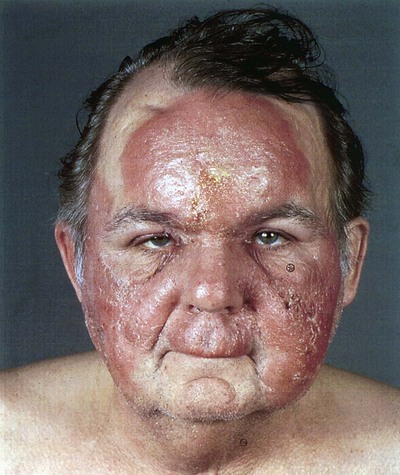
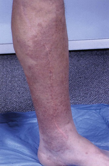
Erysipelas is a superficial cutaneous process, usually restricted to the dermis but with prominent lymphatic involvement. It is distinguished clinically from other forms of cutaneous infection by three features: the lesions are raised above the level of the surrounding skin, there is a clear line of demarcation between involved and uninvolved tissue, and the lesions are a brilliant salmon red. This disorder is more common in infants, young children, and older adults. It is almost always caused by β-hemolytic streptococci. In most cases the infecting agent is the group A streptococci, but similar lesions can be caused by streptococci of group C or G. Rarely, group B streptococci or S. aureus may be the culprit. In older reports, erysipelas was described as characteristically involving the butterfly area of the face (Fig. 199-5), but at present the lower extremities are more frequently involved (Fig. 199-6). In patients with facial erysipelas, there is frequently a history of preceding streptococcal sore throat, although the exact mode of spread to the skin is unknown. When erysipelas involves the extremities, breaks in the cutaneous barrier serve as portals of entry; these include surgical incisions, trauma or abrasions, dermatologic diseases such as psoriasis, or local fungal infections.
The cutaneous lesion begins as a localized area of erythema and swelling and then spreads rapidly with advancing red margins, which are raised and well demarcated from adjacent normal tissue. There is marked edema, often with bleb formation, and in facial erysipelas the eyes are frequently swollen shut. The lesion may demonstrate central resolution while continuing to extend on the periphery. The cutaneous inflammation is accompanied by chills, fever, and toxicity.
The differential diagnosis is limited. Early on, the lesions of facial herpes zoster, contact dermatitis, or giant urticaria may be confused with erysipelas. Lesions resembling erysipelas may occur in patients with familial Mediterranean fever. Cutaneous lesions similar in appearance to those of erysipelas may occur on the hands of patients who sustain cuts or abrasions while handling fish or meats. This entity, known as erysipeloid of Rosenbach and caused by Erysipelothrix rhusiopathiae, is usually unaccompanied by fever or systemic symptoms.
With early diagnosis and treatment, the prognosis is excellent. Rarely, however, the process may spread to deeper levels of the skin and soft tissues. Penicillin, either parenterally or orally, depending on clinical severity, is the treatment of choice. If staphylococcal infection is suspected, a penicillinase-resistant semisynthetic penicillin or cephalosporin should be selected. In a randomized, prospective multicenter trial,178 roxithromycin, a macrolide antimicrobial agent, was equivalent to penicillin. Increased levels of macrolide resistance among group A streptococci, however, have been detected in certain areas of the United States.127,128
Streptococcal Cellulitis
Streptococcal cellulitis, an acute spreading inflammation of the skin and subcutaneous tissues, results from infection of burns, wounds, or surgical incisions but may also follow mild trauma. Clinical findings include local pain, tenderness, swelling, and erythema. The process may extend rapidly to involve large areas of skin. Systemic manifestations include fever, chills, and malaise, and there may be associated lymphangitis, bacteremia, or both. In contrast to erysipelas, the lesion is not raised, the demarcation between involved and uninvolved skin is indistinct, and lesions are more pink than salmon red. Often, however, the clinical differentiation between these entities is not clear cut.
Two predisposing causes of streptococcal cellulitis deserve special mention. One is the parenteral injection of illicit drugs.179-181 These cases are often associated with bacteremia and deep tissue infections such as septic thrombophlebitis, suppurative arthritis, osteomyelitis, and, occasionally, infective endocarditis. Second, patients who have impaired lymphatic drainage from upper or lower extremities are prone to recurrent episodes of streptococcal cellulitis. Examples include individuals with filariasis and women who have undergone radical mastectomy with axillary node dissection.182 It is speculated that repetitive infection further damages local lymphatics and worsens lymphatic stasis.183
Recurrent episodes of severe cellulitis have also been reported in certain patients who have undergone coronary artery bypass grafting.184 The lesion invariably occurs in the extremity from which the saphenous vein was removed, and at times it may exhibit features of erysipelas (see Fig. 199-6). Patients with tinea pedis of the venectomy limb appear to be particularly at risk.185-187 As with other forms of cellulitis, pathogenic bacteria are difficult to recover during these episodes. The appearance of the lesions and the response to penicillin therapy suggest, however, a streptococcal cause. The few β-hemolytic streptococci that have been recovered and characterized often belong to serogroups other than A.188
Disruption of the cutaneous barrier (leg ulcers, wounds, dermatophytosis) is a risk factor for the development of cutaneous streptococcal infection.189 There is suggestive evidence that local dermatophyte infection (i.e., athlete's foot) may serve as a reservoir for β-hemolytic streptococci that initiate episodes of erysipelas or cellulitis of the lower extremities.185,190 Thus, care should be taken to eradicate such fungal infections in patients who experience recurrent bouts of erysipelas or cellulitis. Another potential reservoir is anal streptococcal colonization.191 Other risk factors include venous insufficiency, edema, and obesity.189 Not surprisingly, an increased risk for recurrent cellulitis has also been reported in homeless persons.192
Cellulitis may be caused by infection with a variety of bacterial pathogens (see Chapter 95), but most cases are caused by S. pyogenes (or, occasionally, streptococci of groups B, C, or G) or by S. aureus. S. aureus infection of the skin is usually associated with a pyogenic, fluctuant focus with surrounding erythema and is referred to as “purulent cellulitis.” In the absence of positive blood cultures, which are present in only 5% of cases of nonpurulent cellulitis, a specific microbiologic diagnosis is often not possible. Aspirate or biopsy samples from sites of active cellulitis are helpful when positive on smear or culture, but unfortunately such specimens are usually negative in adult patients.193-195
It is often impossible to differentiate streptococcal from staphylococcal cellulitis on initial presentation confidently. In this case a semi-synthetic penicillinase-resistant penicillin should be used. In penicillin-allergic patients, a first-generation cephalosporin may be used if the hypersensitivity is not of the immediate type. Clindamycin or vancomycin may be used in patients who manifest anaphylactic hypersensitivity to β-lactam antibiotics, and the latter should be administered if there is reason to suspect infection with methicillin-resistant S. aureus. Patients with milder cases of streptococcal cellulitis may be switched to oral medications after an initial favorable response to parenteral therapy.
The role of continuous antimicrobial prophylaxis196-198 in patients prone to frequent recurrences remains unsettled. At present, such prophylaxis seems justified only for patients with very frequent or severe episodes, and the optimal regimen has not been established.
Necrotizing Fasciitis (Streptococcal Gangrene)
Necrotizing fasciitis is an infection of the deeper subcutaneous tissues and fascia, characterized by extensive and rapidly spreading necrosis and by gangrene of the skin and underlying structures. As detailed in Chapter 95, this entity may arise in several distinct epidemiologic settings, may be caused by multiple aerobic and anaerobic microorganisms, and may vary in clinical manifestations. The present discussion is limited to necrotizing fasciitis caused by group A streptococci199 and described by Meleney200 in 1924 as hemolytic streptococcal gangrene. Characteristically, streptococcal gangrene begins at a site of trivial or even inapparent trauma or in an operative incision. The initial lesion may appear only as an area of mild erythema but over the next 24 to 72 hours undergoes a rapid evolution. The inflammation becomes more pronounced and extensive, the skin becomes dusky and then purplish, and bullae containing yellow or hemorrhagic fluid appear. Bacteremia is frequently present, and metastatic abscesses may occur. By the fourth to fifth day, frank gangrenous changes are evident in the affected skin,201 followed by extensive sloughing. The process may march inexorably over large areas of the body unless measures are taken to contain it. The patient with streptococcal gangrene appears perilously ill, with high fever and extreme prostration. Mortality rates are high, even with appropriate treatment.201 Fournier's gangrene, a form of necrotizing fasciitis involving the male genital area, may rarely be caused by group A streptococci.
The course of necrotizing fasciitis today appears to be much more fulminant than that described by Meleney. Specifically, ecchymoses and bullae may appear within 2 to 3 days and associated myonecrosis is more common. In addition, the mortality rate in 1924 was 20%, whereas mortality rates of 20% to 70% have been reported in the current era. This difference is even more remarkable because antibiotics, intravenous fluids, ventilators, and dialysis were not available in 1924.
Diagnosis and Differential Diagnosis
Successful management of necrotizing fasciitis is dependent on early recognition, yet early in their course, patients may present with fever and toxicity when the cutaneous lesion may appear relatively benign.202 Fever and severe pain are the first manifestations of disease. In those with a defined portal of entry such as a surgical incision, burn, insect bite, or varicella lesion, there is redness of the skin, pain, and swelling. In the 50% of patients who develop necrotizing fasciitis without a defined portal of entry, the infection begins deep to the skin, frequently at the site of a hematoma, muscle strain, or traumatic joint injury. In these, crescendo pain is the most reliable clinical clue.
Routine radiographs, computed tomography (CT), and magnetic resonance imaging (MRI) may show localized swelling of the deep structures but characteristically do not show frank abscess formation or gas in the tissue and thus are not definitive procedures. This is particularly problematic in those patients without a portal of entry who have deep infection at the site of recent trauma such as muscle tear, hematoma, or prior surgery, in whom the clinician cannot distinguish the cause of the deep swelling. Unfortunately, imaging studies often serve to delay rather than facilitate a diagnosis. Clinical judgment is crucial, and initiation of aggressive medical and surgical treatment may be caused by the time it takes for imaging studies. Fever and increasingly severe pain are the best and earliest signs of infection. Some patients do not present with fever,202 and others may have taken nonsteroidal anti-inflammatory drugs (NSAIDs) that mask fever and reduce pain. Unexplained tachycardia, marked left-sided shift, and an elevated creatine phosphokinase level are also important clues to the diagnosis of necrotizing fasciitis, and their presence should prompt surgical inspection of the deep tissues. Gram stains of aspirated fluid reveal chains of gram-positive cocci that contain few, if any, white blood cells. Similarly, a biopsy with frozen section may aid in the diagnosis of necrotizing fasciitis.203,204
Myositis and Myonecrosis
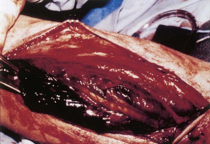
Most cases of purulent muscle infection occur in the tropics, and S. aureus is the predominant causative agent. Myositis caused by group A streptococci has been rare but occurs in many patients with necrotizing fasciitis and strep TSS. Most of these cases occur after blunt nonpenetrating trauma or occur spontaneously. Most likely, bacteria are translocated to the deep tissue hematogenously from the throat. Systemic toxicity is common, and mortality as high as 80% has been reported.205 Destruction of tissue is poorly understood, but infection and inflammation within the confined muscle compartment space may result in pressures exceeding arterial pressure, necessitating emergent fasciotomy and débridement (Fig. 199-7). There is much overlap in the clinical features of necrotizing fasciitis and myonecrosis,201,205 and the differentiation must be made by surgical inspection or biopsy.
Streptococcal Toxic Shock Syndrome
Strep TSS is defined as described in Table 199-2 but, simply put, it is any streptococcal infection associated with the sudden onset of shock and organ failure. Such cases were first described in the mid to late 1980s, and reports of strep TSS have subsequently emanated from North America, Europe, Australia, and Asia.172,177,178,206-213,214 Most cases have occurred sporadically. The highest incidence of invasive streptococcal disease occurred in a small Minnesota community, where 26 cases/100,000 population were recorded.213 In addition, outbreaks of invasive group A streptococcal infections have occurred in closed environments such as nursing homes215-219 and hospitals.220 Secondary cases of strep TSS are unusual, but transmission to family members220,221 or health care workers220,222 has been well documented by demonstrating identical pulsed-field gel electrophoresis patterns from cross-infecting strains. Although many of the studies cited earlier described strep TSS in adults, several reports have documented that this disorder also occurs in children.207,212,213,223,224 Thus, persons of all ages can be afflicted and, although some have underlying medical conditions such as diabetes and alcoholism,207,209,225-229 many have no predisposing medical condition and are not immunocompromised. This contrasts sharply to reviews of group A streptococcal bacteremia from several decades ago225-227 that found the disease to occur primarily among the very young, the very old, or patients with predisposing conditions such as cancer, renal failure, leukemia, severe burns, or iatrogenic immunosuppression.
TABLE 199-2
I. Isolation of group A streptococci (Streptococcus pyogenes) A. From a normally sterile site (e.g., blood, cerebrospinal, pleural, or peritoneal fluid, tissue biopsy, surgical wound) B. From a nonsterile site (e.g., throat, sputum, vagina, superficial skin lesion) II. Clinical signs of severity A. Hypotension: systolic blood pressure ≤90 mm Hg in adults or below fifth percentile for age in children B. Two or more of the following signs: 1. Renal impairment: creatinine ≥177 µmol/L (≥2 mg/dL) for adults or ≥2 times the upper limit of normal for age; in patients with preexisting renal disease, a twofold or greater elevation over the baseline level 2. Coagulopathy: platelets ≤100 × 109/L (≤100,000/mm3) or disseminated intravascular coagulation defined by prolonged clotting times, low fibrinogen level, and the presence of fibrin degradation products 3. Liver involvement: serum aspartate aminotransferase, alanine aminotransferase, or total bilirubin levels greater than or equal to two times the upper limit of normal for age; in patients with preexisting liver disease, a twofold or greater elevation over the baseline level 4. Adult respiratory distress syndrome defined by acute onset of diffuse pulmonary infiltrates and hypoxemia in the absence of cardiac failure, or evidence of diffuse capillary leak manifested by acute onset of generalized edema, or pleural or peritoneal effusions with hypoalbuminemia 5. A generalized erythematous macular rash that may desquamate 6. Soft tissue necrosis, including necrotizing fasciitis or myositis, or gangrene |
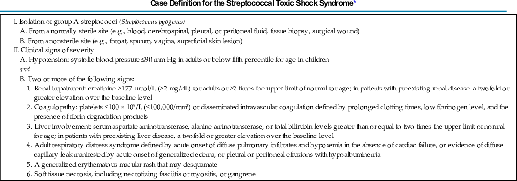
*An illness fulfilling criteria IA and II (A and B) can be defined as a definite case. An illness fulfilling criteria IB and II (A and B) can be defined as a probable case if no other cause for the illness is identified.
From The Working Group on Severe Streptococcal Infections. Defining the group A streptococcal toxic shock syndrome: rationale and consensus definition. JAMA. 1993;269:390-391.
The portals of entry for streptococci are the vagina, pharynx, mucosa, and skin in 50% of cases.176 Surgical procedures such as suction lipectomy, hysterectomy, vaginal delivery, bunionectomy, reduction mammoplasty, hernia repair, bone pinning, and vasectomy have provided portals in other cases (Table 199-3). Rarely, infection occurs secondary to streptococcal pharyngitis.230-232 Virus infections such as varicella and influenza have provided portals of entry in other cases.176,212,230
TABLE 199-3
| Age (neonates and older adults) |
| Diabetes |
| Alcoholism |
| Surgical procedures |
| Trauma |
| Penetrating (insect bites, lacerations, slivers, abrasions, burns) |
| Nonpenetrating (hematoma, bruise, muscle strain, hemarthrosis) |
| Varicella |
| Contact with a case |
| High prevalence of invasive strains in the community |
| Nonsteroidal anti-inflammatory drugs* |
*Based on limited evidence.
Additional factors that increase the risk for invasive group A streptococcal infection, including bacteremia, strep TSS, and necrotizing fasciitis, are listed in Table 199-3. Three studies have demonstrated that a high or increasing prevalence of M-1 or M-3 strains among throat isolates may signal an increased incidence of strep TSS in a community.212,228,233 NSAIDs taken for problems such as muscle strain, trauma, or postpartum pain may mask the early signs and symptoms of streptococcal infection or possibly predispose to more severe infection, such as necrotizing fasciitis or strep TSS.176,234,235
Pathogenesis
Entry of group A streptococci into the deeper tissues and bloodstream may occur as a result of breach of a barrier, or the organism itself may penetrate intact mucous membranes such as the pharyngeal mucosa. Although bacteremia is a very uncommon phenomenon in streptococcal pharyngitis, transient bacteremia must occur in those 50% of patients who develop invasive infections without a portal of entry. In either case, group A streptococci avoid phagocytosis largely because of the antiphagocytic properties of M protein.8 Adherence of group A streptococci to pharyngeal mucosal cells is a prerequisite to colonization or infection and has been related to surface structures, such as lipoteichoic acid and fibronectin-binding proteins. Penetration or translocation of group A streptococci through respiratory epithelial cells has been demonstrated for M-1 types of group A streptococci. Some have suggested that those M-1 strains possessing an invasin (inv+) gene penetrate more efficiently.236 If penetration of mucosal barriers occurs commonly, it does not result in clinically detectable bacteremia in the vast majority of patients, because the incidence of invasive infection is very low, that is, 3.5 cases/100,000 population.237 Thus, clearance of group A streptococci must be highly efficient in the vast majority of humans either because of preexisting type-specific immunity or nonspecific clearance mechanisms in the reticuloendothelial system. Recent studies have suggested that after colonization of mucosa or skin, SpeB may play a role in attenuation of the local host response because of its proteolytic activity. Later in the growth cycle, group A streptococcal production of SpeB is curtailed through alteration of the control of virulence regulator (CovR), allowing these particular strains to bind plasminogen, evade the immune system, and switch to an invasive phenotype.238
The enigma is how and why group A streptococcus causes deep infection of traumatized muscle and fascia in the absence of penetrating injury. Recent studies have demonstrated that injured muscle cells express vimentin on their surface during the healing and repair process and that group A streptococci bind vimentin.239 In addition, intravenously injected group A streptococci home to the site of muscle injury, and this process is augmented in the presence of NSAIDs.240
Mechanisms of Shock and Organ Failure: Cytokine Induction
Within the deeper tissues and bloodstream, the induction of cytokine synthesis plays a critically important role in the production of shock and organ failure. Pyrogenic exotoxins (scarlatina toxins, erythrotoxins) have the ability to cause fever, enhance susceptibility to endotoxin, suppress IgM antibody synthesis, and act as superantigens. These toxins, like the staphylococcal enterotoxins (A, B, C) and toxic shock syndrome toxin 1 (TSST-1), can stimulate T-cell responses through their ability to bind to both the major histocompatibility complex class II antigen-presenting cells and the V-β region of the T-cell receptor.241 The net effect is the induction of monocyte cytokines (tumor necrosis factor-α [TNF-α], interleukin [IL]-1β, and IL-6) as well as the lymphokines (TNF-β, IL-2, and interferon-γ).242-248 There is evidence that M protein fragments may also act as superantigens.246 Pyrogenic exotoxins C and MF, as well as SSA and several new streptococcal pyrogenic exotoxins, are also capable of inducing massive quantities of proinflammatory cytokines, as well as lymphokines,247 that contribute to shock and organ failure. Some clinical studies have suggested that variation in human leukocyte antigen haplotype may predispose worse outcomes in some patients with strep TSS.248
Other streptococcal virulence factors are also capable of inducing proinflammatory cytokines such as TNF-α and IL-1β. Specifically, SpeB, a potent cysteine protease, causes release of IL-1β from preformed intracellular pools.249 Streptolysin O also stimulates mononuclear cells to produce TNF-α and IL-1β and, in the presence of SpeA, has synergistic effects on IL-1β production.250 Heat-killed group A streptococci, as well as peptidoglycan and lipoteichoic acid, are also potent inducers of TNF-α and IL-1β.251,252 Noncytokine mechanisms of shock may also play a role. A cysteine protease produced by group A streptococci was shown to release bradykinin from a high-molecular-weight kininogen.253 Bradykinin is a potent vasodilator of systemic and pulmonary vasculature and could be responsible, at least in part, for the early hypotension observed in strep TSS.253
Thus, there are likely many streptococcal and host factors that contribute to the shock and organ failure characteristic of strep TSS. That TNF plays a central role is supported by two observations. First, high levels of TNF were observed in a baboon model of group A streptococcal bacteremia at a point in time when profound hypotension was manifest.254 Second, in that model, a neutralizing monoclonal antibody against TNF restored normal blood pressure and reduced mortality by 50%.254 Diffuse capillary leak contributes to volume depletion and hypotension and is likely related to cytokine release but may also be related to circulating M protein–fibrinogen complexes.255
Clinical Manifestations
The first phase of strep TSS begins with an influenza-like prodrome characterized by fever, chills, myalgias, nausea, vomiting, and diarrhea that precedes hypotension by 24 to 48 hours.176 Confusion and/or combativeness is present in 55% of patients. Where there is a defined or superficial portal of entry such as a laceration, suspicion of streptococcal infection or frank evidence of infection may be present at this phase of infection. In contrast, in patients without a portal of entry (50% of cases) and who subsequently develop necrotizing fasciitis or frank myonecrosis, postpartum infection, peritonitis, or joint space infection, pain that progressively increases in severity is the most common initial symptom that prompts patients to seek medical care and, interestingly, precedes clinical evidence of localized infection by 12 to 24 hours.176 In both children212 and adults176 the soft tissues are the most common primary site of infection. In the remaining cases, pneumonia, meningitis, endophthalmitis, meningitis, peritonitis, myocarditis, joint infection, and intrauterine infection have been described.176,212
The second phase of strep TSS is characterized by tachycardia, tachypnea, persistent fever, and, in patients who subsequently have necrotizing fasciitis or myonecrosis, increasingly severe pain at the site of infection. In others, fever and severe pain are the best early clinical clues.256 In children, toxicity during varicella or persistence of fever longer than 4 days should also prompt careful evaluation. Many patients are seen in emergency departments at this stage and frequently sent home on one or two occasions with mistaken diagnoses, such as deep vein thrombophlebitis, muscle strain, viral gastroenteritis, dehydration, and sprained ankle.202 High fever and excruciating pain, particularly in individuals with no risk factors for deep vein thrombosis, should arouse suspicion of a deep-seated infection. The laboratory tests described later are helpful, and CT and MRI may be useful to define the level of tissue injury but are not specific (see earlier discussion of necrotizing fasciitis).
The third phase of strep TSS is characterized by the symptoms and signs mentioned but with the sudden onset of shock and organ failure. Many patients are in florid shock at the time of admission, but in almost 50% of patients hypotension is apparent during the first 4 to 8 hours after admission. Clinical evidence of necrotizing fasciitis is frequently a late finding, often occurring after hypotension is present. The appearance of purple bullae and dusky-appearing skin is a bad prognostic sign and should prompt emergent surgical exploration (see earlier discussion of necrotizing fasciitis). It should be noted that currently the progression of necrotizing fasciitis from red skin to purple bullae may take place within a 24-hour period, whereas that described by Meleney in 1924 took 7 to 10 days. In addition, the rapidity with which shock and multiorgan failure can progress is impressive, and many patients die within 24 to 48 hours of hospitalization.176
Laboratory tests should be performed in patients with aggressive soft tissue infections or in patients with severe pain and fever who appear toxic. The serum creatinine measurement is particularly useful because renal impairment (creatinine level more than twice normal) is apparent even during the second phase, before hypotension is apparent. In addition, creatine phosphokinase levels in serum are markedly elevated in those with necrotizing fasciitis and myonecrosis. The white blood cell count is usually normal or elevated at admission but with a profound left shift that includes myelocytes and metamyelocytes. Finally, serum albumin and calcium levels are usually low on admission and drop precipitously as a diffuse capillary leak syndrome develops. Thrombocytopenia does not develop until later in the course but is the earliest sign of disseminated coagulopathy.176 Profound metabolic acidosis develops early in the third phase, and serum bicarbonate, lactate, and blood gas pH determinations are crucial tests to follow therapeutic progress. Because the acute respiratory distress syndrome develops in 55% of patients with strep TSS, pulse oximetry and, later, blood gas levels are necessary to evaluate the need for intubation and ventilation.
Management
Source Control
Prompt and aggressive surgical exploration and débridement of suspected deep-seated streptococcal infection are mandatory. It is as important to establish the cause of the infection as it is to determine the extent of necrosis. CT and MRI are helpful to locate the primary site of infection, but because the group A streptococci do not form gas or frank abscess, radiologic interpretations are frequently not definitive. Such findings in a patient with extreme pain and fever or who is toxic should prompt surgical consultation. Once necrosis is established, extensive débridement is necessary, because shock and organ failure continue to progress if devitalized tissue remains. Although necrosis of the fascia may be present, it is important to know that necrosis of muscle, skin, and subcutaneous tissue also is commonly present.
Fluid Resuscitation
If several liters of crystalloid intravenous fluid challenge do not rapidly improve blood pressure (mean arterial pressure >60 mm Hg) or tissue perfusion, then invasive monitoring is indicated. The goal should be to maintain a pulmonary artery occlusion pressure of 12 to 16 mm Hg.257 If this goal is reached but hypotension persists, the serum albumin concentration and hematocrit should be checked, because profoundly low albumin levels are common and because hemolysins produced by group A streptococci can cause dramatic drops in circulating red cell mass. Thus, transfusion with packed red blood cells, with or without albumin, may be useful to improve blood pressure and preserve tissue perfusion.
Because of intractable hypotension and diffuse capillary leak, massive amounts of intravenous fluids (10 to 20 L/day) may be required in an adult. Albumin replacement may be necessary because many patients' serum albumin levels decrease to lower than 2.0 g/dL.
Antimicrobial Therapy
Prompt antimicrobial therapy is mandatory, and empirical broad-spectrum coverage for septic shock should be instituted initially. Once the streptococcal cause is confirmed, high-dose penicillin and clindamycin should be given. This recommendation is based on the following information: (1) all strains of group A streptococci remain sensitive to penicillin; (2) resistance to erythromycin is currently found in less than 5% of group A streptococci in the United States, but the rate is higher in certain locales, and there have been rare reports of resistance to clindamycin; (3) clindamycin and erythromycin are more active in experimental models of necrotizing fasciitis and myonecrosis; (4) penicillin-binding proteins are not expressed during stationary-phase growth of group A streptococci, and thus penicillin is ineffective in severe deep infections in which large numbers of bacteria are present; (5) clindamycin suppresses exotoxin and M protein production by group A streptococci; (6) clindamycin has a much longer half-life; (7) combinations of penicillin and clindamycin have indifferent interaction against group A streptococci in vitro at clinically relevant concentrations of antibiotics (no antagonistic effects were found)258; and (8) clindamycin and azithromycin suppress cytokine production by human mononuclear cells.259,260
Management in the Intensive Care Unit
In patients with persistent hypotension, monitoring of cardiac outputs, pulmonary artery occlusion pressure, and mean arterial pressure is important. Intubation and ventilator support are usually required because of the high incidence of acute respiratory distress syndrome (55%) in patients with strep TSS. Pressors such as dopamine are used frequently, although no controlled trials have been performed in strep TSS. In patients with intractable hypotension, high doses of dopamine, epinephrine, or phenylephrine have been used, but caution should be exercised in those with evidence of disseminated intravascular coagulation and in particular in those with cold, cyanotic digits. Symmetrical gangrene involving all 20 digits and toes, the tip of the nose, ear lobes, and the breast areola have been described. In addition, we have observed amputation of one, two, three, and even four extremities. In these cases, both excessive pressors and disseminated intravascular coagulation likely contributed to symmetrical gangrene.
Dialysis and Hemoperfusion
Either of these methodologies may be necessary because more than 50% of patients develop acute renal failure. Both dialysis and hemoperfusion may also nonspecifically reduce the concentrations of circulating toxins. It is interesting that a study from Sweden that used hemofiltration achieved the lowest mortality rate in patients with strep TSS ever recorded.229 Finally, a polystyrene superantigen absorbing device was developed in Japan and shown to be highly efficacious in absorbing pyrogenic exotoxin A or TSST-1 from plasma and, when used extracorporeally in animals infused with TSST-1 and lipopolysaccharide, mortality improved from 100% to 50%.261
Intravenous Immune Globulin
The rationale for the use of intravenous immune globulin (IVIG) in the treatment of strep TSS is based on the data implicating extracellular exotoxins as mediators of shock and organ failure. George and Gladys Dick, in 1924, demonstrated that convalescent sera from patients with scarlet fever neutralized scarlatina toxins and, when passively administered, attenuated the course of severe scarlet fever.262 Just as penicillin was becoming available, anti–scarlatina toxin horse serum became commercially available in the United States; however, because of the availability of penicillin and the decline in the severity of scarlet fever, it was never used. Several reports have described the successful use of IVIG in patients with strep TSS.263-265 The largest treatment group (15 patients) showed a significant reduction in mortality compared with matched historical controls.266 The mortality rate of 70% in the control group was among the highest ever reported, whereas the rate was 30% in the IVIG group. This rate is similar to that of some series that did not use IVIG.176 A double-blind clinical trial was undertaken in northern Europe comparing IVIG with albumin in patients with strep TSS. All patients received clindamycin. The mortality rate in the IVIG group was 16%, whereas that in the albumin group was 32%.267 Unfortunately, the study was stopped because of low enrollment and only seven or eight patients with proved group A streptococcal infections were in each group. Thus, the differences were not significant. A retrospective study in patients with strep TSS has also shown no benefit of IVIG on mortality.268 It is hoped that further double-blind studies with sufficient numbers of cases will resolve the continuing dilemma regarding the potential efficacy of IVIG.269 It is clear that if IVIG were to be used, it should be given early and probably more than one dose should be given, because batches of IVIG have variable neutralizing activity against streptococcal exotoxins.247,270
There have been no comparative trials describing the efficacy of hyperbaric oxygen treatment in strep TSS, although some state that such treatment reduces mortality and the need for further débridements.271 Certainly, use of this modality should not delay or be used in preference to surgical débridement when the latter is indicated.
Bacteremia
Group A streptococcal bacteremia has been relatively uncommon in the antibiotic era.272 Before the mid-1980s, bacteremia occurred predominantly at the extremes of life and was usually community acquired. Occasional cases were seen in young and middle-aged adults associated with surgical wound infections and endometritis.
During the past decade, however, there has been an increase in the number of reported cases of group A streptococcal bacteremia, reflecting the changing epidemiology and clinical patterns of invasive streptococcal infection as noted earlier. Many of the patients were previously healthy adults between the ages of 20 and 50 years. There has been an apparent increase in cases associated with parenteral injection of illicit drugs,81,176,227 as well as nosocomial outbreaks in nursing homes.215-219
Bacteremia in children may emanate from an upper respiratory tract infection, but it is more commonly associated with cutaneous foci, including burns and varicella.222 Older patients with streptococcal bacteremia present with a variety of chronic illnesses; their relation to the bacteremia is often unclear. Diabetes mellitus, cirrhosis, and peripheral vascular disease do appear, however, to be predisposing factors in older adults and, as in children, the portal of entry is usually the skin. Malignancy and immunosuppression are risk factors in both age groups.201,273 Although group A streptococcal bacteremia may at times be transient and relatively benign,274 it is more often fulminant. The onset is abrupt, with chills, high fever, and prostration. Rarely, patients may present with acute abdominal pain.274,275 Mortality in five modern series225,274-277 has ranged from 27% to 38%.
Other Streptococcal Infections
Lymphangitis may accompany cellulitis or may occur after clinically minor or inapparent skin infection. Lymphangitis is readily recognized by the presence of red, tender, linear streaks directed toward enlarged, tender, regional lymph nodes. It is accompanied by systemic symptoms such as chills, fever, malaise, and headache. Puerperal sepsis follows abortion or delivery when streptococci colonizing the patient herself or transmitted from medical personnel invade the endometrium and surrounding structures, lymphatics, and bloodstream. The resulting endometritis and septicemia may be complicated by pelvic cellulitis, septic pelvic thrombophlebitis, peritonitis, or pelvic abscess. This disease was associated with high mortality in the preantibiotic era. Although endocarditis caused by S. pyogenes was relatively common in the preantibiotic era, it is now rarely reported.278,279 Meningitis caused by S. pyogenes usually follows upper respiratory tract infection, including sinusitis or otitis,280 or neurosurgical conditions.281 It is indistinguishable clinically from other forms of acute pyogenic meningeal infection.282
Pneumonia caused by S. pyogenes is frequently associated with preceding viral infections such as influenza, measles, or varicella or with chronic pulmonary disease. Numerous epidemics have been described in military recruit populations.283,284 An increased number of cases has been reported over the past few years in association with the resurgence of invasive streptococcal infections. In one third or fewer of the cases, there was a history of preceding streptococcal upper respiratory tract infection. The onset is typically abrupt, and the disease is characterized by chills, fever, dyspnea, cough productive of blood-streaked sputum, pleuritic chest pain, and, in more severe cases, cyanosis. The pulmonary picture is that of bronchopneumonia, with consolidation being uncommon. Empyema develops in 30% to 40% of cases, tends to appear early in the disease, and typically consists of copious amounts of thin serosanguineous fluid. Bacteremia occurs in 10% to 15% of cases. Complications include mediastinitis, pericarditis, pneumothorax, and bronchiectasis; and the clinical course of the disease is often prolonged. Mortality has generally been low with penicillin therapy and adequate drainage of empyema, perhaps reflecting its occurrence in healthy military recruits. However, in a recent Canadian report of 222 cases of community-acquired pneumonia among adults (median age, 56 years), the case-fatality rate was 38%.285 Interestingly, a recent review of deaths in the 1918 pandemic of influenza has demonstrated that the major cause of death was secondary bacterial pneumonia.286 Whereas S. pneumoniae was the most common, group A streptococcus was second and S. aureus was third. Among patients with empyema complicating pneumonia, group A streptococcus was the most common cause. Investigators have demonstrated in a mouse model that a nonlethal influenza infection greatly enhanced the severity and mortality of secondary respiratory tract infection after challenge with group A streptococcus.287
Group A streptococcal perianal cellulitis and vulvovaginitis are symptomatic but benign disorders primarily affecting children.288,289 Asymptomatic carriage of group A streptococci in the vagina, anus, scalp, or, rarely, upper respiratory tract of adults has, however, been the source of outbreaks of nosocomial streptococcal infection.290
Prophylaxis and Risk for Secondary Cases of Streptococcal Toxic Shock Syndrome
Strep TSS is most commonly community acquired and sporadic, yet clusters of invasive cases have been described in nursing homes,215-219 families,220,221 and hospital workers.222,291 In San Francisco, 23 hospital workers became colonized or infected with group A streptococci as a result of contact from a single case of strep TSS.291 This example, as well as many historical studies in schools, military posts, and nursing homes, shows that group A streptococci are highly contagious. Luckily, mere contact or colonization is usually not sufficient to cause a secondary case of invasive group A streptococcal infection. Epidemiologic studies by the Centers for Disease Control and Prevention found one secondary case of invasive infection among more than 1,500 contacts.146 This would extrapolate to 66/100,000 population/year for secondary cases.292 As noted, the current incidence of primary cases of invasive group A streptococcal infections in the United States is 3.5/100,000 population/year. Thus, the risk to contacts is roughly 20 times greater than that for the general population but is still very low. Given the relative infrequency of these infections and the lack of a clearly effective chemoprophylactic regimen, routine screening for and prophylaxis against streptococcal infection are not recommended for household contacts of index patients. In deciding who should receive prophylaxis, the clinician needs to factor in the duration of contact, intimacy of contact, and underlying host factors of individual contacts. Specifically, contacts with open wounds, recent surgery, recent childbirth, concurrent viral infections such as varicella or influenza, or immunodeficiency diseases should receive prophylaxis. In a multicenter study of adults aged 18 to 45 years, human immunodeficiency virus infection and injecting drug use were independently associated with an increased risk for invasive group A streptococcal disease. In those aged 45 years of age or older, diabetes, cardiac disease, cancer, and corticosteroid use were significant risk factors.293 Moreover, adults 65 years or older are at increased risk for mortality should they contract invasive disease. Thus, it may be prudent to initiate prophylaxis in households with older adults or those with the just-mentioned risk factors.
Lacking firm data on which to base antimicrobial prophylaxis, it seems reasonable to choose those agents that have achieved highest rates of pharyngeal eradication in asymptomatic individuals; among these are clindamycin and azithromycin. Specific regimens have been published elsewhere.146