Chapter 49 Acrocyanosis
The term acrocyanosis is derived from the Greek words akron (meaning “extremity”) and kyanos (meaning “blue”). As a medical condition, acrocyanosis is an uncommon functional vasospastic disorder characterized by persistent bluish discoloration, primarily of the hands and feet. The original description of acrocyanosis is credited to Crocq in 1896, thus the eponym Crocq’s disease. Classically, acrocyanosis affects young asthenic females in regions with cooler climates. In contrast to Raynaud’s syndrome, the bluish discoloration of acrocyanosis is persistent rather than episodic and is not associated with discomfort.1 Primary acrocyanosis is not associated with an identifiable cause and is usually a benign disorder with a good prognosis; skin ulceration and tissue loss are extremely rare. Patients with primary disease usually do well with behavioral measures such as avoidance of exposure to cold; pharmacological therapy is rarely necessary. Secondary acrocyanosis occurs in association with another underlying disorder. An acrocyanotic appearance may be seen in conjunction with any disease that causes central cyanosis or markedly decreases blood flow to the extremity. Treatment and prognosis of secondary acrocyanosis depends on the underlying condition.1
Epidemiology
Limited information is available on many of the epidemiological aspects of primary acrocyanosis, including its precise incidence and prevalence. Acrocyanosis is believed to occur more frequently with a low body mass index (BMI), outdoor occupations, and in areas with cooler climates.2 Despite early reports suggesting similar gender predilection, acrocyanosis seems to affect women more frequently than men, with a female-to-male ratio of 6:1.3 It is more common in younger individuals, the typical age range being 20 to 45 years.4 In addition, occurrence of acrocyanosis seems to decrease with increasing age, regardless of the regional climate.5 It is seen frequently in patients with anorexia nervosa, affecting up to 20% of women with this disorder.6 It has been suggested that as many as 10% of patients have a family history of acrocyanosis in one of the first-degree relatives, suggesting a genetic basis.4
Etiology
As already noted, acrocyanosis is commonly a primary or idiopathic disorder that is not associated with an identifiable cause. It also occurs as a secondary condition in association with connective tissue disorders, some hematological conditions, anorexia nervosa, neurovascular disorders, drugs, toxins, infections, heritable metabolic diseases, and some malignancies.7 An acrocyanotic appearance also occurs in conjunction with any disease that causes central cyanosis or markedly decreases blood flow to the extremities (Box 49-1).
 Box 49-1
Pathophysiology
Acrocyanosis is due to a decrease in the amount of oxygen delivered to the tissues of the extremities. Several mechanisms for primary acrocyanosis have been proposed, but the precise mechanism remains elusive. Potential pathophysiological disturbances include abnormal arteriolar tone, alteration of microvascular responsiveness with capillary and venular dilation and stasis, and abnormal sympathetic nervous system activity (Box 49-2).
The essential abnormality in primary acrocyanosis seems to be peripheral cutaneous vasoconstriction due to increased tone of the arterioles, associated with secondary vasodilation of capillaries and subpapillary venous plexi.8 Arteriolar vasoconstriction produces the cyanotic discoloration, and compensatory venular dilation in the postcapillary sphincter leads to excessive sweating. Persistent vasoconstriction at the precapillary sphincter creates a local hypoxic environment that may cause increased release of vasodilatory mediators such as adenosine in the capillary beds.9 This in turn leads to dilation of postcapillary venules. The difference in the vessel tone may create a countercurrent exchange system in an attempt to retain heat, and this leads to sweating. Potential mediators that may contribute to the pathogenesis of acrocyanosis include serotonin, adrenaline, noradrenaline, and endothelin (ET)-1. Levels of these mediators have been shown to be increased in acrocyanosis.3,10
The exact cause of the disordered arteriolar tone in acrocyanosis is unknown. Most evidence points to local sensitivity of the arterioles to cold and an exaggerated sympathetic nervous system response. Earlier studies suggested an exaggerated digital arteriolar vasoconstrictive response to cold stimuli,11 whereas subsequent studies have implicated the sympathetic nervous system.1 An interesting case described normalization of cutaneous thermal-induced vasoreactivity in the hands of a young girl with acrocyanosis during phenobarbital-induced sleep. In this state, her hands responded in the same manner as the rest of body to heat and cold exposure. This finding supports the hypothesis that the abnormal vascular tone in acrocyanosis may be of central origin.12 Other mechanisms hypothesized include increased blood viscosity,13 decreased distal blood flow at low temperatures, and persistently elevated vasoconstrictive mediators such as endothelin-1 levels,14 but convincing supporting evidence for these is not readily available.
Clinical Presentation
Primary acrocyanosis typically occurs in thin young women who are not physically active. They commonly present with persistent bluish discoloration of the hands and feet. The distribution of the discoloration is symmetrical.5 It less frequently involves forearms, ears, lips, nose, or nipples. It is the discoloration that usually prompts patients to seek medical care. Patients may also describe a sensation of coolness of the affected areas, but generally will not have pain. There may be slight swelling of the digits and increased perspiration. Although the discoloration is persistent, emotion and cold temperature can intensify the acrocyanotic appearance, and warmth can diminish it.1 Discoloration and coolness are worse in cold weather, but typically persist even during the summer months.
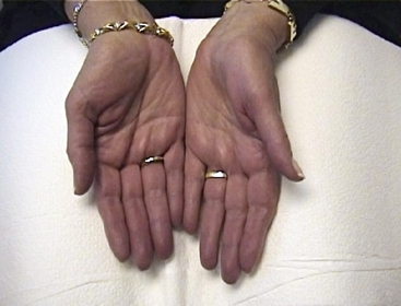
Examination reveals bluish discoloration of the hands and feet, which are cool and often sweaty (Fig. 49-1). The color has also been described as bluish-pink or orange-like tinged with red, and differing hues may be seen at various times. A brownish-yellow color on the dorsum of hands and feet has been described and attributed to exposure to the warmth of the sun because it is most obvious in summer. Pressure on the discolored skin causes pallor, and color returns slowly and irregularly. Fingers may be puffy, but trophic changes do not occur.
In patients with primary disease, there are no other abnormal physical findings. Arterial pulses are normal, suggesting there is no large-vessel arterial obstruction.8 Elevation of a cyanotic limb above heart level produces pallor, suggesting venous obstruction is not present. Patients with secondary acrocyanosis may have clinical features of the underlying condition. Episodes of digital pallor or rubor do not occur, although patients may have both acrocyanosis and Raynaud’s phenomenon. This is especially true in patients with scleroderma or systemic lupus erythematosus (SLE) as an underlying cause.
Diagnosis
Primary acrocyanosis is diagnosed clinically based on a careful history and physical examination. Laboratory or imaging studies typically are not necessary.15 Cyanosis is symmetrical in distribution and will be present at the time of examination, since it is persistent. In addition, there are usually no associated trophic skin changes, localized pain, or ulcerations. Peripheral pulse examination is normal, excluding significant peripheral artery occlusive disease. Pulse oximetry reveals normal oxygen saturation.
Secondary acrocyanosis is suggested by asymmetrical distribution of the discoloration, presence of pain, or trophic changes of the fingers or toes. Hypoxemia or absent pulses suggest central cyanosis or arterial obstructive disease, respectively. Selected laboratory tests are necessary to confirm the diagnosis of secondary acrocyanosis. Arterial blood gas is appropriate to evaluate for hypoxemia. Antinuclear antibodies (ANAs) are typically absent, but if present suggest an underlying connective tissue disorder. If the history and physical examination are suggestive, directed testing for cryoglobulins, antiphospholipid antibodies, cold agglutinins, or antibodies to Epstein-Barr virus should be performed.
Nailfold capillaroscopy is often abnormal in acrocyanosis. Features include abnormal capillary morphology, pericapillary edema, and even hemorrhage. Findings on capillaroscopy are often nondiagnostic and may be associated with secondary disease. It has been shown that mean capillary density is decreased in patients with acrocyanosis compared with normal subjects, but not to the extent seen in patients with Raynaud’s phenomenon due to scleroderma.16 Other capillary abnormalities include capillary loops of diameter larger than normal. The presence of megacapillaries and areas of sparse or absent capillaries suggest underlying scleroderma or mixed connective tissue, whereas bushy capillaries suggest underlying systemic lupus erythematosus. Other findings such as decreased oscillations over the radial artery and decreased digital vessel sounds when assessed by Doppler have been described but are nonspecific.17
Histopathology
Cutaneous biopsy is usually unnecessary for the diagnosis of acrocyanosis. Findings described on biopsy are thickening of the medial coat of arterioles and dilation of papillary and subpapillary capillaries. Other features include mild perivascular infiltration, mild dermal edema, and skin fibrosis with formation of new blood vessels.4
Differential Diagnosis
Primary acrocyanosis should be distinguished from other cold-related vasospastic conditions such as Raynaud’s syndrome, pernio, and livedo reticularis.15 This determination is based upon general appearance, distribution, and persistence of the cyanosis. Then the finding of acrocyanosis requires consideration of other local or systemic features that would suggest an underlying secondary disorder.18
Systemic hypoxemia with increased deoxyhemoglobin is a common etiology of an acrocyanotic appearance. Presence of central cyanosis with arterial hypoxemia should prompt a search for underlying cardiopulmonary disease.19 Peripheral cyanosis may result from an alteration in hemoglobin (Hb), with accumulation of the oxidized form, methemoglobin.20 This may be seen with exposure to oxidizing agents such as aniline, benzocaine, dapsone, phenazopyridine, nitrates, and naphthalene. Cold agglutinins and cryoglobulinemia may cause acrocyanosis.21,22 Cold agglutinins may be associated with infectious mononucleosis and Mycoplasma pneumonia and also occur in lymphoproliferative disorders. Acrocyanosis has been reported as a presenting feature of Hodgkin lymphoma.23 Acrocyanosis is also caused by cryoglobulinemia associated with hepatitis C infection and several other diseases.24
Acrocyanosis is well described in anorexia nervosa.25 It tends to occur in the more severely ill and malnourished patients. Typically these patients have pallor of the face and trunk, slower pulse rates, and high fasting plasma glucose levels. These patients also have decreased distal blood flow and impaired vasodilation to a heat stimulus.6
Numerous connective tissue disorders may manifest acrocyanosis, but other signs of these diseases are usually present. Patients with scleroderma and SLE may have both acrocyanosis and episodic attacks of Raynaud’s phenomenon.16,26 Antinuclear antibodies may be detected in patients with connective tissue disorders. Acrocyanosis is also present in up to one third of patients with antiphospholipid antibodies, and may be seen in conjunction with other cutaneous manifestations such as dermatographism, urticaria, livedo reticularis, cutaneous nodules, ulceration, and purpura.27,28
Several medications and toxins are associated with acrocyanosis, including tricyclic antidepressants, interferon alpha (IFN-α)-2a, amphotericin B, arsenic, and butyl nitrite.29–33 Therefore, it is important to consider medications or toxic substances as a potential cause.
Acrocyanosis has been observed in several neurological disorders with suspected neurovascular instability. An acrocyanotic appearance may be seen following spinal cord injuries or with complex regional pain syndrome.34 This later disorder often occurs following trauma and is typified by pain and autonomic dysregulation. Acrocyanosis and lower-extremity edema has also been described in postural orthostatic tachycardia syndrome and is ascribed to venous pooling.35
Acrocyanosis secondary to inherited metabolic disorders is more likely to present during childhood or adolescence. These disorders are suspected when other characteristic associated manifestations are present. Infants with ethylmalonic aciduria have petechiae, diarrhea, pyramidal signs, and mental retardation.36 Children with mitochondrial disease may have hair abnormalities, pigmentation disorders, and hypertrichosis.37
Acrocyanosis may also be present in patients with microemboli, microthrombi, or atheromatous embolism. Patients with atheromatous emboli are typically older with other signs of atherosclerosis, and they have other cutaneous signs such as petechiae, livedo reticularis, and painful skin lesions on the toes (so-called blue toe syndrome).38 Microthrombi and/or microemboli may occur in acutely ill patients with disseminated intravascular coagulation (DIC), heparin-induced thrombocytopenia (HIT), or septic embolism due to endocarditis.39
Treatment
Primary acrocyanosis is a benign condition that usually requires only conservative measures; there is no effective curative medical or surgical treatment (Box 49-3). Treatments typically focus on symptom relief or more often cosmetic appearance. Some patients are so affected they avoid social contact.15 Reassurance and behavioral measures, such as avoidance of exposure to cold and use of protective clothing, often improve the discoloration.40 In addition, psychophysiological measures such as biofeedback training, conditioning of reflexes, and hypnosis may give partial relief.
Pharmacological intervention is rarely necessary. Various drugs have been advocated, although no controlled studies have been performed. Calcium channel blockers, nicotinic acid derivatives, adrenergic blocking agents (rauwolfia, guanethidine, reserpine, and α-blockers), cyclandelate, topical minoxidil, and rutin compounds have been claimed to provide symptomatic improvement.18 Bromocriptine has been reported to relieve acrocyanosis in a few days, but may induce Raynaud’s phenomenon in about one third of patients.41 Sympathectomy or disrupting the fibers of the sympathetic nervous system to the area usually will alleviate acrocyanosis. However, such a drastic procedure is rarely appropriate for a benign disease.42
Treatment of secondary acrocyanosis depends on the underlying cause.
Prognosis
Although there is no cure, the prognosis of primary acrocyanosis is very good. It is not associated with increased risk of death, amputation, or other complications. Apart from the discoloration, patients with primary acrocyanosis usually have no other signs or symptoms such as pain, skin ulcerations, or tissue loss. Patients can expect to lead normal lives. In contrast, the prognosis of secondary acrocyanosis seems to vary widely depending upon the underlying condition.
1 Coffman J.D. Acrocyanosis and livedo reticularis. In Raynaud’s phenomenon. New York: Oxford University Press; 1989. pp 167–170
2 Carpentier P.H. Definition and epidemiology of vascular acrosyndromes. Rev Prat. 1998;48:1641–1646.
3 Davis E. Clinical aspects of acrocyanosis. Adv Microcirc. 1982;10:101–106.
4 Stern E.S. The aetiology and pathology of acrocyanosis. Br J Derm. 1937;49:100–108.
5 Creager M.A., Dzau V.J. Vascular disease of the extremity. In: Kasper D.L., Fauci A.S., Lango D.L. Harrison’s principles of internal medicine. ed 16. New York: McGraw-Hill; 2005:1490–1495.
6 Hediger C., Rost B., Itin P. Cutaneous manifestations in anorexia nervosa. Schweiz Med Wochenschr. 2000;130:565–575.
7 Brown P.J., Zirwas M.J., English J.C. The purple digit an algorithmic approach to diagnosis. Am J Clin Dermatol. 2010;11:103–116.
8 Mansilha A., Sampaio S. Vasospastic disorders of the upper extremities. In: Liapis C.D., Balzer K., Benedetti-Valentini F., Fernandes J. European manual of medicine: vascular surgery. Berlin: Springer-Verlag; 2007:237–246. Part 3
9 Bollinger A. Function of the precapillary vessels in peripheral vascular disease. J Cardiovasc Pharmacol. 1985;7:S147–S151.
10 Kurklinsky A.K., Miller V.M., Rooke T.W. Acrocyanosis: the Flying Dutchman. Vasc Med. 2011;16:288–301.
11 Lottenbach K. Vascular response to cold in acrocyanosis. Helvetia Medica Acta. 1967;33:437–444.
12 Day R., Klingman W.O. The effect of sleep on the skin temperature reactions in a case of acrocyanosis. J Clin Invest. 1939;8:271–276.
13 Coperman P.W. Acrocyanosis: a blood disease? Proc R Soc Med. 1973;66:741–742.
14 Mangiafico R.A., Malatino L.S., Santonocito M., et al. Plasma endothelin-1 concentrations during cold exposure in essential acrocyanosis. Angiology. 1996;47:1033–1038.
15 Heidrich H. Functional vascular diseases: Raynaud’s syndrome, acrocyanosis and erythromelalgia. Vasa. 2010;39:33–41.
16 Monticone G., Colonna L., Palmeri G., et al. Quantitative nailfold capillary microscopy findings in patients with acrocyanosis compared with patients having systemic sclerosis and normal subjects. J Am Acad Dermatol. 2000;42:787–790.
17 Davis E. Oscillometry of radial artery in acrocyanosis and cold sensitivity. J Mal Vasc. 1992;17:214–217.
18 Nousari H.C., Kimyai-Asadi A., Anhalt G.J. Chronic idiopathic acrocyanosis. J Am Acad Dermatol. 2001;45:S207–S208.
19 Golombek S.G., Ally S., Woolf P.K. A newborn with cardiac failure secondary to a large vein of Galen malformation. South Med J. 2004;97:516–518.
20 Jiminez M.A., Polena S., Coplan N.L., et al. Methemoglobinemia and transesophageal echo. Proc West Pharmacol Soc. 2007;50:134–135.
21 Cholongitas E., Ioannidou D. Acrocyanosis due to cold agglutinins in a patient with rheumatoid arthritis. J Clin Rheumatol. 2009;15:375.
22 Sinha A., Richardson G., Patel R.T. Cold agglutinin related acrocyanosis and paroxysmal haemolysis. Eur J Vasc Endovasc Surg. 2005;30:563–565.
23 Solak Y., Aksoy S., Kilickap S., et al. Acrocyanosis as a presenting symptom of Hodgkin lymphoma. Am J Hematol. 2006;81:151–152.
24 Trejo O., Ramos-Casals M., Garcia-Carrasco M., et al. Cryoglobulinemia: study of etiological factors and clinical and immunologic features in 443 patients from a single center. Medicine. 2001;80:252–262.
25 Strumia R. Skin signs in anorexia nervosa. Dermatoendocrinol. 2009;1:268–270.
26 Richter J.G., Sander O., Schneider M., et al. Diagnostic algorithm for Raynaud’s phenomenon and vascular skin lesions in systemic lupus erythematosus. Lupus. 2010;19:1087–1095.
27 Vayssairat M., Abauf N., Baudot N., et al. Abnormal IgG cardiolipin antibody titers in patients with Raynaud’s phenomenon and/or related disorders: prevalence and clinical significance. J Am Acad Dermatol. 1998;38:555–558.
28 Diógenes M.J., Diógenes P.C., de Morais Carneiro R.M., et al. Cutaneous manifestations associated with antiphospholipid antibodies. Int J Dermatol. 2004;43:632–637.
29 Anderson R.P., Morris B.A. Acrocyanosis to imipramine. Arch Dis Child. 1988;63:204–205.
30 Ozaras R., Yemisen M., Mete B., et al. Acrocyancosis developed with amphotericin B deoxycholate but not amphotericin B lipid complex. Mycoses. 2007;50:242.
31 Campo-Voegeli A., Estrach T., Marti R.M., et al. Acrocyanosis induced by interferon alpha (2a). Dermatology. 1998;196:361–363.
32 Hall A.H. Chronic arsenic poisoning. Toxicol Lett. 2002;128:69–72.
33 Hoegl L., Thoma-Greber E., Poppinger J., et al. Butyl nitrite-induced acrocyanosis in an HIV infected patient. Arch Dermatol. 1999;135:90–92.
34 Glazer E., Pacanowski J.P., Leon L.R. Asymptomatic lower extremity acrocyanosis: report of two cases and review of the literature. Vascular. 2011;19:105–110.
35 Steward J.M., Gewitz M.H., Weldon A., et al. Patterns of orthostatic intolerance: the orthostatic tachycardia syndrome and adolescent chronic fatigue. J Pediatr. 1999;135:218–221.
36 Burlina A.B., Dionisi-Vici C., Bennett M.J., et al. A new syndrome with ethylmalonic aciduria and normal fatty acid oxidation in fibroblasts. J Pediatr. 1994;124:79–86.
37 Boderman C., Rötig A., Rustin P., et al. Hair and skin disorders as signs of mitochondrial diseases. Paediatrics. 1999;103:428–433.
38 Yücel A.E., Kart-Köseoglu H., Demirhan B., et al. Cholesterol crystal embolization mimicking vasculitis: success with corticosteroid and cyclophosphamide therapy in two cases. Rheumatol Int. 2006;26:454–460.
39 Sharma B.D., Kabra S.R., Gupta B. Symmetrical peripheral gangrene. Trop Doct. 2004;34:2–4.
40 Planchon B., Becker E., Carpentier P.H., et al. Acrocyanosis: changing concepts and nosological limitations. J Mal Vasc. 2001;26:5–15.
41 Morrish D.W., Crockford P.M. Acrocyanosis treated with bromocriptine. Lancet. 1976;2:851.
42 Grima M.R. Nursing case study: bilateral cervical sympathectomy for acrocyanosis. Nurs Times. 1975;71:1850–1852.