Chapter 154 Scleroderma and Raynaud Phenomenon
Juvenile scleroderma encompasses a range of conditions unified by the presence of fibrosis of the skin. Juvenile scleroderma is divided into 2 major categories, localized scleroderma (LS, also known as morphea), which is largely limited to the skin, and systemic sclerosis (SSc), with organ involvement. Although localized disease is the predominant type seen in pediatric populations, systemic sclerosis is associated with severe morbidity and mortality.
Etiology and Pathogenesis
The etiology of scleroderma is unknown, but the mechanism of disease appears to be a combination of a vasculopathy, autoimmunity, immune activation, and fibrosis. Triggers, including trauma, infection, and, possibly, subclinical graft versus host reaction from persistent maternal cells (microchimerism), injure vascular endothelial cells, resulting in increased expression of adhesion molecules. These molecules entrap platelets and inflammatory cells, resulting in vascular changes with manifestations such as Raynaud phenomenon and pulmonary hypertension. Inflammatory cells infiltrate the area of initial vascular damage, causing further vascular damage and resulting in thickened artery walls and reduction in capillary numbers. Macrophages and other inflammatory cells then migrate into affected tissues and secrete cytokines that induce fibroblasts to reproduce and synthesize excessive amounts of collagen, resulting in fibrosis and subsequent lipoatrophy, dermal fibrosis, and loss of sweat glands and hair follicles. In late stages, the entire dermis may be replaced by compact collagen fibers.
Autoimmunity is believed to be a key process in the pathogenesis of both localized and systemic scleroderma, given the high percentage of affected children with autoantibodies. Children with localized disease often have a positive ANA test result (42%), and 47% of this subgroup have antihistone antibodies. Other autoantibodies seen include rheumatoid factor (RF) (16%) and antiphospholipid antibodies (12%). The relationship between specific autoantibodies and the various forms of scleroderma is not well understood, and all antibody test results may be negative in a child, especially one who has LS.
Classification
Localized scleroderma is distinct from systemic scleroderma and rarely progresses to systemic disease. Within the category of LS there are several subtypes that are differentiated by both the distribution of the lesions and the depth of involvement (Table 154-1). Up to 15% of children have a combination of 2 or more subtypes.
Table 154-1 CLASSIFICATION OF PEDIATRIC SCLERODERMA (MORPHEA)
LOCALIZED SCLERODERMA
Plaque Morphea
Generalized Morphea
Bullous Morphea
Bullous lesions that can occur with any of the subtypes of morphea
Linear Scleroderma
Linear lesions can extend through the dermis, subcutaneous tissue, and muscle to underlying bone; more likely unilateral
Deep Morphea
SYSTEMIC SCLEROSIS
Diffuse
Limited
Epidemiology
Juvenile scleroderma is rare, with an estimated prevalence of 1/100,000. Localized scleroderma is far more common than SSc in children, by a 10 : 1 ratio, with plaque morphea and linear scleroderma being the most common subtypes. Linear scleroderma is predominantly a pediatric condition, with 65% of patients diagnosed before age 18. After age 8 yr the female : male ratio for both LS and SSc is approximately 3 : 1, whereas in patients younger than 8 yr there is no sex predilection.
Clinical Manifestations
Localized Scleroderma
The onset of scleroderma is generally insidious, and manifestations vary according to disease subtype. The initial skin manifestations of localized disease usually include erythema or a bluish hue seen around an area of waxy induration; subtle erythema may be the only presenting sign (Fig. 154-1). Early edema and erythema are followed by indurated, hypopigmented or hyperpigmented, atrophic lesions (Fig. 154-2). Linear scleroderma varies in size from a few centimeters to the entire length of the extremity, with varying depth. Patients sometimes present with arthralgias, synovitis, or flexion contractures (Fig. 154-3). Children also experience limb length discrepancies as a result of growth impairment due to involvement of muscle and bone. Children with en coup de sabre (Fig. 154-4) may have symptoms unique to central nervous system (CNS) involvement, such as seizures, hemifacial atrophy, ipsilateral uveitis, and learning/behavioral changes.
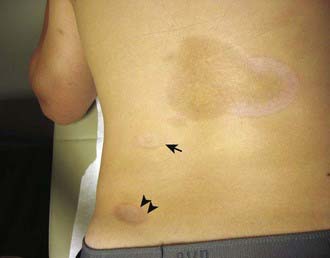
Figure 154-1 Boy with generalized morphea. Note the active circular lesion (arrowheads) with a surrounding rim of erythema. The largest lesion has areas of postinflammatory hyperpigmentation and depression with an area of erythema on the right. The small lesion (arrow) demonstrates depression due to lipoatrophy.
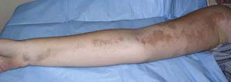
Figure 154-2 Inactive linear scleroderma demonstrating hyperpigmented lesion with areas of normal skin (skip lesions).
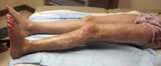
Figure 154-3 Child with untreated linear scleroderma resulting in knee contracture, immobility of ankle, chronic skin breakdown of scar on the lateral knee, and areas of hypopigmentation and hyperpigmentation. The affected leg is 1 cm shorter.
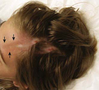
Figure 154-4 Child with en coup de sabre lesion on scalp extending down to forehead. Prior to treatment the skin on the scalp was bound down with chronic skin breakdown. Note the area of hypopigmentation extending down the forehead (arrows).
Up to 25% of children with LS have extracutaneous manifestations, most commonly arthritis (47%) and neurologic symptoms (17%) associated with en coup de sabre.
Systemic Scleroderma
Systemic scleroderma also has an insidious onset with a prolonged course characterized by periods of remission and exacerbation, ending in either remission or, more commonly, chronic disability and death.
The skin manifestations of SSc include an early phase of edema that spreads proximally from the dorsum of the hands and fingers and includes the face. An eventual decrease in edema is followed by induration and fibrosis of skin, ultimately resulting in loss of subcutaneous fat, sweat glands, and hair follicles. Later, atrophic skin becomes shiny and waxy in appearance. As lesions spread proximally, flexion contractures develop at the elbows, hips, and knees associated with secondary muscle weakness and atrophy. In the face, this process results in a small oral stoma with decreased mouth aperture. Skin ulceration over pressure points, such as the elbows, may be associated with subcutaneous calcifications. Severe Raynaud phenomenon causes ulceration of the fingertips with subsequent loss of tissue pulp and tapered fingers (sclerodactyly) (Fig. 154-5). Resorption of the distal tufts of the distal phalanges may occur (acro-osteolysis). Hyperpigmented postinflammatory changes surrounded by atrophic depigmentation gives a salt-and-pepper appearance. Over a period of years, remodeling of lesions sometimes results in focal improvement in skin thickening.
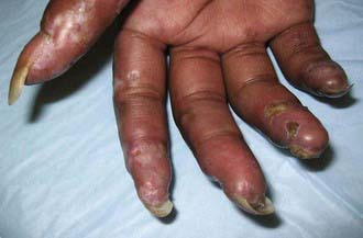
Figure 154-5 Sclerodactyly and finger ulcerations in a patient with systemic sclerosis who is poorly compliant with treatment.
Pulmonary disease is the most common visceral manifestation of SSc and includes both arterial and interstitial involvement (alveolitis). Symptoms range from asymptomatic disease to exercise intolerance, dyspnea at rest, and right-sided heart failure. Pulmonary arterial hypertension (PAH) is a poor prognostic sign, developing either as a consequence of lung disease or independently as part of the vasculopathy. Clinical manifestations of PAH in children appear late in the course, are subtle, and include cough and dyspnea on exertion. Pulmonary evaluation should include pulmonary function testing (PFT), bronchioalveolar lavage, and high resolution chest CT. PFTs reveal decreased vital capacity and decreased diffusion of carbon monoxide capacity (DLCO), while neutrophilia and/or eosinophilia on bronchioalveolar lavage suggest active alveolitis. Chest CT is much more sensitive than chest radiographs, which are often normal, showing typical basilar ground-glass abnormalities, reticular linear opacities, nodules, honey combing, and mediastinal adenopathy.
Other organ systems are involved in SSc. Gastrointestinal tract disease is seen in 25% of children with the disease. Common manifestations include esophageal and intestinal dysmotility resulting in dysphagia, reflux, dyspepsia, gastroparesis, bacterial overgrowth, dilated bowel loops and pseudo-obstruction, dental caries, as well as malabsorption and failure to thrive. Renal arterial disease can cause chronic or severe episodic hypertension; unlike in adult disease, renal crisis is rare. Cardiac fibrosis is associated with arrhythmias, ventricular hypertrophy, and decreased cardiac function. Mortality from juvenile systemic sclerosis is most commonly a result of cardiopulmonary disease.
Raynaud Phenomenon
Raynaud phenomenon (RP) is the most frequent initial symptom in pediatric systemic sclerosis, present in 70% of affected children months to years before other manifestations. Raynaud phenomenon refers to the classic triphasic sequence of blanching, cyanosis, and erythema of the digits induced by cold exposure and/or emotional stress. Raynaud phenomenon is most commonly independent of an underlying rheumatic disease (Raynaud disease), but it can be a consequence of other diseases as well as scleroderma, such as systemic lupus erythematosus and mixed connective tissue disease (Table 154-2). The color changes are brought about by (1) initial arterial vasoconstriction, resulting in hypoperfusion and pallor (blanching), (2) venous stasis (cyanosis), and (3) reflex vasodilatation caused by the factors released from the ischemic phase (erythema). The color change is classically reproduced by immersing the hands in iced water and reversed by warming. During the blanching phase, there is inadequate tissue perfusion in the affected area, associated with pain and paresthesias and resulting in ischemic damage only when associated with a rheumatic disease. The blanching usually affects the distal fingers but may also involve thumbs, toes, ears, and tip of the nose. The affected area is usually well demarcated and uniformly white.
Table 154-2 CLASSIFICATION OF RAYNAUD PHENOMENON
From Firestein GS, Budd RC, Harris ED Jr, et al, editors: Kelley’s textbook of rheumatology, ed 8, vol II, Philadelphia, 2009, Saunders/Elsevier.
Raynaud phenomenon often begins in adolescence and is characterized by symmetric occurrence, the absence of tissue necrosis and gangrene, and the lack of manifestations of an underlying rheumatic disease. Children have normal nail-fold capillaries (absence of periungual telangiectasias). Raynaud phenomenon should be distinguished from acrocyanosis and chilblains. Acrocyanosis is a vasospastic disorder resulting in cool, painless, bluish discoloration in the hands and sometimes feet despite normal tissue perfusion. It may be exacerbated by stimulant medications used to treat attention deficit disorder. Chilblains is a condition with episodic color changes and the development of nodules related to severe cold exposure and spasm-induced vessel and tissue damage; this condition has been associated with systemic lupus erythematosus.
Diagnosis
The diagnosis of localized scleroderma is based on the distribution and depth of characteristic lesions. Biopsy is helpful to confirm the diagnosis. Classification criteria for juvenile systemic sclerosis were recently devised, reflecting differences in presentation and course compared with adult onset disease. The new classification requires proximal sclerosis/induration of the skin as well as the presence of 2 of 20 minor criteria (Table 154-3).
Table 154-3 PROVISIONAL CRITERIA FOR THE CLASSIFICATION OF JUVENILE SYSTEMIC SCLEROSIS (SSC)
MAJOR CRITERION (REQUIRED)
Proximal skin sclerosis/induration of the skin
MINOR CRITERIA (AT LEAST 2 REQUIRED)
From Zulian F, Woo P, Athreya BH, et al: The Pediatric Rheumatology European Society/American College of Rheumatology/European League against Rheumatism provisional classification criteria for juvenile systemic sclerosis, Arthritis Rheum 57:203–212, 2007.
Differential Diagnosis
The most important condition to differentiate from LS is SSc. Contractures and synovitis from juvenile arthritis can be differentiated from those due to linear scleroderma by the absence or presence of skin changes. Other conditions to consider include chemically induced scleroderma-like disease, diabetic cheiroarthropathy, pseudoscleroderma, and scleredema. Pseudoscleroderma is composed of a group of unrelated diseases characterized by patchy or diffuse cutaneous fibrosis without the other manifestations of scleroderma. These include phenylketonuria, syndromes of premature aging, and localized idiopathic fibrosis. Scleredema is a transient, self-limited disease of both children and adults that has sudden onset after a febrile illness (especially streptococcal infections) and is characterized by patchy sclerodermatous lesions on the neck and shoulders and extending to the face, trunk, and arms.
Laboratory Findings
There are no laboratory studies diagnostic of either localized or systemic scleroderma. Although the results of complete blood counts, serum chemistry analyses, and urinalysis are normal, children may have elevated erythrocyte sedimentation rate, eosinophilia, or hypergammaglobulinemia, all of which normalize with treatment. Elevations of muscle enzymes, particularly aldolase, can be seen with muscle involvement. Patients with SSc may have anemia, leukocytosis, and eosinophilia and are more likely to have a high-titer positive ANA test result and to test positive for anti-Scl 70 antibody (anti-topoisomerase I). Imaging studies delineate the affected area and can be used to follow disease progression. MRI is useful in en coup de sabre and Parry Romberg syndrome for determination of CNS or orbital involvement. Infrared thermography utilizes the temperature variation between areas of active and inactive cutaneous disease to help differentiate active disease from damage. The role of ultrasound to look at lesion activity is evolving. High-resolution CT, pulmonary function tests, echocardiography, and manometry are useful tools for diagnosing and monitoring visceral involvement in SSc.
Treatment
Treatment for scleroderma varies according to the subtype and severity. Superficial morphea may benefit from topical corticosteroids or ultraviolet (UV) therapy. For lesions involving deeper structures, systemic therapy is recommended. A combination of methotrexate and corticosteroids is effective in treating LS by preventing lesion extension and resulting in significant skin softening and improved range of motion of affected joints. Treatment regimens include 3 months of either monthly high-dose intravenous corticosteroids (30 mg/kg, max dose 1000 mg) for 3 consecutive days a month, or high daily oral corticosteroids (0.5-2 mg/kg/day). In addition, methotrexate is given at 1-mg/kg weekly (max dose 25 mg), usually via subcutaneous administration to optimize bioavailability in doses over 0.5 mg/kg or 20 mg weekly. Physical and occupational therapy are important adjuncts to pharmacologic treatment. Eosinophilic fasciitis often responds well to corticosteroids but may also benefit from methotrexate.
Treatments for juvenile systemic sclerosis target specific disease manifestations. Raynaud phenomenon is treated with cold avoidance. Pharmacologic interventions are generally reserved for more severe disease. Calcium channel blockers (nifedipine 30-60 mg of sustained-release form daily, amlodipine 2.5-10 mg daily) are the most common pharmacologic interventions. Additional potential therapies for Raynaud phenomenon include losartan, prazosin, bosentan, and sildenafil. Angiotensin-converting enzyme inhibitors (captopril, enalapril) are recommended for hypertension associated with renal disease. Methotrexate or mycophenolate mofetil may be beneficial for skin manifestations. Cyclophosphamide is used to treat pulmonary alveolitis and prevent fibrosis. Corticosteroids should be used cautiously in systemic sclerosis due to an association with renal crisis.
Prognosis
Localized scleroderma is usually self-limited, with the initial inflammatory stage followed by a period of stabilization and then softening for an average disease duration of 3-5 yr; there are reports of active disease lasting up to 20 yr. Prolonged disease activity is associated primarily with linear and deep disease subtypes. Localized scleroderma can result in significant morbidity, disfigurement, and disability, especially with the linear and deep subtypes.
Juvenile systemic sclerosis has a more variable prognosis. Although many children have a slow, insidious course, others demonstrate a rapidly progressive form with early organ failure and death. Skin manifestations reportedly soften years after disease onset. Overall, the prognosis of juvenile systemic sclerosis is better than that of the adult form, with 5-, 10-, and 15-year survival rates, respectively, in children of 89%, 80-87%, and 74-87%. The most common cause of death is heart failure due to myocardial and pulmonary fibrosis.
Charles C, Clements P, Furst DE. Systemic sclerosis: hypothesis-driven treatment strategies. Lancet. 2006;367:1683-1690.
Fitch PG, Rettig P, Burnham JM, et al. Treatment of pediatric localized scleroderma with methotrexate. J Rheumatol. 2006;33:609.
Gabrielli A, Avvedimento EV, Krieg T. Scleroderma. N Engl J Med. 2009;360:1989-2002.
Gu YS, Kong J, Cheema GS, et al. The immunobiology of systemic sclerosis. Semin Arthritis Rheum. 2008;38:132-160.
Hudson M, Fritzler MJ, Baron M, et al. Systemic sclerosis: establishing diagnostic criteria. Medicine. 2010;89(3):159-165.
Kreuter A, Gambichler T, Breuckmann F, et al. Pulsed high-dose corticosteroids combined with low-dose methotrexate in severe localized scleroderma. Arch Dermatol. 2005;141:847.
Laxer RM, Zulian F. Localized scleroderma. Curr Opin Rheumatol. 2006;18:606-613.
Martini G, Foeldvari I, Russo R, et al. Systemic sclerosis in childhood: clinical and immunologic features of 153 patients in an international database. Arthritis Rheum. 2006;54:3971-3978.
Nigrovic PA, Fuhlbrigge RC, Sundel RP. Raynaud’s phenomenon in children: a retrospective review of 123 patients. Pediatrics. 2003;111:715-721.
Peterson LS, Nelson AM, Su WPD, et al. The epidemiology of morphea (localized scleroderma) in Olmsted county 1960–1993. J Rheumatol. 1997;24:73-80.
Scalapino K, Arkachaisri T, Lucas M, et al. Childhood onset systemic sclerosis: classification, clinical and serologic features, and survival in comparison with adult onset disease. J Rheumatol. 2006;33:1004-1013.
Tashkin DP, Elashoff R, Clements PJ, et al. Cyclophosphamide versus placebo in scleroderma lung disease. N Engl J Med. 2006;354:2655-2666.
Uziel Y, Feldman BM, Krafchik BR, et al. Methotrexate and corticosteroid therapy for pediatric localized scleroderma. J Pediatr. 2000;136:91.
Zulian F, Martini G. Childhood systemic sclerosis. Curr Opin Rheumatol. 2007;19:592-597.
Zulian F, Vallongo C, Woo P, et al. Localized scleroderma in childhood is not just a skin disease. Arthritis Rheum. 2005;52:2873-2881.