Chapter 309 Diagnostic Radiology in Dental Assessment
The panoramic radiograph provides a single tomographic image of the upper and lower jaw, including all the teeth and supporting structures. The x-ray tube rotates about the patient’s head with reciprocal movement of the film or image receptor during the exposure. The panoramic image shows the mandibular bodies, rami, and condyles; maxillary sinuses; and a majority of the facial buttresses. Such images are used to show abnormalities of tooth number, development and eruption pattern, cystic and neoplastic lesions, bone infections, and fracture, as well as dental caries and periodontal disease (see  Fig. 309-1 on the Nelson Textbook of Pediatrics website at www.expertconsult.com).
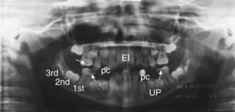
Figure 309-1 A panoramic radiograph of a 10 yr old child showing extensive dental caries of the 1st permanent molars (arrows), as well as normal structures: erupted 1st permanent molar, unerupted 2nd molar, and unerupted 3rd molar; erupted incisors (EI), unerupted permolars (UP), and erupted primary canines (pc).
Cephalometric radiographs are posteroanterior and lateral skull films that are taken using a cephalostat (head positioner) and employ techniques that clearly demonstrate the facial skeleton and soft facial tissues. Similar protocols for positioning children are used throughout the world. From these images, cranial and facial points and planes can be determined and compared with standards derived from thousands of images. A child’s facial growth can be assessed serially when cephalometric radiographs are taken sequentially. Relationships among the maxilla, mandible, cranial base, and facial skeleton can be determined in a quantitative manner. Additionally, the alignment of the teeth and the relation of the teeth to the supporting bone can be serially measured.
Intraoral dental radiographs are highly detailed, direct-exposure films that demonstrate sections of the child’s teeth and supporting bone structures. The film or image receptor is placed lingual to the teeth, and the x-ray beam is directed through the teeth and supporting structures. The resulting images are used to detect dental caries, loss of alveolar bone (periodontal disease), abscesses at the roots of the teeth, and trauma to the teeth and alveolar bone and to demonstrate the developmental status of permanent teeth within the bone.