Chapter 382 Neoplasms of the Larynx, Trachea, and Bronchi
382.1 Vocal Nodules
Vocal nodules are the most common cause of chronic hoarseness in children. They are not true neoplasms. Chronic vocal abuse or misuse produces nodules at the junction of the anterior and middle thirds of the phonating edge of the vocal cords. These symmetric, bilateral swellings interfere with voice production and cause children to strain the voice. Vocal nodules can occur in infants and are exacerbated by laryngopharyngeal reflux.
Voice therapy may be effective in the cooperative child, but for most toddlers and young children, behavioral therapy is necessary. Nodules usually resolve by early teenage years as the child matures and vocal abuse is moderated. Surgical excision is rarely indicated but may be necessary if the child is unable to communicate adequately, becomes aphonic, or requires tension and straining to make any utterance whatsoever.
Laryngopharyngeal reflux commonly exacerbates vocal abuse, adding to swelling of the vocal cords. When vocal abuse is the main factor, the voice is worse in the evenings; with laryngopharyngeal reflux, hoarseness is worse in the morning. An antireflux regimen is indicated (Chapter 315.1).
382.2 Recurrent Respiratory Papillomatosis
Papillomas are the most common respiratory tract neoplasms in children, occurring in 4.3/100,000. They are simply warts—benign tumors—caused by the human papillomavirus (HPV) (Chapter 258); the same pathology is found in condylomata acuminata (vaginal warts). HPV types 6 and 11 are most commonly associated with laryngeal disease. Fifty percent of recurrent respiratory papillomatosis (RRP) cases occur in children <5 yr, but the diagnosis may be made at any age; 67% of children with RRP are born to mothers who had condylomata during pregnancy or parturition. The risk for transmission is ∼1/500 vaginal births in mothers with active condylomata. Neonates have been reported to have RRP, suggesting intrauterine transmission of HPV.
Clinical Manifestations
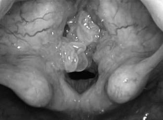
These benign squamous lesions can produce chronic hoarseness in the infant. Most occur in the larynx, specifically on the vocal cords, but in 31% these lesions occur in other areas of the respiratory tract: nose, pharynx (especially the uvula and posterior soft palate), trachea, bronchi, and lungs. As growth of the lesions on the vocal cords progresses, hoarseness increases and communication becomes difficult. Respiratory distress develops. Surgical excision is a quality of life issue that warrants removal to improve the voice. Intervention becomes a medical necessity when airway obstruction progresses (Fig. 382-1). Symptoms often occur first during sleep with symptoms typical of obstructive sleep apnea. Progressive respiratory distress during sleep, exertion, daily activities, and finally at rest indicates the need for surgical intervention.
382.3 Congenital Subglottic Hemangioma
Clinical Manifestations
Typically, congenital subglottic hemangiomas are symptomatic within the 1st 2 mo of life, almost all occurring before 6 mo of age. Stridor is biphasic but usually more prominent during inspiration. The infant may be hoarse, have a barking cough, and present with croup. Fifty percent of congenital subglottic hemangiomas are associated with facial lesions. Radiographs classically delineate an asymmetric subglottic narrowing. The diagnosis is made by direct laryngoscopy.
Treatment
Medical management includes systemic steroids. Prednisone 2-4 mg/kg/day is given orally for 4-6 wk, less if the lesions stabilize sooner. The dosage is then tapered. If there is no response, the drug is discontinued. Propranolol 2-3 mg/kg/day has also been effective.
Although tracheostomy establishes a safe airway, every effort should be made to find an alternative. Corticosteroids can also be injected directly into the lesion. Endoscopic excision with the CO2 laser is effective. Combining several modalities increases the possibility of avoiding tracheotomy. External surgical excision is suitable for some lesions.
382.4 Vascular Anomalies
Vascular malformations are not true neoplastic lesions. They have a normal rate of endothelial turnover and various channel abnormalities. They are categorized by their predominant type (capillary, venous, arterial, lymphatic, or a combination thereof). Slow-flow malformations have capillary, lymphatic, or venous components. These incorrectly were called capillary hemangiomas, cystic hygromas or lymphangiomas, and cavernous hemangiomas, respectively.
Lymphatic malformations (cystic hygromas) rarely occur in the larynx. When they do, they are invariably an extension of disease from elsewhere in the head and neck. Airway obstruction can necessitate tracheostomy. The lesion can be debulked with the CO2 laser.
382.5 Other Laryngeal Neoplasms
Neurofibromatosis rarely involves the larynx. When children are affected, limited local resection is undertaken to maintain an airway and optimize the voice. Complete surgical extirpation is virtually impossible without debilitating resection of vital laryngeal structures. Most surgeons select the option of less-aggressive symptomatic surgery because of the poorly circumscribed and infiltrative nature of these fibromas. Rhabdomyosarcoma and other malignant tumors of the larynx are rare. Symptoms of hoarseness and progressive airway obstruction prompt initial evaluation by flexible laryngoscopy in the office.
382.6 Tracheal Neoplasms
Tracheal tumors include malignant and benign neoplasms. The two most common benign tumors are inflammatory pseudotumor and hamartoma. The inflammatory pseudotumor is probably a reaction to a previous bronchial infection or traumatic insult. Growth is slow and the tumor may be locally invasive. Hamartomas are tumors of primary tissue elements that are abnormal in proportion and arrangement.
Tracheal neoplasms manifest with stridor, wheezing, cough, or pneumonia and are rarely diagnosed until 75% of the lumen has been obstructed (Fig 382-2). Symptoms mimic asthma and are often misdiagnosed as such. Chest radiographs or airway films can identify the obstruction. Pulmonary function studies demonstrate an abnormal flow-volume loop. A mild response to bronchodilator therapy may be misleading. Rational treatment is based upon the histopathology.
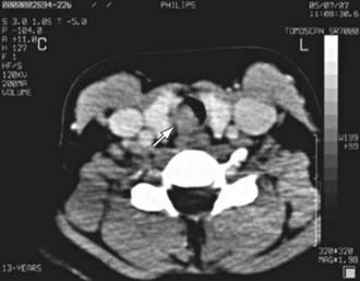
Figure 382-2 A CT scan of the trachea with a circumscribed intraluminal tracheal mass (arrow) in the tracheal wall.
(From Venizelos I, Papathomas T, Anagnostou E, et al: Pediatric inflammatory myofibroblastic tumor of the trachea: a case report and review of the literature, Pediatr Pulmonol 43:831–835, 2008.)
382.7 Bronchial Tumors
Bronchial tumors are rare. Two thirds are malignant. Bronchial “adenomas” are the most common, representing 30% of all lung tumors. Bronchogenic carcinoma is the second most common and occurs in approximately 20% of cases. Bronchial carcinoid also occurs. The diagnosis is confirmed at bronchoscopy and biopsy; treatment depends on the histopathology.