Chapter 401 Pulmonary Embolism, Infarction, and Hemorrhage
401.1 Pulmonary Embolus and Infarction
Venous thromboembolic disease (VTE) is well described in children and adolescents with or without risk factors (Table 401-1). Improvements in therapeutics for childhood illnesses and increased survival with chronic illness may contribute to the larger number of children presenting with thromboembolic events, which can be a significant source of morbidity and mortality.
Table 401-1 RISK FACTORS FOR PULMONARY EMBOLISM
ENVIRONMENTAL
WOMEN’S HEALTH
MEDICAL ILLNESS
SURGICAL
THROMBOPHILIA
NONTHROMBOTIC
Modified from Goldhaber SZ: Pulmonary embolism, Lancet 363:1295–1305, 2004.
Etiology
Commonly appreciated risk factors for thromboembolic disease in adults include immobility, malignancy, pregnancy, infection, and hypercoagulability; up to 20% of adults with this disorder may have no identifiable risk factor (see Table 401-1). Children with deep venous thrombosis (DVT) and pulmonary embolism (PE) are much more likely to have 1 or more identifiable conditions or circumstances placing them at risk. In a large Canadian registry, 96% of pediatric patients were found to have 1 risk factor and 90% had 2 or more risk factors.
Embolic disease has variable etiologies in children. An embolus can contain thrombus, air, amniotic fluid, septic material, or metastatic neoplastic tissue; thromboemboli are most commonly encountered. A commonly encountered risk factor for DVT and PE in the pediatric population is the presence of a central venous catheter. The presence of a catheter in a vessel lumen as well as instilled medications can induce endothelial damage and favor thrombus formation.
Children with malignancies are also at considerable risk. The risk of PE is more significant in children with solid rather than hematologic malignancies. PE has been described in children with Wilms tumor (tumor embolism) as well as leukemia. A child with malignancy may have numerous risk factors related to the primary disease process and the therapeutic interventions. Infection from chronic immunosuppression may interact with hypercoagulability of malignancy and chemotherapeutic effects on the endothelium.
In the neonatal period, thromboembolic disease and PE are often related to indwelling catheters used for parenteral nutrition and medication delivery. Emboli in neonates may occasionally reflect maternal risk factors, such as diabetes and toxemia of pregnancy. Infants with congenitally acquired homozygous deficiencies of antithrombin, protein C, and protein S are also likely to present with thromboembolic disease in the neonatal period.
Prothrombotic disease can also manifest in older infants and children. Disease can be congenital or acquired; DVT/PE may be the initial presentation. Factor V Leiden mutation (Chapter 472), hyperhomocysteinemia (Chapter 79.3), prothrombin 20210A mutation (Chapter 472), anticardiolipin antibody, and elevated values of lipoprotein A have all been linked to thromboembolic disease. Children with sickle cell disease are also at high risk for pulmonary embolus and infarction. Acquired prothrombotic disease is represented by nephrotic syndrome (Chapter 521) and antiphospholipid antibody syndrome. From one quarter to one half of children with systemic lupus erythematosus (Chapter 152) have thromboembolic disease.
Other risk factors include infection, cardiac disease, recent surgery, and trauma. Surgical risk is thought to be more significant when immobility will be a prominent feature of the recovery. Use of oral contraceptives confers additional risk, although the level of risk in patients taking these medications appears to be decreasing, perhaps as a result of the lower amounts of estrogen in current formulations.
Septic emboli are rare in children but may be caused by osteomyelitis, cellulitis, urinary tract infection, jugular vein or umbilical thrombophlebitis, and right-sided endocarditis.
Epidemiology
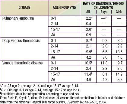
Younger age appears to be somewhat protective in thromboembolic disease. The DVT incidence in 1 study of hospitalized children was 5.3/10,000 admissions. A study that analyzed data from 1979 through 2001 found 0.9 cases of PE per 100,000 children per yr, 4.2 cases of DVT per 100,000 children per yr, and 4.9 cases of VTE per 100,000 children per yr (Table 401-2).
Pediatric autopsy reviews have estimated the incidence of thromboembolic disease in children as between 1% and 4%. Not all of these embolic findings were clinically significant. Thromboembolic pulmonary disease is often unrecognized, and antemortem studies may underestimate the true incidence. Pediatric deaths from isolated pulmonary emboli are rare. Most thromboemboli are related to central venous catheters. The source of the emboli may be lower or upper extremity veins as well as the pelvis and right heart. The most common location for an embolus unassociated with the presence of an indwelling venous catheter is the lower extremity.
Pathophysiology
Favorable conditions for thrombus formation include injury to the vessel endothelium, hemostasis, and hypercoagulability. Once an embolus develops and travels to the pulmonary circulation, symptoms of presentation are largely attributable to unequal ventilation and perfusion. The occlusion of the involved vessel prevents perfusion of distal alveolar units, thereby creating an increase in dead space and hypoxia with an elevated alveolar-arterial oxygen tension difference, or a-AO2 (PAO2 - PaO2). Most patients are hypocarbic secondary to hyperventilation, which often persists even when oxygenation is optimized. Abnormalities of oxygenation and ventilation are likely to be less significant in the pediatric population, possibly owing to less underlying cardiopulmonary disease and greater reserve. The vascular supply to lung tissue is abundant, and pulmonary infarction is unusual with pulmonary embolus but may result from distal arterial occlusion and alveolar hemorrhage. In rare instances of death from massive pulmonary embolus, marked increases in pulmonary vascular resistance and heart failure are usually present. Most of these severe outcomes are expectedly found in those with pre-existing cardiopulmonary disease.
Clinical Manifestations
Presentation is variable, and many pulmonary emboli are silent. Rarely, a massive PE may manifest as cardiopulmonary failure. Children are more likely to have underlying disease processes or risk factors but might still present asymptomatically with small emboli. Common symptoms and signs of PE include hypoxia (cyanosis), tachypnea, dyspnea, cough, diaphoresis, and chest pain. Localized crackles may occasionally be appreciated on examination. These are nonspecific complaints and may be attributed to an underlying disease process or an unrelated/incorrect diagnosis in many cases. A high level of clinical suspicion is required because the diagnosis of pulmonary embolus is infrequently considered in children. A large number of diagnoses can cause similar symptoms. Therefore, confirmatory testing should follow a clinical diagnosis of PE. In addition, clinical risk scoring systems may help raise the likelihood of a PE. High-risk findings include surgery or lower extremity fractures, malignancy, hemoptysis, lower extremity signs of a DVT (pain, swelling), tachycardia, and hypoxia.
Laboratory Findings and Diagnosis
Radiographic images of the chest are often normal in a child with PE. Any abnormalities found on chest radiographs are likely to be nonspecific. Patients with septic emboli may have multiple areas of nodularity and cavitation, which are typically located peripherally in both lung fields. Many patients with PE have hypoxemia. The a-AO2 gradient is more sensitive in detecting gas exchange derangements. A review of results of a complete blood count (CBC), urinalysis, and coagulation profile is warranted. Prothrombotic diseases should be highly suspected on the basis of past medical or family history; therefore, additional laboratory evaluations include fibrinogen assays, protein C, protein S, and antithrombin III studies, and analysis for factor V Leiden mutation as well as evaluation for lupus anticoagulant and anticardiolipin antibodies.
Electrocardiographs may reveal ST segment changes or evidence of pulmonary hypertension with right ventricular failure (cor pulmonale). These changes are nonspecific and nondiagnostic. Echocardiograms may be warranted to assess ventricular size and function. A transAmerican Thoracic Society/European Respiratory Society International Multidisciplinary Consensus echocardiogram is required if there is any suspicion of intracardiac thrombi or endocarditis.
Noninvasive venous ultrasound testing with Doppler flow can be used to confirm DVT in the lower extremities; ultrasonography may not detect thrombi in the upper extremities or pelvis. In patients with significant venous thrombosis, D dimers are usually elevated. When a high level of suspicion exists, confirmatory testing with venography should be pursued. DVT can be recurrent and multifocal and may lead to repeated episodes of pulmonary embolism.
Although a ventilation-perfusion ( ) radionuclide scan is a noninvasive and potentially sensitive method of pulmonary embolus detection, the interpretation of
  scans can be problematic. Helical or spiral CT with an intravenous contrast agent is valuable and the diagnostic test of choice to detect a PE. Specificity exceeds 90%. CT studies detect emboli in lobar and segmental vessels with acceptable sensitivities. Poorer sensitivities may be encountered in the evaluation of the subsegmental pulmonary vasculature. Pulmonary angiography is the gold standard for diagnosis of PE, but with current availability of multidetector spiral CT angiography, it is not necessary except in unusual cases.
Treatment
Initial treatment should always be directed toward stabilization of the patient. Careful approaches to ventilation, fluid resuscitation, and inotropic support are always indicated, because improvement in one area of decompensation can often exacerbate coexisting pathology.
After the patient with a PE has been stabilized, the next therapeutic step is anticoagulation. Evaluations for prothrombotic disease must precede anticoagulation. Treatment is generally initiated with heparin. Anticoagulation is usually achieved when the activated partial thromboplastin time (PTT) is 1.5-2 times the control. Long-term therapy with heparin should be avoided whenever possible. Complications of heparin include bleeding and acquired thrombocytopenia.
Heparin therapy continues for several days before oral therapy with warfarin is begun. Anticoagulation may be required for 3-6 mo in the setting of acute thromboembolic disease. Longer treatment is indicated in patients with ongoing thrombotic disease. Coagulation profiles are obtained regularly to guide warfarin and heparin therapies.
In adults, therapy with low molecular weight (LMW) heparin is considered equivalent to that with heparin. The longer half-life of LMW heparin allows discontinuous dosing, and effectiveness appears comparable. Minimal monitoring is involved with drug administration. Other advantages may include decreased risks of both osteoporosis and thrombocytopenia. At this time, LMW heparin is being used in many pediatric patients. Studies of LMW heparin use in children and neonates have suggested similarities to its use in adults in terms of success of thrombolysis, rate of recurrence, and complications.
Thrombolytic agents can be combined with anticoagulants in the early stages of treatment. Thrombolytic agents include urokinase, streptokinase, and, most often, recombinant tissue plasminogen activator. Combined therapy may reduce the incidences of progressive thromboembolism, pulmonary embolus, and postphlebitic syndrome. Mortality rate appears to be unaffected by additional therapies; nonetheless, the additional theoretic risk of hemorrhage limits the use of combination therapy in all but the most compromised patients. The use of thrombolytic agents in patients with active bleeding, recent cerebrovascular accidents, or trauma is contraindicated.
Surgical embolectomy is invasive and is associated with significant mortality. Its application should be limited to those with persistent hemodynamic compromise refractory to standard therapy.
Prognosis
Mortality in pediatric patients with PE is likely to be attributable to an underlying disease process rather than to the embolus itself. Conditions associated with a poorer prognosis include malignancy, infection, and cardiac disease. The mortality rate in children from PE is 2.2%. Recurrent thromboembolic disease may complicate recovery. The practitioner must conduct an extensive evaluation for underlying pathology so as to prevent progressive disease. Postphlebitic syndrome is another recognized complication of pediatric thrombotic disease. Venous valvular damage can be initiated by the presence of DVT, leading to persistent venous hypertension with ambulation and valvular reflux. Symptoms include edema, pain, increases in pigmentation, and ulcerations. Affected pediatric patients may suffer lifelong disability.
Askegard-Giesmann JR, Caniano DA, Kenney BD. Rare but serious complications of central line insertion. Semin Pediatr Surg. 2009;18:73-83.
Baird JS, Killinger JS, Kalkbrenner KJ, et al. Massive pulmonary embolism in children. J Pediatr. 2010;156:148-151.
Bonduel M, Hepner M, Sciuccati G, et al. Prothrombotic abnormalities in children with venous thromboembolism. J Pediatr Hematol Oncol. 2000;22:66-72.
Chan AK, Deveber G, Monagle P, et al. Venous thrombosis in children. J Thromb Haemost. 2003;1:1443-1455.
Holzer R, Peart I, Ciotti G, et al. Successful treatment with rTPA. Pediatr Cardiol. 2002;23:548-552.
Hull RD. Diagnosing pulmonary embolism with improved certainty and simplicity. JAMA. 2006;295:213-215.
Johnson AS, Bolte RG. Pulmonary embolism in the pediatric patient. Pediatr Emerg Care. 2004;20:555-560.
Kyrle PA, Eichinger S. New diagnostic strategies for pulmonary embolism. Lancet. 2008;371:1312-1314.
Manco-Johnson MJ, Nuss R, Hays T, et al. Combined thrombolytic and anticoagulant therapy for venous thrombosis in children. J Pediatr. 2000;136:446-453.
Nowak-Gottl U, Bidlingmaier C, et al. Pharmacokinetics, efficacy, and safety of LMWHs in venous thrombosis and stroke in neonates, infants and children. Br J Pharmacol. 2008;153:1120-1127.
Perrier A, Roy PM, Sanchez O, et al. Multidetector-row computed tomography in suspected pulmonary embolism. N Engl J Med. 2005;352:1760-1768.
Stein P, Kayali F, Olson R. Incidence of venous thromboembolism in infants and children: data from the National Hospital Discharge Survey. J Pediatr. 2004;145:563-565.
Stein PD, Fowler SE, Goodman LR. Multidetector computed tomography for acute pulmonary embolism. N Engl J Med. 2006;354:2317-2326.
Tapson VF. Acute pulmonary embolism. N Engl J Med. 2008;358:1037-1052.
van Belle A, Buller HR, Huisman MV, et al. Effectiveness of managing suspected pulmonary embolism using an algorithm combining clinical probability, D-Dimer testing, and computed tomography. Christopher Study Investigators. JAMA. 2006;295:172-179.
Wong KS, Lin TY, Huang YC, et al. Clinical and radiographic spectrum of septic pulmonary embolism. Arch Dis Child. 2002;87:312-315.
401.2 Pulmonary Hemorrhage and Hemoptysis
Pulmonary hemorrhage is relatively uncommon but potentially fatal in children. Diffuse, slow bleeding in the lower airways may become severe and manifest as anemia, fatigue, or respiratory compromise without the patient ever experiencing episodes of hemoptysis. Hemoptysis must always be separated from episodes of hematemesis or epistaxis, all of which have similar presentations in the young patient.
Etiology
Conditions that can manifest as pulmonary hemorrhage or hemoptysis in children are found in Table 401-3. The chronic (opposed to an acute) presence of a foreign body can lead to inflammation and/or infection, thereby inducing hemorrhage. Hemorrhage most commonly reflects chronic inflammation and infection such as that seen in cystic fibrosis with bronchiectasis or in tuberculosis with cavitary disease, although it may also reflect an acute condition such as bronchitis and bronchopneumonia. Other relatively common etiologies are congenital heart disease and trauma. Traumatic irritation or damage of the airway is often accidental in nature. Bleeding can also be related to instrumentation of the airway as is commonly seen in a child with a tracheostomy. It is important to note, however, that children who have been victims of nonaccidental trauma or deliberate suffocation can also be found to have blood in the mouth or airway. Less commonly, syndromes associated with vasculitic, autoimmune, and idiopathic disorders can be associated with diffuse alveolar hemorrhage (DAH). The mechanisms of DAH are multiple and are discussed further in Chapter 400.
Table 401-3 ETIOLOGY OF PULMONARY HEMORRHAGE (HEMOPTYSIS)
FOCAL HEMORRHAGE
DIFFUSE HEMORRHAGE
Acute idiopathic pulmonary hemorrhage (AIPH) occurs in young infants as a distinct entity, but the disorders in Table 401-3 must also be considered.
Epidemiology
The frequency with which pulmonary hemorrhage occurs in the pediatric population is difficult to define. This difficulty is largely related to the variability in disease presentation. Chronic bronchiectasis as seen in cystic fibrosis or ciliary dyskinesia can cause hemoptysis but usually in children >10 yr of age. The incidence of pulmonary hemorrhage may be significantly underestimated because many children and young adults swallow rather than expectorate mucus, a behavior that may prevent recognition of hemoptysis, the primary presenting symptom of the disorder.
AIPH is defined as evidence of blood in the airway in a child age ≤1 yr, with no medical conditions predisposing to pulmonary hemorrhage and severe respiratory distress leading to respiratory failure. This entity may be more common than previously thought. Most cases are idiopathic, but some have been associated with von Willebrand disease. There is no association between the disorder and house contamination with molds.
Pathophysiology
Pulmonary hemorrhage can be localized or diffuse. Focal hemorrhage from an isolated bronchial lesion is often secondary to infection or chronic inflammation. Erosion through a chronically inflamed airway into the adjacent bronchial artery is a mechanism for potentially massive hemorrhage. Bleeding from such a lesion is more likely to be bright red, brisk, and secondary to enlarged bronchial arteries and systemic arterial pressures. The severity of more diffuse hemorrhage can be difficult to ascertain. The rate of blood loss may be insufficient to reach the proximal airways. Therefore, the patient may present without hemoptysis. The diagnosis of pulmonary hemorrhage is generally achieved by finding evidence of blood or hemosiderin in the lung. Within 48-72 hr of an episode of bleeding, alveolar macrophages convert the iron from erythrocytes into hemosiderin. It may take weeks to clear these hemosiderin-laden macrophages completely from the alveolar spaces. This fact may allow differentiation between acute and chronic hemorrhage. Hemorrhage is often followed by the influx of neutrophils and other proinflammatory mediators. With repeated or chronic hemorrhage, pulmonary fibrosis can become a prominent pathologic finding.
Clinical Manifestations
The severity of presentation in patients with hemoptysis and pulmonary hemorrhage is highly variable. Older children and young adults with a focal hemorrhage may complain of warmth or a “bubbling” sensation in the chest wall. This can occasionally aid the clinician in locating the area involved. Rapid and large-volume blood loss manifests as symptoms of cyanosis, respiratory distress, and shock. Chronic, subclinical blood loss may manifest as anemia, fatigue, dyspnea, or altered activity tolerance. Less commonly, patients present with persistent infiltrates on chest radiograph or symptoms of chronic illness such as failure to thrive.
Laboratory Findings and Diagnosis
Every patient with suspected hemorrhage should have a laboratory evaluation with CBC and coagulation studies. The CBC result may demonstrate a microcytic, hypochromic anemia. Other laboratory findings are highly dependent on the underlying diagnosis. A urinalysis may show evidence of nephritis in patients with concomitant pulmonary and renal diseases. The classic finding, which defines pulmonary hemorrhage, is that of hemosiderin-laden macrophages in pulmonary secretions. These can be detected by sputum analysis with Prussian blue staining. Chest radiographs may demonstrate fluffy bilateral densities, as seen in AIPH of infancy (Fig. 401-1) or the patchy consolidation seen in idiopathic pulmonary hemosiderosis (Fig. 401-2). Alveolar infiltrates seen on chest radiograph may be regarded as a representation of recent bleeding, but their absence does not rule out a hemorrhage. Infiltrates, when present, are often symmetric and diffuse. CT may be indicated to assess for underlying disease processes.
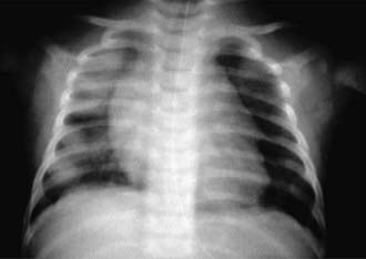
Figure 401-1 Radiographic appearance of acute idiopathic pulmonary hemorrhage in infancy.
(From Brown CM, Redd SC, Damon SA; Centers for Disease Control and Prevention [CDC]: Acute idiopathic pulmonary hemorrhage among infants: recommendations from the Working Group for Investigation and Surveillance, MMWR Recomm Rep 53:1–12, 2004.)
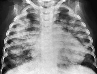
Figure 401-2 Diffuse pulmonary hemorrhage that was thought to be the result of idiopathic pulmonary hemosiderosis in a 3 yr old boy. Frontal radiograph reveals bilateral airspace consolidation that is patchy. Tracheal washing contained large numbers of macrophages filled with hemosiderin. Ten days later, most of the consolidative changes in the lungs had cleared. The patient’s anemia was successfully treated with blood transfusion.
(From Slovis T, editor: Caffey’s pediatric diagnostic imaging, ed 11, Philadelphia, 2008, Mosby/Elsevier; courtesy of Bertram Girdany, MD, Pittsburgh, PA.)
Flexible bronchoscopy with bronchoalveolar lavage is frequently utilized to obtain pulmonary secretions in a child or young adult who is not able to expectorate secretions. Lung biopsy is rarely necessary unless bleeding is chronic or an etiology cannot be determined with other methods. Pulmonary function testing, including a determination of gas exchange, is important to assess the severity of the ventilatory defect. In older children, spirometry may demonstrate evidence of predominantly obstructive disease in the acute period. Restrictive disease secondary to fibrosis is typically seen with more chronic disease. DLCO measurements are typically elevated in the setting of pulmonary hemorrhage because of the strong affinity of hemoglobin for carbon monoxide.
Treatment
In the setting of massive blood loss, volume resuscitation and transfusion of blood products are necessary. Maintenance of adequate ventilation and circulatory function is crucial. Rigid bronchoscopy may be utilized for removal of debris or the application of topical vasoconstrictive agents. Ideally, treatment is directed at the specific pathologic process responsible for the hemorrhage. When bronchiectasis is a known entity and a damaged artery can be localized, bronchial artery embolization is often the therapy of choice. If embolization fails, total or partial lobectomy may be required. In circumstances of diffuse hemorrhage, corticosteroids and other immunosuppressive agents have been shown to be of benefit. Prognosis depends largely on the underlying disease process.
Bartyik K, Bede O, Tiszlavicz L, et al. Pulmonary capillary haemangiomatosis in children and adolescents: report of a new case and a review of the literature. Eur J Pediatr. 2004;163:731-737.
Brown CM, Redd SC, Damon SA, Centers for Disease Control and Prevention (CDC). Acute idiopathic pulmonary hemorrhage among infants: Recommendations from the Working Group for Investigation and Surveillance. MMWR Recomm Rep. 2004;53:1-12.
Centers for Disease Control and Prevention (CDC). Investigation of acute idiopathic pulmonary hemorrhage among infants—Massachusetts, December 2002-June 2003. MMWR Morbid Mortal Wkly Rep. 2004;53:817-820.
Coss-Bu JA, Sachdeva RC, Bricker JT, et al. Hemoptysis: a 10-year retrospective study. Pediatrics. 1997;100:E7.
Cottin V, Chinet T, Lavolé A, et al. Pulmonary arteriovenous malformations in hereditary hemorrhagic telangiectasia: a series of 126 patients. Medicine. 2007;86:1-17.
Godfrey S. Pulmonary hemorrhage/hemoptysis in children. Pediatr Pulmonol. 2004;37:476-484.
Lazor R, Bigay-Game L, Cottin V, et al. Alveolar hemorrhage in anti-basement membrane antibody disease: a series of 28 cases. Medicine. 2007;86:181-193.