Chapter 433 Diseases of the Myocardium
The extremely heterogeneous groups of heart muscle diseases that are associated with structural and/or functional cardiac dysfunction (cardiomyopathy) are important causes of morbidity and mortality in the pediatric population. Certain anatomic and physiologic conditions such as congenital heart disease, hypertension, and coronary artery disease may result in heart muscle dysfunction, but are distinct from the conditions presented in this chapter. Several classification schemes have been formulated in an effort to provide logical, useful, and scientifically based etiologies for the cardiomyopathies. Insight into the molecular genetic basis of cardiomyopathies has increased exponentially and it is likely that etiologic classification schemes will continue to evolve.
Table 433-1 classifies the cardiomyopathies based on their anatomic (ventricular morphology) and functional pathophysiology. Dilated cardiomyopathy, the most common form of cardiomyopathy, is characterized predominantly by left ventricular dilation and decreased left ventricular systolic function (Fig. 433-1). Hypertrophic cardiomyopathy demonstrates increased ventricular myocardial wall thickness, normal or increased systolic function, and often, diastolic (relaxation) abnormalities (Table 433-2). Restrictive cardiomyopathy is characterized by nearly normal ventricular chamber size and wall thickness with preserved systolic function, but dramatically impaired diastolic function leading to elevated filling pressures and atrial enlargement (see Fig. 433-3). Arrhythmogenic right ventricular cardiomyopathy and left ventricular non-compaction are characterized by specific morphologic abnormalities and heterogeneous functional disturbances.
Table 433-1 ETIOLOGY OF PEDIATRIC MYOCARDIAL DISEASE
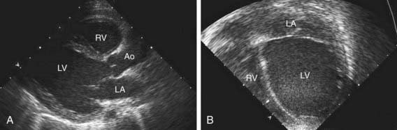
Figure 433-1 Echocardiogram of a patient with dilated cardiomyopathy. A, Parasternal long-axis view showing the enlarged left ventricle. B, Apical four-chamber view showing the large left ventricle compressing the right ventricle. Ao, ascending aorta; LA, left atrium; LV, left ventricle; RV, right ventricle.
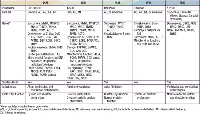

Figure 433-3 Echocardiogram of a patient with restrictive cardiomyopathy. The apical four-chamber view shows the markedly enlarged right and left atria, compared to the normal sized left and right ventricular chambers. LA, left atrium; LV, left ventricle; RA, right atrium; RV, right ventricle.
Bibliography
Cox GF, Sleeper LA, Lowe AM, et al: Factors associated with establishing a causal diagnosis for children with cardiomyopathy, Pediatrics 4:1519–1531, 2006.
Elliott P, Andersson B, Arbustini E, et al: Classification of the cardiomyopathies: a position statement from the European Society Of Cardiology Working Group on Myocardial and Pericardial Diseases, Eur Heart J 29:270–276, 2008.
Hershberger RE, Lindenfeld J, Mestroni L, et al: Genetic evaluation of cardiomyopathy—a Heart Failure Society of America practice guideline, J Card Fail 15:83–97, 2009.
Judge DP: Use of genetics in the clinical evaluation of cardiomyopathy, JAMA 302:2471–2476, 2009.
Maron BJ, Roberts WC, Arad M, et al: Clinical outcome and phenotypic expression in LAMP2 cardiomyopathy, JAMA 301:1253–1258, 2009.
Maron BJ, Towbin JA, Thiene G, et al: Contemporary definitions and classification of the cardiomyopathies: an American Heart Association Scientific Statement from the Council on Clinical Cardiology, Heart Failure and Transplantation Committee; Quality of Care and Outcomes Research and Functional Genomics and Translational Biology Interdisciplinary Working Groups; and Council on Epidemiology and Prevention, Circulation 113:1807–1816, 2006.
Mocumbi AO, Fereira MB, Sidi D, et al: A population study of endomyocardial fibrosis in a rural area of Mozambique, N Engl J Med 359:43–48, 2008.
Morita H, Rehm HL, Menesses A, et al: Shared genetic caused of cardiac hypertrophy in children and adults, N Engl J Med 358:1899–1908, 2008.
Paul M, Zumhagen S, Stallmeyer B, et al: Genes causing inherited forms of cardiomyopathies: a current compendium, Herz 34:98–109, 2009.
Scaglia F, Towbin JA, Craigen WJ, et al: Clinical spectrum, morbidity, and mortality in 113 pediatric patients with mitochondrial disease, Pediatrics 114:925–931, 2004.
433.1 Dilated Cardiomyopathy
Etiology and Epidemiology
Dilated cardiomyopathy (DCM), the most common form of cardiomyopathy in children, is the cause of significant morbidity and mortality as well as a common indication for cardiac transplantation. The etiologies are diverse. Unlike adult patients with dilated cardiomyopathy, ischemic etiologies are rare in children, although these include anomalous origin of the left coronary artery from the pulmonary artery, premature coronary atherosclerosis (homozygous type II hypercholesterolemia), and coronary inflammatory diseases, such as Kawasaki disease. It is estimated that up to 50% of cases are genetic, including some with metabolic causes (see Table 433-1). Although the most common etiology of dilated cardiomyopathy remains idiopathic, it is likely that undiagnosed familial/genetic conditions and myocarditis predominate. The annual incidence of DCM in children younger than 18 yr is 0.57 cases per 100,000 per year. Incidence is higher in males, African Americans, and in infants less than 1 yr old.
Pathogenesis
The pathogenesis of the ventricular dilation and altered contractility seen in dilated cardiomyopathy varies depending on the underlying etiology. Genetic abnormalities of several components of the cardiac muscle including sarcomere protein, the cytoskeleton, and the proteins that bridge the contractile apparatus to the cytoskeleton, have been identified in autosomal dominant and X-linked inherited disorders. Dilated cardiomyopathy can occur following viral myocarditis and, although the primary pathogenesis varies from direct myocardial injury to viral-induced inflammatory injury, the resulting myocardial damage, ventricular enlargement, and poor function likely occur by a final common pathway similar to that which occurs in genetic disorders.
In 20-50% of cases, the DCM is familial with autosomal dominant inheritance most common (see Table 433-2). Duchenne and Becker muscular dystrophies (Chapter 601.1) are X-linked cardiomyopathies that account for 5-10% of familial dilated cardiomyopathy cases. These dystrophinopathies result in an abnormal sarcomere-cytoskeleton connection, causing impaired myocardial force generation, myocyte damage/scarring, chamber enlargement, and altered function.
Mitochondrial myopathies, like the muscular dystrophies, may present clinically with a predominance of extra cardiac findings and are inherited in a recessive or mitochondrial pattern. Disorders of fatty acid oxidation present with systemic derangements of metabolism (hypoketotic hypoglycemia, acidosis, liver dysfunction), some with peripheral myopathy and neuropathy, and others with sudden death or life-threatening cardiac arrhythmias
Anthracycline cardiotoxicity (doxorubicin [Adriamycin]) on rare occasion causes acute inflammatory myocardial injury, but more classically results in dilated cardiomyopathy and occurs in up to 30% of patients given a cumulative dose of doxorubicin exceeding 550 mg/m2. The risk of toxicity appears to be exacerbated by concomitant radiation therapy.
Clinical Manifestations
Although more prevalent in patients less than 1 yr of age, all age groups may be affected. Clinical manifestations of dilated cardiomyopathy are most commonly those of congestive heart failure, but can also include palpitations, syncope, and sudden death. Irritability or lethargy can be accompanied by additional nonspecific complaints of failure to thrive, nausea, vomiting, or abdominal pain. Respiratory symptoms (tachypnea, wheezing, cough, or dyspnea on exertion) are often present. Uncommonly, patients may present acutely with pallor, altered mentation, hypotension, and shock. Patients can be tachycardic with narrow pulse pressure and have hepatic enlargement, and rales or wheezing. The precordial cardiac impulse is increased and the heart may be enlarged to palpation or percussion. Auscultation may reveal a gallop rhythm in addition to tachycardia and occasionally murmurs of mitral or, less commonly, tricuspid insufficiency may be present. The presence of hypoglycemia, acidosis, hypotonia, or signs of liver failure suggests an inborn error of metabolism. Neurologic or skeletal muscle deficits are associated with mitochondrial disorders or muscular dystrophies.
Laboratory Findings
Electrocardiographic screening reveals atrial or ventricular hypertrophy, nonspecific T-wave abnormalities, and occasionally atrial or ventricular arrhythmias. The chest x-ray demonstrates cardiomegaly and may reveal pulmonary vascular prominence or pleural effusions. The echocardiogram is often diagnostic, demonstrating the characteristic findings of left ventricular enlargement, decreased ventricular contractility, and occasionally a globular (remodeled) left ventricular contour (see Fig. 433-1). Right ventricular enlargement and depressed function are occasionally noted. Echo Doppler studies can reveal evidence of pulmonary hypertension, mitral regurgitation, or other structural cardiac or coronary abnormalities.
Additional testing should include CBC, renal and liver function tests, CPK, cardiac troponin I, lactate, plasma amino acids, urine organic acids, and an acylcarnitine profile. Additional genetic and enzymatic testing may be useful (see Table 433-2). Cardiac catheterization and endomyocardial biopsy are not routine but may be useful in patients with acute dilated cardiomyopathy. Biopsy samples can also be assessed for the presence of mononuclear cell infiltrates, myocardial damage, storage abnormalities, and viral infection or genomes. It is important to consider screening of 1st-degree family members utilizing echocardiography and ECG.
Prognosis and Management
The 1- and 5-yr rates of death or need for transplantation in patients diagnosed with DCM is 31% and 46%, respectively. Independent risk factors at DCM diagnosis for subsequent death or transplantation include older age, congestive heart failure, lower left ventricular fractional shortening z score, and underlying etiology. Dilated cardiomyopathy is the most common cause for cardiac transplantation in pediatric and adult studies.
The therapeutic approach to patients with dilated cardiomyopathy includes a careful assessment to uncover possible treatable etiologies, screening of family members, and rigorous pharmacologic therapy. Decongestive therapy may improve symptoms of heart failure, prolong survival, and occasionally results in complete resolution of dysfunction. Patients are often treated with diuretics and angiotensin-converting enzyme (ACE) inhibitors. The use of digitalis and angiotensin receptor blockers may be of additional benefit. β-Adrenergic blockade with carvedilol or metoprolol is often utilized in patients with chronic congestive heart failure although pediatric specific outcome data have failed to show effectiveness. In patients presenting with extreme degrees of heart failure or circulatory collapse, intensive care measures are often required, including intravenous inotropes and diuretics, mechanic ventilatory support, and on occasion, mechanical circulatory support, which may include ECMO, ventricular assist devices, and ultimately cardiac transplantation. In patients with dilated cardiomyopathy and atrial or ventricular arrhythmias, specific antiarrhythmic therapy should be instituted.
Burkett EL, Hershberger RE. Clinical and genetic issues in familial dilated cardiomyopathy. J Am Coll Cardiol. 2005;45:969-981.
Jefferies JL, Towbin JA. Dilated cardiomyopathy. Lancet. 2010;375:752-760.
Mordente A, Meucci E, Silvestrini A, et al. New developments in anthracycline-induced cardiotoxicity. Curr Med Chem. 2009;16:1656-1672.
Towbin JA, Lowe AM, Colan SD, et al. Incidence, causes, and outcomes of dilated cardiomyopathy in children. JAMA. 2006;296:1867-1876.
Shekhawat PS, Matern D, Strauss AW. Fetal fatty acid oxidation disorders, their effect on maternal health and neonatal outcome: impact of expanded newborn screening on their diagnosis and management. Pediatr Res. 2005;57:78R-86R.
433.2 Hypertrophic Cardiomyopathy
Etiology and Epidemiology
Hypertrophic cardiomyopathy (HCM) is a heterogeneous, relatively common, and potentially severe form of cardiomyopathy. The causes of hypertrophic cardiomyopathy are heterogeneous and include inborn errors of metabolism, neuromuscular disorders, syndromic conditions, and genetic abnormalities of the structural components of the cardiomyocyte (see Table 433-1). Both the age of onset and associated features are helpful in identifying the underlying etiology.
HCM is a genetic disorder and frequently occurs as a result of mutations in sarcomere or cytoskeletal components of the cardiomyocyte. Mutations of the genes encoding cardiac β-myosin heavy-chain (MYH7) and myosin-binding protein C (MYBPC3) are the most common (see Table 433-2). Mutations are inherited in an autosomal dominant pattern with widely variable penetrance; many cases represent de novo mutations. Some patients have mutations in more than 1 gene; this may result in early onset and more severe symptoms. Additional genetic causes for HCM include nonsarcomeric protein mutations, such as the γ-2-regulatory subunit of AMP-activated protein kinase (PRKAG2) and the lysosome-associated membrane protein 2α-galactosidase (Danon disease, a form of glycogen storage disease). Syndromic conditions, such as Noonan syndrome, may manifest with hypertrophic cardiomyopathy at birth and recognition of extracardiac manifestations is important in making the diagnosis.
Glycogen storage disorders such as Pompe disease often present in infancy with a heart murmur, abnormal ECG, systemic signs and symptoms, and occasionally heart failure. The characteristic electrocardiogram in Pompe disease demonstrates prominent P waves, a short P-R interval, and massive QRS voltages; the echocardiogram confirms severe, often concentric, left ventricular hypertrophy.
Pathogenesis
Hypertrophic cardiomyopathy is characterized by the presence of an increased left ventricular wall thickness in the absence of structural heart disease or hypertension. Often the interventricular septum is disproportionately involved, leading to the previous designation of idiopathic hypertrophic subaortic stenosis (IHSS) or the current term of asymmetric septal hypertrophy. In the presence of a resting or provocable outflow tract gradient, the term hypertrophic obstructive cardiomyopathy (HOCM) is used. Although the left ventricle is predominantly affected, the right ventricle may be involved, particularly in infancy. The mitral valve can demonstrate systolic anterior motion of the mitral valve and mitral insufficiency. Left ventricular outflow tract obstruction occurs in 25% of patients, is dynamic in nature, and may in part be secondary to the abnormal position of the mitral valve as well as the obstructing subaortic hypertrophic cardiac muscle. The cardiac myofibrils and myofilaments demonstrate disarray and myocardial fibrosis.
Typically, systolic pump function is preserved or even hyperdynamic, though systolic dysfunction may occur late. Outflow tract obstruction with or without mitral insufficiency may be provoked by physiologic manipulations such as the Valsalva maneuver, positional changes, and physical activity. Frequently, the hypertrophic and fibrosed cardiac muscle demonstrates relaxation abnormalities (diminished compliance) and left ventricular filling may be impaired (diastolic dysfunction).
Clinical Manifestations
Many patients are asymptomatic, and 50% of cases present with a heart murmur or during screening when another family member has been diagnosed with HCM. Symptoms of HCM may include palpitations, chest pain, easy fatigability, dyspnea, dizziness, and syncope. Sudden death is a well-recognized but uncommon manifestation that often occurs during physical exertion.
Characteristic physical examination findings include an overactive precordial impulse with a lift or heave, abnormal peripheral pulses (hyperdynamic or diminished), a systolic ejection murmur in the aortic region not associated with an ejection click, and an apical blowing murmur of mitral insufficiency.
Diagnosis
The electrocardiogram typically demonstrates left ventricular hypertrophy with ST segment and T-wave abnormalities. Intraventricular conduction delays and signs of ventricular preexcitation (Wolff-Parkinson-White syndrome) may be present and should raise the possibility of Danon disease or Pompe disease. Chest radiography demonstrates normal or mildly increased heart size with a prominence of the left ventricle. Echocardiography is diagnostic in identifying, localizing, and quantifying the degree of myocardial hypertrophy (Fig. 433-2). Doppler interrogation defines, localizes, and quantifies the degree of ventricular outflow tract obstruction and also demonstrates and quantifies the degree of mitral insufficiency. Diastolic dysfunction can be confirmed by M-mode, flow, and tissue Doppler techniques.
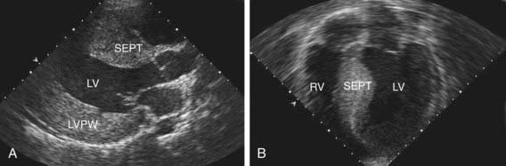
Figure 433-2 Echocardiograms demonstrating hypertrophic cardiomyopathy. A, Parasternal long-axis view of a patient with severe concentric left ventricular hypertrophy. B, Four-chamber view of a patient with asymmetric septal hypertrophy. LV, left ventricle; LVPW, left ventricular posterior wall; RV, right ventricle; SEPT, septum.
Cardiac catheterization may be indicated in some cases of hypertrophic cardiomyopathy to define the left ventricular outflow gradient with and without pharmacologic provocation, to perform electrophysiologic investigation for assessment of arrhythmia risk, or in rare cases, endomyocardial biopsy.
Additional diagnostic studies include metabolic testing, genetic testing for specific syndromic conditions, or genetic testing for mutations in genes known to cause isolated HCM (see Table 433-2). The clinical availability of these tests is expanding. In adults, where isolated HCM is a common genetic diagnosis, it has been possible to identify a subset of mutations that confer an increased risk for arrhythmia or sudden death. As identification of the molecular basis of disease in children increases, similar correlations are expected to emerge. In addition, genetic diagnosis is useful to identify at risk family members that require ongoing surveillance.
Prognosis and Management
Children under 1 yr of age or with inborn errors of metabolism or malformation syndromes have a significantly poorer prognosis. The risk of sudden death in older patients is greater in those with a history of cardiac arrest, ventricular tachycardia, exercise hypotension, syncope, excessive (>3 cm) ventricular wall thickness, and a ventricular obstruction gradient over 30 mm Hg. Although intrafamilial variability in symptoms occurs, a family history of sudden death is a highly significant predictor of risk.
Competitive sports and strenuous physical activity should be prohibited as most sudden deaths in patients with HCM occur during or immediately after vigorous physical exertion. β-Adrenergic blocking agents (propranolol, atenolol) or calcium channel blocking agents (verapamil) may be useful in diminishing ventricular outflow tract obstruction, modifying ventricular hypertrophy, and improving ventricular filling. Although significant symptomatic improvement occurs in some patients, the risk for development of heart failure or sudden death has not been lessened. In patients with atrial or ventricular arrhythmias, specific antiarrhythmic therapy should be utilized. Patients with documented ventricular arrhythmias, strong family histories of arrhythmias or sudden death, or patients with syncope should be treated with an implantable cardioverter defibrillator (ICD).
Innovative interventional procedures to anatomically or physiologically reduce the degree of LV outflow tract obstruction have been utilized. Dual chamber pacing, alcohol septal ablation, surgical septal myomectomy, and mitral valve replacement have all met with limited success.
First-degree relatives of patients identified as having HCM should be screened with electrocardiography and echocardiography. Genetic testing is available clinically. It is important to first test the affected individual in the family rather than “at risk” individuals since 20-40% of cases of HCM will not demonstrate mutations in currently available panels of genes. If a causative mutation is identified, “at risk” members of the family can be effectively tested. In families with HCM without demonstrable gene mutations, repeat noninvasive cardiac screening with ECG and echo should be undertaken in at risk individuals every 3-5 yr for patients under 12 yr of age and yearly throughout the teenage years and young adulthood. The clinical course of other affected family members and the results of genetic testing may be of some use in stratifying risk in an affected child.
Colan SD, Lipshultz SE, Lowe AM, et al. Epidemiology and cause-specific outcome of hypertrophic cardiomyopathy in children: findings from the Pediatric Cardiomyopathy Registry. Circulation. 2007;115:773-781.
Ho CY, Lopez B, Coelho-Filho OR, et al. Myocardial fibrosis as an early manifestation of hypertrophic cardiomyopathy. N Engl J Med. 2010;363(6):552-562.
Kaski JP, Esteban MTT, Lowe M, et al. Outcomes after implantable cardioverter-defibrillator treatment in children with hypertrophic cardiomyopathy. Heart. 2007;93:372-374.
Lupsa BC, Sachdev V, Lungu AO, et al. Cardiomyopathy in congenital and acquired generalized lipodystrophy. Medicine. 2010;89(4):245-250.
Maron BJ, Spirito P, Shen WK, et al. Implantable cardioverter-defibrillators and prevention of sudden cardiac death in hypertrophic cardiomyopathy. JAMA. 2007;298:405-412.
Morita H, Rehm HL, Menesses A, et al. Shared genetic causes of cardiac hypertrophy in children and adults. N Engl J Med. 2008;358:1899-1908.
Nugent AW, Daubeney PE, Chondros P, et al. National Australian Childhood Cardiomyopathy Study. Clinical features and outcomes of childhood hypertrophic cardiomyopathy: results from a national population-based study. Circulation. 2005;112:1332-1338.
Pandit B, Sarkozy A, Pennacchio LA, et al. Gain-of-function RAF1 mutations cause Noonan and LEOPARD syndromes with hypertrophic cardiomyopathy. Nat Genet. 2007;39:1007-1012.
433.3 Restrictive Cardiomyopathy
Etiology and Epidemiology
RCM accounts for <5% of cardiomyopathy cases. Incidence increases with age, and it is more common in females. In equatorial Africa, RCM accounts for a large number of deaths. Infiltrative myocardial causes and storage disorders frequently result in associated LV hypertrophy and may represent HCM with restrictive physiology. Noninfiltrative causes include mutations in genes encoding sarcomeric or cytoskeletal proteins. The majority of RCM cases are considered idiopathic.
Pathogenesis
Restrictive cardiomyopathy is characterized by normal ventricular chamber dimensions, normal myocardial wall thickness, and preserved systolic function. The abnormal myocardium demonstrates impaired ventricular compliance and filling. Filling pressures in the ventricle are typically elevated and are transmitted to the atria which become dilated. Autosomal dominant inheritance has been demonstrated for families with mutations in sarcomeric and cytoskeletal genes.
Clinical Manifestations
Abnormal ventricular filling, sometimes referred to as diastolic heart failure, is manifest in the systemic venous circulation with edema, hepatomegaly, or ascites. Elevation of left-sided filling pressures result in cough, dyspnea, or pulmonary edema. With activity, patients may experience chest pain, shortness of breath, syncope/near syncope, or even sudden death. Pulmonary hypertension and pulmonary vascular disease develop and may progress rapidly. Heart murmurs are typically absent, but a gallop rhythm may be prominent. In the presence of pulmonary hypertension, an overactive right ventricular impulse and pronounced pulmonary component of the second heart sound are present.
Diagnosis
The characteristic electrocardiographic finding of prominent P waves is usually associated with normal QRS voltages and nonspecific ST and T-wave changes. Right ventricular hypertrophy occurs in patients with pulmonary hypertension. The chest x-ray may be normal or demonstrate a prominent atrial shadow and pulmonary vascular redistribution. The echocardiogram is often diagnostic, demonstrating normal-sized ventricles with preserved systolic function and dramatic enlargement of the atria (Fig. 433-3). Flow and tissue Doppler interrogation reveal abnormal filling parameters. Differential diagnosis from constrictive pericarditis is critical, as the latter can be treated surgically. Magnetic resonance imaging may be necessary to demonstrate the thickened or calcified pericardium often present in constrictive pericardial disease.
Prognosis and Management
Pharmacologic treatment modalities are of limited use and the prognosis of patients with restrictive cardiomyopathy is generally poor with rapid clinical deterioration. Sudden death is common, with a 2-yr survival of 50%. When signs of heart failure exist, judicious use of diuretics can result in clinical improvement. As a result of the dramatic atrial enlargement, these patients are predisposed to the development of atrial tachyarrhythmias and thromboemboli. As a result, antiarrhythmic agents may be necessary and anticoagulation with platelet inhibitors or coumadin is indicated.
Cardiac transplantation is the treatment of choice in many centers for patients with restrictive cardiomyopathy, and the results are excellent in patients without pulmonary hypertension, pulmonary vascular disease, or severe congestive heart failure.
Kaski JP, Syrris P, Burch M, et al. Idiopathic restrictive cardiomyopathy in children is caused by mutations in cardiac sarcomere protein genes. Heart. 2008;94:1478-1484.
Rivenes SM, Kearney DL, Smith EO, et al. Sudden death and cardiovascular collapse in children with restrictive cardiomyopathy. Circulation. 2000;102:876-882.
433.4 Left Ventricular Noncompaction, Arrhythmogenic Right Ventricular Cardiomyopathy, and Endocardial Fibroelastosis
Left ventricular noncompaction (LVNC) was initially believed to be a rare disorder found only in children, but is now known to affect individuals of all ages. LVNC is characterized by a distinctive trabeculated or spongy appearing left ventricle (Fig. 433-4) commonly associated with left ventricular hypertrophy and/or dilation, and at times, systolic or diastolic dysfunction. LVNC may be isolated or associated with structural congenital cardiac defects. Patients may present with signs of heart failure, arrhythmias, syncope, sudden death, or as an asymptomatic finding during screening of family members.
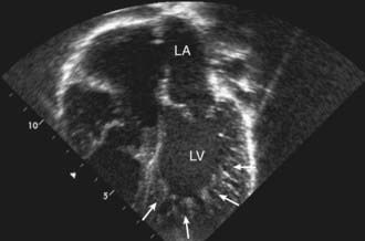
Figure 433-4 Echocardiogram of a patient with left ventricular noncompaction cardiomyopathy. Apical view showing the abnormal trabeculations of the left ventricle at the apex (arrows). For comparison, see the smooth walled LV in Fig. 433-1. LA, left atrium; LV, left ventricle.
Imaging studies utilizing ultrasound or magnetic resonance can demonstrate the characteristic pattern of deeply trabeculated LV myocardium, most characteristically within the apex of the left ventricle. ECG findings are nonspecific and include chamber hypertrophy, ST and T wave changes, or arrhythmias. In some patients, preexcitation is notable and giant voltages occur in approximately 30% of younger children. Metabolic screening should be considered, especially in young children. Elevated serum lactate and urine 3-methylglutaconic acid may be seen in Barth syndrome, an X-linked disorder of phospholipid metabolism caused by a mutation in the tafazzin (TAZ) gene. Clinical testing for TAZ mutations is available and should be considered, especially in males. Patients with mitochondrial disorders frequently demonstrate signs of LVNC. These children are at risk for atrial or ventricular arrhythmias and thromboembolic complications. Treatment includes anticoagulation, antiarrhythmic therapy if needed, and treatment of heart failure if present. In patients refractory to medical therapy, cardiac transplantation has been used successfully.
Arrhythmogenic right ventricular cardiomyopathy (ARVC) has been thought to be uncommon in North America but is among the most common forms of cardiomyopathy in Europe, especially Italy. Autosomal dominant inheritance is common. In addition, recessive forms associated with severe ARVC and skin manifestations are known. Comprehensive genetic screening has been reported to identify a cause in up to 50% of cases. ARVC is typically characterized by a dilated right ventricle (RV) with fibrofatty infiltration of the RV wall; increasingly, LV involvement is being recognized. Global and regional right and left ventricular dysfunction and ventricular tachyarrhythmias are the major clinical findings. Syncope or aborted sudden death can occur and should be treated with antiarrhythmic medications and placement of a defibrillator. In patients with ventricular dysfunction, heart failure management as indicated for patients with dilated cardiomyopathy may be of use.
Endocardial fibroelastosis (EFE), at one time an important cause of heart failure in children, has become uncommon. The decline in primary EFE is likely related to the abolition of mumps virus infections by immunization practices. Rare familial cases exist, but the causative genes are unknown. In secondary EFE, severe congenital heart disease of the left-sided obstructive type (aortic stenosis or atresia, forms of hypoplastic left heart syndrome, or severe coarctation of the aorta) is present. EFE is characterized by an opaque, white, fibroelastic thickening on the endocardial surface of the ventricle, which leads to systolic or diastolic dysfunction. Standard heart failure management or cardiac transplantation has been utilized in the management of EFE.
Left Ventricular Noncompaction
Fazio G, Pipitone S, Iacona MA, et al. The noncompaction of the left ventricular myocardium: our paediatric experience. J Cardiovasc Med. 2007;8:904-908.
Klaassen S, Probst S, Oechslin E, et al. Mutations in sarcomere protein genes in left ventricular noncompaction. Circulation. 2008;117:2893-2901.
Murphy RT, Thaman R, Blanes JG, et al. Natural history and familial characteristics of isolated left ventricular non-compaction. Eur Heart J. 2005;26:187-192.
Pantazis AA, Elliott PM. Left ventricular noncompaction. Curr Opin Cardiol. 2009;24:209-213.
Pignatelli RH, McMahon CJ, Dreyer WJ, et al. Clinical characterization of left ventricular noncompaction in children: a relatively common form of cardiomyopathy. Circulation. 2003;108:2672-2678.
Arrhythmogenic Right Ventricular Cardiomyopathy
Asimaki A, Tandri H, Huang H, et al. A new diagnostic test for arrhythmogenic right ventricular cardiomyopathy. N Engl J Med. 2009;360:1075-1084.
Basso C, Corrado D, Marcus FI, et al. Arrhythmogenic right ventricular cardiomyopathy. Lancet. 2009;373:1289-1300.
Dalal D, Nasir K, Bomma C, et al. Arrhythmogenic right ventricular dysplasia: a United States experience. Circulation. 2005;112:3823-3832.
Hulot JS, Jouven X, Empana JP, et al. Natural history and risk stratification of arrhythmogenic right ventricular dysplasia/cardiomyopathy. Circulation. 2004;11:1879-1884.
433.5 Myocarditis
Acute or chronic inflammation of the myocardium is characterized by inflammatory cell infiltrates, myocyte necrosis, or myocyte degeneration and may be caused by infectious, connective tissue, granulomatous, toxic, or idiopathic processes. There may be associated systemic manifestations of the disease and on occasion the endocardium or pericardium is involved, though coronary pathology is uniformly absent. Patients may be asymptomatic, have nonspecific prodromal symptoms, or present with overt congestive heart failure, compromising arrhythmias, or sudden death. It is thought that viral infections are the most common etiology though myocardial toxins, drug exposures, hypersensitivity reactions, and immune disorders may also lead to myocarditis (Table 433-3).

Etiology and Epidemiology
Viral Infections
Coxsackievirus and other enteroviruses, adenovirus, parvovirus, Epstein-Barr virus, and cytomegalovirus are the most common causative agents in children, though most known viral agents have been reported. In Asia, hepatitis C virus appears to be significant as well. The true incidence of viral myocarditis is unknown as mild cases likely very often go undetected. The disease is typically sporadic but may be epidemic. Manifestations are, to some degree, age dependent: in infants, viral myocarditis can be fulminant; in children, it often will occur as an acute, myopericarditis with congestive heart failure; and in older children and adolescents, it may present with signs and symptoms of acute or chronic congestive heart failure.
Bacterial Infections
Bacterial myocarditis has become far less common with the advent of advanced public health measures, which have minimized infectious causes such as diphtheria. Diphtheritic myocarditis (Chapter 180) is unique as bacterial toxin may produce circulatory collapse and toxic myocarditis characterized by atrioventricular block, bundle branch block, or ventricular ectopy. Any overwhelming systemic bacterial infection can manifest with circulatory collapse and shock with evidence of myocardial dysfunction characterized by tachycardia, gallop rhythm, and low cardiac output. Additional nonviral infectious causes of myocarditis include rickettsia, protozoa, parasitic infections, and fungal disease.
Pathophysiology
Myocarditis is characterized by myocardial inflammation, injury or necrosis, and ultimately fibrosis. Cardiac enlargement and diminished systolic function occur as a direct result of the myocardial damage. Typical signs of congestive heart failure occur and may progress rapidly to shock, atrial or ventricular arrhythmias, and sudden death. Viral myocarditis may also become a chronic process with persistence of viral nucleic acid in the myocardium, and the perpetuation of chronic inflammation secondary to altered host immune response including activated T lymphocytes (cytotoxic and natural killer cells) and antibody-dependent cell mediated damage. Additionally, persistent viral infection may alter the expression of major histocompatibility complex antigens with resultant exposure of neoantigens to the immune system. Some viral proteins share antigenic epitopes with host cells, resulting in autoimmune damage to the antigenically related myocyte. Cytokines such as tumor necrosis factor-α and interleukin-1 are inhibitors of myocyte response to adrenergic stimuli and result in diminished cardiac function. The final result of viral-associated inflammation can be dilated cardiomyopathy.
Clinical Manifestations
Manifestations of myocarditis range from asymptomatic or nonspecific generalized illness to acute cardiogenic shock and sudden death. Infants and young children more often have a fulminant presentation with fever, respiratory distress, tachycardia, hypotension, gallop rhythm, and cardiac murmur. Associated findings may include a rash or evidence of end organ involvement such as hepatitis or aseptic meningitis.
Patients with acute or chronic myocarditis may present with chest discomfort, fever, palpitations, easy fatigability, or syncope/near syncope. Cardiac findings include overactive precordial impulse, gallop rhythm, and an apical systolic murmur of mitral insufficiency. In patients with associated pericardial disease, a rub may be noted. Hepatic enlargement, peripheral edema, and pulmonary findings such as wheezes or rales may be present in patients with decompensated congestive heart failure.
Diagnosis
Electrocardiographic changes are nonspecific and may include sinus tachycardia, atrial or ventricular arrhythmias, heart block, diminished QRS voltages, and nonspecific ST and T-wave changes, often suggestive of acute ischemia. Chest roentgenograms in severe, symptomatic cases reveal cardiomegaly, pulmonary vascular prominence, overt pulmonary edema, or pleural effusions. Echocardiography often shows diminished ventricular systolic function, cardiac chamber enlargement, mitral insufficiency, and occasionally, evidence of pericardial infusion.
Endomyocardial biopsy may be useful in identifying inflammatory cell infiltrates or myocyte damage and performing molecular viral analysis using polymerase chain reaction (PCR) techniques. Catheterization and biopsy, although not without risk (perforation and arrhythmias), should be performed by experienced personnel in patients suspected to have myocarditis or if there is strong suspicion for unusual forms of cardiomyopathy such as storage diseases or mitochondrial defects. Supportive but nonspecific tests include sedimentation rate, CPK isoenzymes, cardiac troponin I, and brain natriuretic peptide (BNP) levels.
Differential Diagnosis
The predominant diseases mimicking acute myocarditis include carnitine deficiency, other metabolic disorders of energy generation, hereditary mitochondrial defects, idiopathic dilated cardiomyopathy, pericarditis, EFE, and anomalies of the coronary arteries (see Table 433-1).
Treatment
Primary therapy for acute myocarditis is supportive (Chapter 436). Acutely, the use of inotropic agents, preferably milrinone, should be entertained but used with caution because of their proarrhythmic potential. Diuretics are often required as well. If in extremis, mechanical ventilatory support and mechanical circulatory support with ventricular assist device implantation or ECMO may be needed to stabilize the patient’s hemodynamic status and act as a bridge to recovery or cardiac transplantation. Diuretics, angiotensin-converting enzyme inhibitors, and angiotensin receptor blockers are of use in patients with compensated congestive heart failure in the outpatient setting, but may be contraindicated in those presenting with fulminant heart failure and cardiovascular collapse. In patients manifesting with significant atrial or ventricular arrhythmias, specific antiarrhythmic agents (for example, amiodarone) should be administered and ICD placement considered.
Immunomodulation of patients with myocarditis is controversial. Intravenous immune globulin may have a role in the treatment of acute or fulminant myocarditis and corticosteroids have been reported to improve cardiac function, but the data are not convincing in children. Relapse has been noted in patients receiving immunosuppression who have been weaned from support. There are no studies to recommend specific antiviral therapies for myocarditis.
Prognosis
The prognosis of symptomatic acute myocarditis in newborns is poor, and 75% mortality has been reported. The prognosis is better for children and adolescents, although patients who have persistent evidence of dilated cardiomyopathy often progress to need for cardiac transplantation. Recovery of ventricular function has been reported in 10-50% of patients, however.
Bowles NE, Ni J, Kearney DL, et al. Detection of viruses in myocardial tissues by polymerase chain reaction: evidence of adenovirus as a common cause of myocarditis in children and adults. J Am Coll Cardiol. 2003;42:466-472.
Cooper LT. Myocarditis. New Engl J Med. 2009;360:1526-1538.
Costello JM, Alexander ME, Greco KM, et al. Lyme carditis in children: presentation, predictive factors, and clinical course. Pediatrics. 2009;123:e835-e841.
Freedman SB, Haladyn JK, Floh A, et al. Pediatric myocarditis: emergency department clinical findings and diagnostic evaluation. Pediatrics. 2007;120:1278-1285.
Freund MW, Kleinveld G, Krediet TG, et al. Prognosis for neonates with enterovirus myocarditis. Arch Dis Child Fetal Neonatal Ed. 2010;95:F206-F212.
Hrobon P, Kuntz KM, Hare JM. Should endomyocardial biopsy be performed for detection of myocarditis? A decision analytic approach. J Heart Lung Transplant. 1998;17:479-486.
Pinkert S, Westermann D, Wang X, et al. Prevention of cardiac dysfunction in acute coxsackievirus B3 cardiomyopathy by inducible expression of a soluble coxsackievirus-adenovirus receptor. Circulation. 2009;120:2358-2366.
Thanjan MT, Ramaswamy P, Lai WW, et al. Acute myopericarditis after multiple vaccinations in an adolescent: case report and review of the literature. Pediatrics. 2007;119:e1400-1403.
Weber MA, Ashworth MT, Risdon RA, et al. Clinicopathological features of paediatric deaths due to myocarditis: an autopsy series. Arch Dis Child. 2008;93:594-598.