Chapter 494 Soft Tissue Sarcomas
The annual incidence of soft tissue sarcomas is 8.4 cases/million white children <14 yr of age. Rhabdomyosarcoma accounts for >50% of soft tissue sarcomas. The prognosis most strongly correlates with age and extent of disease at diagnosis, primary tumor site, and histology.
Rhabdomyosarcoma
Epidemiology
The most common pediatric soft tissue sarcoma, rhabdomyosarcoma, accounts for approximately 3.5% of childhood cancers. These tumors may occur at virtually any anatomic site but are usually found in the head and neck (25%), orbit (9%), genitourinary tract (24%), and extremities (19%); retroperitoneal and other sites account for the remainder of primary sites. The incidence at each anatomic site is related to both patient age and tumor type. Extremity lesions are more likely to occur in older children and to have alveolar histology. Rhabdomyosarcoma occurs with increased frequency in patients with neurofibromatosis and has been associated with maternal breast cancer in the Li-Fraumeni syndrome, suggesting a genetic influence.
Pathogenesis
Rhabdomyosarcoma is thought to arise from the same embryonic mesenchyme as striated skeletal muscle. On the basis of light microscopic appearance, it belongs to the general category of small round cell tumors that includes Ewing sarcoma, neuroblastoma, and non-Hodgkin lymphoma. Definitive diagnosis of a pathologic specimen requires immunohistochemical studies using antibodies to skeletal muscle (desmin, muscle-specific actin, myogenin) and reverse transcription polymerase chain reaction (RT-PCR) or fluorescent in situ hybridization (FISH) for PAX-FKHR (PAX-FOX01A) transcript in the case of alveolar tumors.
Determination of the specific histologic subtype is important in treatment planning and assessment of prognosis. There are four recognized histologic subtypes. The embryonal type accounts for about 60% of all cases and has an intermediate prognosis. The botryoid type, a variant of the embryonal form in which tumor cells and an edematous stroma project into a body cavity like a bunch of grapes, is found most often in the vagina, uterus, bladder, nasopharynx, and middle ear. The alveolar type accounts for about 25-40% of cases and often is characterized by 2;13 or 1;13 chromosomal translocations. The tumor cells tend to grow in nests that often have cleftlike spaces resembling alveoli. Alveolar tumors occur most often in the trunk and extremities and carry the poorest prognosis. The pleomorphic type (adult form) is rare in childhood, accounting for <1% of cases.
Clinical Manifestations
The most common presenting feature of rhabdomyosarcoma is a mass that may or may not be painful. Symptoms are caused by displacement or obstruction of normal structures (Table 494-1). Origin in the nasopharynx may be associated with nasal congestion, mouth breathing, epistaxis, and difficulty with swallowing and chewing. Regional extension into the cranium can produce cranial nerve paralysis, blindness, and signs of increased intracranial pressure with headache and vomiting. When the tumor develops in the face or cheek, there may be swelling, pain, trismus, and, as extension occurs, paralysis of cranial nerves. Tumors in the neck can produce progressive swelling with neurologic symptoms after regional extension. Orbital primary tumors are usually diagnosed early in their course because of associated proptosis, periorbital edema, ptosis, change in visual acuity, and local pain. When the tumor arises in the middle ear, the most common early signs are pain, hearing loss, chronic otorrhea, or a mass in the ear canal; extensions of tumor produce cranial nerve paralysis and signs of an intracranial mass on the involved side. An unremitting croupy cough and progressive stridor can accompany rhabdomyosarcoma of the larynx. Because most of these signs and symptoms also are associated with common childhood conditions, clinicians must be alert to the possibility of tumor.
Table 494-1 COMMON CLINICAL SYMPTOMS OF RHABDOMYOSARCOMA
| REGION | SYMPTOMS |
|---|---|
| Head and neck | Asymptomatic mass, may mimic enlarged lymph node |
| Orbit | Proptosis, chemosis, ocular paralysis, eyelid mass |
| Nasopharynx | Snoring, nasal voice, epistaxis, rhinorrhea, local pain, dysphagia, cranial nerve palsies |
| Paranasal sinuses | Swelling, pain, sinusitis, obstruction, epistaxis, cranial nerve palsies |
| Middle ear | Chronic otitis media, hemorrhagic discharge, cranial nerve palsies, extruding polypoid mass |
| Larynx | Hoarseness, irritating cough |
| Trunk | Asymptomatic mass (usually) |
| Biliary tract | Hepatomegaly, jaundice |
| Retroperitoneum | Painless mass, ascites, gastrointestinal or urinary tract obstruction, spinal cord symptoms |
| Bladder/prostate | Hematuria, urinary retention, abdominal mass, constipation |
| Female genital tract | Polypoid vaginal extrusion of mucosanguineous tissue, vulval nodule |
| Male genital tract | Painful or painless scrotal mass |
| Extremity | Painless mass, may be very small but with secondary lymph node involvement |
| Metastatic | Nonspecific symptoms, associated with the diagnosis of leukemia |
From McDowell HP: Update on childhood rhabdomyosarcoma, Arch Dis Child 88:354–357, 2003.
Rhabdomyosarcoma of the trunk or extremities often is first noticed after trauma and initially may be regarded as a hematoma. If the swelling does not resolve or increases, malignancy should be suspected. Involvement of the genitourinary tract can produce hematuria, obstruction of the lower urinary tract, recurrent urinary tract infections, incontinence, or a mass detectable on abdominal or rectal examination. Paratesticular tumor usually manifests as a painless, rapidly growing mass in the scrotum. Vaginal rhabdomyosarcoma may manifest as a grapelike mass of tumor tissue bulging through the vaginal orifice, known as sarcoma botryoides, and can cause urinary tract or large bowel symptoms. Vaginal bleeding or obstruction of the urethra or rectum may occur. Similar findings can be noted with uterine primaries.
Tumors in any location may disseminate early and cause symptoms of pain or respiratory distress associated with pulmonary metastases. Extensive bone involvement can produce symptomatic hypercalcemia. In such cases, it may be difficult to identify the primary lesion.
Diagnosis
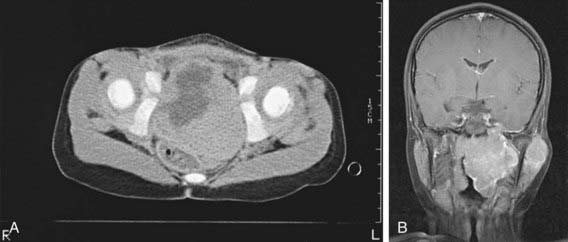
Early diagnosis of rhabdomyosarcoma requires a high index of suspicion. The microscopic appearance that of is of a small round blue cell tumor. Neuroblastoma, lymphoma, and Ewing sarcoma also are small round blue cell tumors. The differential diagnosis depends on the site of presentation. Definitive diagnosis is established by biopsy, microscopic appearance, and results of immunohistochemical stains. A lesion in an extremity may be thought to be a hematoma or hemangioma, an orbital lesion resulting in proptosis may be treated as an orbital cellulitis, or bladder-obstructive symptoms may be missed. Adolescents may ignore paratesticular lesions for a long time. Unfortunately, several months often elapse between the initial symptoms and biopsy. Diagnostic procedures are determined mainly by the area of involvement. CT or MRI is necessary for evaluation of the primary tumor site. With signs and symptoms in the head and neck area, radiographs should be examined for evidence of a tumor mass and for indications of bony erosion. MRI should be performed to identify intracranial extension or meningeal involvement and also to reveal bony involvement or erosion at the base of the skull. For abdominal and pelvic tumors, CT with a contrast agent or MRI can help delineate the tumor (Fig. 494-1). A radionuclide bone scan, chest CT, and bilateral bone marrow aspiration and biopsy should be performed to evaluate the patient for the presence of metastatic disease and to plan treatment. The most critical element of the diagnostic work-up is examination of tumor tissue, which includes the use of special histochemical stains and immunostains. Cytogenetics and molecular genetics may be helpful in detecting specific chromosomal translocations of fusion proteins present in alveolar rhabdomyosarcoma. Lymph nodes also should be sampled for presence of disease spread, especially in tumors of the extremities and in boys >10 yr of age with paratesticular tumors.
Treatment
Patients with completely resected tumors have the best prognosis. Unfortunately, most rhabdomyosarcomas are not completely resectable. At the initial surgery, tumor margins should be carefully defined, and an appropriate search for regional or metastatic disease to adjacent structures or regional lymph nodes should be completed, even if the procedure is limited to biopsy. Treatment is based on the primary tumor location and disease stage, which defines the clinical group. Most patients are given preoperative chemotherapy in an attempt to reduce the extent of surgery required and to preserve vital organs, particularly of the genitourinary tract. Group I tumors are treated with complete local excision followed by chemotherapy to reduce the likelihood of subsequent metastases. Group II tumors (microscopic residual tumor) are treated with surgery followed by local irradiation and systemic chemotherapy. Group III tumors (gross residual tumor) are treated with systemic chemotherapy, irradiation, and possibly surgery. Group IV rhabdomyosarcoma (metastatic) is treated principally with systemic chemotherapy and irradiation. Standard chemotherapeutic agents include vincristine, dactinomycin, and cyclophosphamide (VAC). A trial in intermediate-risk rhabdomyosarcoma randomized patients comparing VAC with VAC alternating with vincristine, topotecan, cyclophosphamide (VTC) and showed no improvement in outcome for patients treated with VAC/VTC over that of standard VAC therapy. Radiation therapy dosing was determined by response to second-look surgery, if performed. Irinotecan, another topoisomerase inhibitor, is currently being evaluated in patients with intermediate-risk. For patients with low-risk disease, clinical trials are investigating the reduction of therapy to maintain the same good outcome while decreasing late effects of treatment. More intensive approaches utilizing mulitagent chemotherapy in conjunction with biologic agents such as insulin-like growth factor inhibitors and other agents are being evaluated in clinical trials for the most high-risk disease.
Prognosis
Prognostic factors include age, stage, histology, and primary site. Among patients with resectable tumor and favorable histology, 80-90% have prolonged disease-free survival. Unresectable tumor localized to certain favorable sites, such as the orbit, also has a high likelihood of cure. About 65-70% of patients with incompletely resected tumor also achieve long-term disease-free survival. Patients with disseminated disease have a poor prognosis; only about 50% achieve remission, and fewer than 50% of these are cured. Older children have a poorer prognosis than younger children. For all patients, surveillance for late effects of cancer treatment (such as impaired bone growth secondary to irradiation, sterility from cyclophosphamide, and second malignancies) is important.
Other Soft Tissue Sarcomas
The non-rhabdomyosarcoma soft tissue sarcomas (NRSTSs) constitute a heterogeneous group of tumors that account for 3% of all childhood malignancies (Table 494-2). Because they are relatively rare in children, much of the information about their natural history and treatment has been derived from studies in adult patients. In children, the median age at diagnosis is 12 yr, with a male:female ratio of 2.3:1. These tumors commonly arise in the trunk or lower extremities. The most common histologic types are synovial sarcoma (42%), fibrosarcoma (13%), malignant fibrous histiocytoma (12%), and neurogenic tumors (10%). Molecular genetic studies often prove useful in diagnosis, because several of these tumors have characteristic chromosomal translocations.
Table 494-2 FEATURES OF MOST COMMON TYPES OF NON-RHABDOMYOCARCOMA SOFT TISSUE SARCOMAS
| TISSUE TYPE | TUMOR | NATURAL HISTORY AND BIOLOGY |
|---|---|---|
| Adipose | Liposarcoma | A very rare tumor. Usually arises in the extremities or retroperitoneum; associated with a nonrandom translocation, t(12;16) (q13;p11). Tends to be locally invasive and rarely metastasizes; wide local excision is the treatment of choice. The role of radiation therapy and chemotherapy in treating gross residual or metastatic disease is not established. |
| Fibrous | Fibrosarcoma | Most common soft tissue sarcoma in children <1 yr. Congenital fibrosarcoma is a low-grade malignancy that commonly arises in the extremities or trunk and rarely metastasizes. Surgical excision is treatment of choice; dramatic responses to preoperative chemotherapy may occur. In children >4 yr, the natural history is similar to that in adults (a 5-yr survival rate of 60%); wide surgical excision and preoperative chemotherapy are commonly used. Associated with t(12;15)(p13;q25) or trisomy 11, also +8, +17, +20. |
| Malignant fibrous histiocytoma | Most commonly arises in the trunk and extremities, deep in the subcutaneous layer. Histologically subdivided into storiform, giant cell, myxoid, and angiomatoid variants. The angiomatoid type tends to affect younger patients and is curable with surgical resection alone. Wide surgical excision is the treatment of choice. Chemotherapy has produced objective tumor regressions. | |
| Vascular | Hemangiopericytoma | Often arises in the lower extremities or retroperitoneum; may manifest as hypoglycemia and hypophosphatemic rickets. Both benign and malignant histology. Nonrandom translocations t(12;19) (q13;q13) and t(13;22) (q22;q13.3) have been described. Complete surgical excision is the treatment of choice. Chemotherapy and radiation therapy may produce responses. |
| Angiosarcoma | Rare in children; 33% arise in skin, 25% in soft tissue, and 25% in liver, breast, or bone. Associated with chronic lymphedema and exposure to vinyl chloride in adults. Survival rate is poor (12% at 5 yr) despite some responses to chemotherapy/radiation therapy. | |
| Hemangioendothelioma | Can occur in soft tissue, liver, and lung. Localized lesions have a favorable outcome; lesions in lung and liver often are multifocal and have a poor prognosis. | |
| Peripheral nerves | Neurofibrosarcoma | Also known as the malignant peripheral nerve sheath tumor. Develops in up to 16% of patients with neurofibromatosis type 1 (NF1); almost 50% occur in patients with NF1. Deletions of chromosome 22q11-q13 or 17q11 and p53 mutations have been reported. Commonly arises in trunk and extremities and is usually locally invasive. Complete surgical excision is necessary for survival; response to chemotherapy is suboptimal. |
| Synovium | Synovial sarcoma | The most common non-rhabdomyosarcoma soft tissue sarcoma in some series. Often manifesting in the 3rd decade, but 33% of patients are <20 yr. Typically arises around the knee or thigh and is characterized by a nonrandom translocation t(X;18) (p11;q11). Wide surgical excision is necessary. Radiation therapy is effective in microscopic residual disease, and ifosfamide-based therapy is active in advanced disease. |
| Unknown | Alveolar soft part sarcoma | Slow-growing tumor; tends to recur or to metastasize to lung and brain years after diagnosis. Often arises in the extremities and head and neck. A myogenic origin has been proposed. Resection of primary and metastatic sites, when possible, is recommended. |
| Smooth muscle | Leiomyosarcoma | Often arises in the gastrointestinal tract and may be associated with a t(12;14) (q14;q23) translocation. Associated with Epstein-Barr virus in immunodeficiency syndromes (including AIDS). Complete surgical excision is the treatment of choice. |
Surgery remains the mainstay of therapy, but a careful search for lung and bone metastases should be undertaken before surgical excision. Chemotherapy and radiation therapy should be considered for large, high-grade, and unresectable tumors. The role of chemotherapy for NRSTSs is not as well defined as for rhabdomyosarcoma. Patients with unresectable or metastatic disease are treated with multiagent chemotherapy in addition to irradiation and/or surgery. Tumor size, stage (clinical group), invasiveness, and histologic grade correlate with survival.
Arndt CAS, Crist WM. Medical progress: common musculoskeletal tumors of childhood and adolescence. N Engl J Med. 1999;341:342-352.
Arndt CAS, Stoner JA, Hawkins DS, et al. Vincristine, actinomycin, and cyclophosphamide compared with vincristine, actinomycin, and cyclophosphamide alternating with vincristine, topotecan, and cyclophosphamide for intermediate-risk rhabdomyosarcoma: children’s oncology group study D9803. J Clin Oncol. 2009;27:5182-5188.
Crist WM, Anderson JR, Meza JL, et al. Intergroup rhabdomyosarcoma study. IV: results for patients with nonmetastatic disease. J Clin Oncol. 2001;19:3091-3102.
Hettmer S, Wagers AJ. Uncovering the origins of rhabdomyosarcoma. Nat Med. 2010;16:171-173.
McDowell HP. Update on childhood rhabdomyosarcoma. Arch Dis Child. 2003;88:354-357.
Meyer WH, Spunt SL. Soft tissue sarcomas of childhood. Cancer Treat Rev. 2004;30:269-280.
Schaaf G, Hamdi M, Zwijnenburg D, et al. Silencing of SPRY1 triggers complete regression of rhabdomyosarcoma tumors carrying a mutated RAS gene. Cancer Res. 2010;70:762-771.
Sinha S, Peach AHS. Diagnosis and management of soft tissue sarcoma. BMJ. 2011;342:157-162.
Spunt SL, Poquette CA, Hurt YS, et al. Prognostic factors for children and adolescents with surgically resected nonrhabdomyosarcoma soft tissue sarcoma: an analysis of 121 patients treated at St. Jude Children’s Research Hospital. J Clin Oncol. 1999;17:3697-3705.
Stevens MCG. Treatment for childhood rhabdomyosarcoma: the cost of cure. Lancet Oncol. 2005;6:77-84.