Chapter 596 Brain Abscess
Brain abscesses can occur in children of any age but are most common in children between 4 and 8 yr and neonates. The causes of brain abscess include embolization due to congenital heart disease with right-to-left shunts (especially tetralogy of Fallot), meningitis, chronic otitis media and mastoiditis, sinusitis, soft tissue infection of the face or scalp, orbital cellulitis, dental infections, penetrating head injuries, immunodeficiency states, and infection of ventriculoperitoneal shunts.
Pathology
Cerebral abscesses are evenly distributed between the 2 hemispheres, and 80% of cases are divided equally between the frontal, parietal, and temporal lobes. Brain abscesses in the occipital lobe, cerebellum, and brainstem account for about 20% of the cases. Most brain abscesses are single, but 30% are multiple and may involve more than 1 lobe. The pathogenesis is undetermined in 10-15% of cases. An abscess in the frontal lobe is often caused by extension from sinusitis or orbital cellulitis, whereas abscesses located in the temporal lobe or cerebellum are frequently associated with chronic otitis media and mastoiditis. Abscesses resulting from penetrating injuries tend to be singular and caused by Staphylococcus aureus, whereas those resulting from septic emboli, congenital heart disease, or meningitis often have several causal organisms.
Etiology
The responsible bacteria include streptococci (Streptococcus milleri, Streptococcus pyogenes group A or B, Streptococcus pneumoniae, Enterococcus faecalis), anaerobic organisms (gram-positive cocci, Bacteroides spp., Fusobacterium spp., Prevotella spp., Actinomyces spp.), and gram-negative aerobic bacilli (Haemophilus aphrophilus, Haemophilus parainfluenzae, Haemophilus influenzae, Enterobacter, Escherichia coli, Proteus spp.). Citrobacter is most common in neonates. One organism is cultured in 70% of abscesses, 2 in 20%, and 3 or more in 10% of cases. Abscesses associated with mucosal infections (sinusitis) frequently have anaerobic bacteria. Fungal abscesses (Aspergillus, Candida) are more common in immunosuppressed patients.
Clinical Manifestations
The early stages of cerebritis and abscess formation are associated with nonspecific symptoms, including low-grade fever, headache, and lethargy. The significance of these symptoms is generally not recognized, and an oral antibiotic is often prescribed with resultant transient relief. As the inflammatory process proceeds, vomiting, severe headache, seizures, papilledema, focal neurologic signs (hemiparesis), and coma may develop. A cerebellar abscess is characterized by nystagmus, ipsilateral ataxia and dysmetria, vomiting, and headache. If the abscess ruptures into the ventricular cavity, overwhelming shock and death usually ensue.
Diagnosis
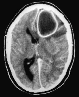
The peripheral white blood cell count can be normal or elevated, and the blood culture is positive in 10% of cases. Examination of the cerebrospinal fluid (CSF) shows variable results; the white blood cells and protein may be minimally elevated or normal, and the glucose level may be low. CSF cultures are rarely positive; aspiration of the abscess is much more likely to establish a bacteriologic diagnosis. Molecular diagnostics with PCR are being evaluated to establish a bacterial etiology in aspirates from brain abscesses. Because examination of the CSF is seldom useful and a lumbar puncture may cause herniation of the cerebellar tonsils, the procedure should not be undertaken in a child suspected of having a brain abscess. The electroencephalogram (EEG) shows corresponding focal slowing, and the radionuclide brain scan indicates an area of enhancement due to disruption of the blood-brain barrier in >80% of cases. CT with contrast and MRI are the most reliable methods of demonstrating cerebritis and abscess formation (Fig. 596-1). MRI is the diagnostic test of choice. The CT findings of cerebritis are characterized by a parenchymal low-density lesion, and MRI T2 weighted images indicate increased signal intensity. An abscess cavity shows a ring-enhancing lesion by contrast CT, and the MRI also demonstrates an abscess capsule with gadolinium administration.
Treatment
The initial management of a brain abscess includes prompt diagnosis and institution of an antibiotic regimen that is based on the probable pathogenesis and the most likely organism. When the cause is unknown, the combination of vancomycin, a 3rd-generation cephalosporin, and metronidazole is commonly used. The same regimen is initiated when otitis media, sinusitis, or mastoiditis is the likely cause. If there is a history of penetrating head injury, head trauma, or neurosurgery, vancomycin plus a 3rd-generation cephalosporin is appropriate. When cyanotic congenital heart disease is the predisposing factor, ampicillin-sulbactam alone or a 3rd-generation cephalosporin plus metronidazole may be used. Meropenem has good activity against gram-negative bacilli, anaerobes, staphylococci, and streptococci, including most antibiotic-resistant pneumococci, and may be used alone to replace the combination of metronidazole and a β-lactam in the previous regimens. Notably, meropenem does not provide activity against methicillin-resistant S. aureus and may have decreased activity against penicillin-resistant strains of S. pneumoniae, indicating that vancomycin should remain a part of the initial regimen when these organisms are suspected. Abscesses secondary to an infected ventriculoperitoneal shunt may be initially treated with vancomycin and ceftazidime. When Citrobacter meningitis (often in neonates) leads to abscess formation, a 3rd-generation cephalosporin is used, typically in combination with an aminoglycoside. Listeria monocytogenes may cause a brain abscess in the neonate and if suspected, ampicillin should be added to the cephalosporin. In immunocompromised patients, broad-spectrum antibiotic coverage is used, and amphotericin B therapy should be considered.
A brain abscess can be treated with antibiotics without surgery if the abscess is <2 cm in diameter, the illness is of short duration (<2 wk), there are no signs of increased intracranial pressure, and the child is neurologically intact. If the decision is made to treat with antibiotics alone, the child should have follow-up neuroimaging studies to ensure the abscess is decreasing in size. An encapsulated abscess, particularly if the lesion is causing a mass effect or increased intracranial pressure, should be treated with a combination of antibiotics and aspiration. Surgical excision of an abscess is rarely required, because the procedure may be associated with greater morbidity compared with aspiration of a cavity. Surgery is indicated when the abscess is >2.5 cm in diameter, gas is present in the abscess, the lesion is multiloculated, the lesion is located in the posterior fossa, or a fungus is identified. Associated infectious processes, such as mastoiditis, sinusitis, or a periorbital abscess, may require surgical drainage. The duration of antibiotic therapy depends on the organism and response to treatment, but is usually 4-6 wk.
Prognosis
Mortality rate associated with brain abscess has decreased significantly to 15-20% with the use of CT or MRI and prompt antibiotic and surgical management. Factors associated with high mortality rate at the time of admission include age <1 yr, multiple abscesses and coma. Long-term sequelae occur in at least 50% of survivors and include hemiparesis, seizures, hydrocephalus, cranial nerve abnormalities, and behavior and learning problems.
Brook I. Aerobic and anaerobic bacteriology of intracranial abscesses. Pediatr Neurol. 1992;8:210-214.
Goodkin HP, Harper MB, Pomeroy SL. Intracranial abscess in children: historical trends at Children’s Hospital Boston. Pediatrics. 2004;113:1765-1770.
Masalma A, Armougom F, Scheld WM, et al. The expansion of the microbiological spectrum of brain abscesses with use of multiple 16S ribosomal DNA sequencing. Clin Infect Dis. 2009;48:1169-1178.
Saez-Lloreus XJ, Umana NA, Odio CN, et al. Brain abscesses in infants and children. Pediatr Infect Dis J. 1989;8:449-458.
Sjolin J, Lilja A, Erikson N, et al. Treatment of brain abscess with cefotaxime and metronidazole: prospective study on 15 consecutive patients. Clin Infect Dis. 1993;17:857-863.
Smith RR. Neuroradiology of intracranial infection. Pediatr Neurosurg. 1992;18:92-104.
Yogev R, Bar-Meir M. Management of brain abscesses in children. Pediatr Infect Dis J. 2004;23:157-159.