Positioning Terminology
Radiographic positioning refers to the study of patient positioning performed for radiographic demonstration or visualization of specific body parts on image receptors. The radiologic technologist must clearly understand the correct use of positioning terminology. This section lists, describes, and illustrates the commonly used terms consistent with the positioning and projection terminology as approved and published by the American Registry of Radiologic Technologists (ARRT).
4
Throughout this text, named positions (i.e., with the proper name of the person who first described a specific position or procedure) are referred to as methods, such as the Towne, Waters, and Caldwell methods. The ARRT concurs regarding the use of the named method in parentheses after the projection or position term. The description of radiographic positions by the proper name method is becoming less common.
General Terms
- Radiograph (ra′-de-o-graf): (1) An image of a patient’s anatomic part(s), as produced by the action of x-rays on an image receptor (Fig. 1.38). If the radiograph is produced with the use of traditional film-screen (analog) technology, the image is captured and displayed on film; if the radiograph is produced via digital technology, the image is viewed and stored on display monitors. (2) The term radiograph refers to the recording medium and the image.
- Radiography (ra″-de-og′-rah-fe): The process and procedures of producing a radiograph.
- Image receptor (IR): The device that responds to the ionizing radiation to create the radiographic image after it exits the patient; refers to both analog (film-based) cassettes and digital acquisition devices.
- Central ray (CR): Refers to the centermost portion of the x-ray beam emitted from the x-ray tube—the portion of the x-ray beam that has the least divergence.
Radiographic Examination or Procedure
A radiologic technologist is shown positioning a patient for a routine chest examination or procedure (Fig. 1.39). A radiographic examination involves five general functions:
-
- 1. Positioning of body part and alignment with the IR and CR
- 2. Application of radiation protection measures and devices
- 3. Selection of exposure factors (radiographic technique)
- 4. Instructions to the patient related to respiration (breathing) and initiation of the x-ray exposure
- 5. Processing of the IR (analog) [chemical processing] or digital processing systems
Anatomic Position
The anatomic (an″-ah-tom′-ik) position is a reference position that defines specific surfaces and planes of the body. It also defines anatomic directional terms such as anterior, posterior, medial, lateral, superior, and inferior regions of the body. The anatomic position is an upright position with arms abducted slightly (down), hands by sides with palms forward, and head and feet together and directed straight ahead (Fig. 1.40).
Viewing Radiographs
A general rule in viewing radiographs is to display them so that the patient is facing the viewer, with the patient in the anatomic position.


Body Planes, Sections, and Lines (Fig. 1.41)
Positioning terms that describe CR angles or relationships between body parts often are related to imaginary planes that pass through the body in the anatomic position. The study of CT, MRI (magnetic resonance imaging), and sonography (diagnostic medical ultrasound) emphasizes sectional anatomy, which also involves the primary body planes and sections as described subsequently.
Plane: Straight Line Surface Connecting Two Points
Four common planes used in medical imaging are the sagittal plane, coronal plane, horizontal (axial) plane, and oblique plane.
Sagittal Plane
A sagittal (saj′-i-tal) plane is any longitudinal plane that divides the body into right and left parts.
The midsagittal plane, sometimes called the median plane, is a midline sagittal plane that divides the body into equal right and left parts. It passes approximately through the sagittal suture of the skull. Any plane parallel to the midsagittal or median plane is called a sagittal plane.
Coronal Plane
A coronal (ko-ro′-nal) plane is any longitudinal plane that divides the body into anterior and posterior parts.
The midcoronal plane divides the body into approximately equal anterior and posterior parts. It is called a coronal plane because it passes approximately through the coronal suture of the skull. Any plane parallel to the midcoronal or frontal plane is called a coronal plane.
Horizontal (Axial) Plane
A horizontal (axial) plane is any transverse plane that passes through the body at right angles to a longitudinal plane, dividing the body into superior and inferior portions.
Oblique Plane
An oblique plane is a longitudinal or transverse plane that is at an angle or slant and is not parallel to the sagittal, coronal, or horizontal plane.
Sectional Image of Body Part
Longitudinal Sections—Sagittal, Coronal, and Oblique
These sections or images run lengthwise in the direction of the long axis of the body or any of its parts, regardless of the position of the body (erect or recumbent). Longitudinal sections or images may be taken in the sagittal, coronal, or oblique plane.
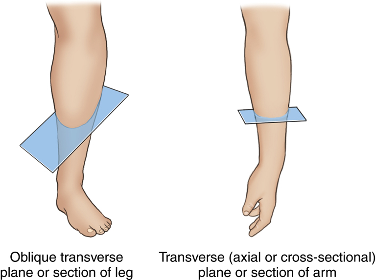
Transverse or Axial Sections (Cross-Sections)
Sectional images are at right angles along any point of the longitudinal axis of the body or its parts (Fig. 1.42)
Sagittal, Coronal, and Axial Images
CT, magnetic resonance imaging (MRI), and sonography images are obtained in these three common orientations or views. These common orientations are sagittal, coronal, and transverse (axial). (MRI sectional images are shown in Figs. 1.43 through 1.45.)


Planes of the Skull (FIG. 1.46)
Base Plane of Skull
This precise transverse plane is formed by connecting the lines from the infraorbital margins (inferior edge of bony orbits) to the superior margin of the external auditory meatus (EAM), the external opening of the ear. This sometimes is called the Frankfort horizontal plane,
1
as used in orthodontics and cranial topography to measure and locate specific cranial points or structures.
Occlusal Plane
This horizontal plane is formed by the biting surfaces of the upper and lower teeth with jaws closed (used as a reference plane of the head for cervical spine and skull radiography).