Body Positions
In radiography, the term position is used in two ways, first as general body positions, as described next, and second as specific body positions, which are described in the pages that follow.
General Body Positions
The eight most commonly used general body positions in medical imaging are as follows:
- 1. Supine (soo′-pine): Lying on back, facing upward (Fig. 1.55)
- 2. Prone (prohn): Lying on abdomen, facing downward (head may be turned to one side) (Fig. 1.56)
- 3. Erect (e-reckt′) (upright): An upright position, to stand or sit erect
- 4. Recumbent (re-kum′-bent) (reclining): Lying down in any position (prone, supine, or on side)
- 5. Trendelenburg 5 (tren-del′-en-berg) position: A recumbent position with the body tilted with the head lower than the feet (Fig. 1.57)
- 6. Fowler 6 (fow′-ler) position: A recumbent position with the body tilted with the head higher than the feet ( Fig. 1.58)
- 7. Sims position (semiprone position): A recumbent oblique position with the patient lying on the left anterior side, with the right knee and thigh flexed and the left arm extended down behind the back. A modified Sims position as used for insertion of the rectal tube for a barium enema is shown in Fig. 1.59 (demonstrated in Chapter 13).
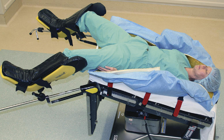
- 8. Lithotomy (li-thot′-o-me) position: A recumbent (supine) position with knees and hip flexed and thighs abducted and rotated externally, supported by ankle supports (Fig. 1.60). This position is seen frequently in the surgical suite for certain urinary studies.


Specific Body Positions
In addition to identifying general body positions, the term position is used in radiography to refer to a specific body position described by the body part closest to the IR (oblique and lateral) or by the surface on which the patient is lying (decubitus).
Lateral Position
Lateral (lat′-er-al) position refers to the side of, or a side view.
Specific lateral positions are described by the side of the body closest to the IR or the body part from which the CR exits. A right lateral position is shown with the right side of the body closest to the IR in the erect position (Fig. 1.61). Fig. 1.62 demonstrates a recumbent left lateral position.
A true lateral position is always 90°, or perpendicular, or at a right angle, to a true AP or PA projection. If it is not a true lateral, it is an oblique position.
Oblique Position 5
Oblique (ob-lek′, or ob-lik′)
7
(oh bleek′, or oh blike′) position refers to an angled position in which neither the sagittal nor the coronal body plane is perpendicular or at a right angle to the IR.
Oblique body positions of the thorax, abdomen, or pelvis are described by the side of the body closest to the IR or the body part from which the CR exits.
Left and Right Posterior Oblique (LPO and RPO) Positions
LPO and RPO describe the specific oblique positions in which the left or right posterior aspect of the body is closest to the IR. A left posterior oblique (LPO) is demonstrated in both the erect (Fig. 1.63 and recumbent (Fig. 1.64) positions.
The CR exits from the left or right posterior aspect of the body.
NOTE: These also can be referred to as AP oblique projections because the CR enters an anterior surface and exits posteriorly. However, this is not a complete description and requires a specific position clarifier such as LPO or RPO position. Therefore, throughout this text, these body obliques are referred to as positions and not projections.
Right and Left Anterior Oblique (RAO and LAO) Positions
RAO and LAO refer to oblique positions in which the right or left anterior aspect of the body is closest to the IR and can be erect or recumbent general body positions. (A right anterior oblique [RAO] is shown in both examples (Figs. 1.65 and 1.66).
NOTE: These also can be described as PA oblique projections if a position clarifier is added, such as an RAO or LAO position.
It is not correct to use these oblique terms or the abbreviations LPO, RPO, RAO, or LAO as projections because they do not describe the direction or path of the CR; rather, these are positions.


Decubitus (Decub) Position
The word decubitus (de-ku′bi-tus) literally means to lie down, or the position assumed in lying down.
This body position, meaning to lie on a horizontal surface, is designated according to the surface on which the body is resting. This term describes a patient who is lying on one of the following body surfaces: back (dorsal), front (ventral), or side (right or left lateral).
In radiographic positioning, decubitus is always performed with the CR horizontal.
Decubitus positions are essential for detecting air-fluid levels or free air in a body cavity such as the chest or abdomen, where the air rises to the uppermost part of the body cavity. Decubitus positions are often performed if the patient cannot assume erect position.
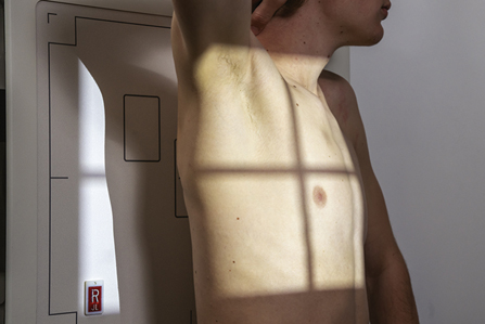
Right or Left Lateral Decubitus Position—AP or PA Projection
In this position, the patient lies on the side, and the x-ray beam is directed horizontally from anterior to posterior (AP) (Fig. 1.67) or from posterior to anterior (PA) (Fig. 1.68).
The AP or PA projection is important as a qualifying term with decubitus positions to denote the direction of the CR.
This position is either a left lateral decubitus (see Fig. 1.67) or a right lateral decubitus (see Fig. 1.68).
NOTE: The decubitus position is identified according to the dependent side (side down) and the AP or PA projection indication. Example: Left lateral decubitus (PA projection) is with the patient lying on left side facing the image receptor. The CR enters the posterior side and exits the anterior side.
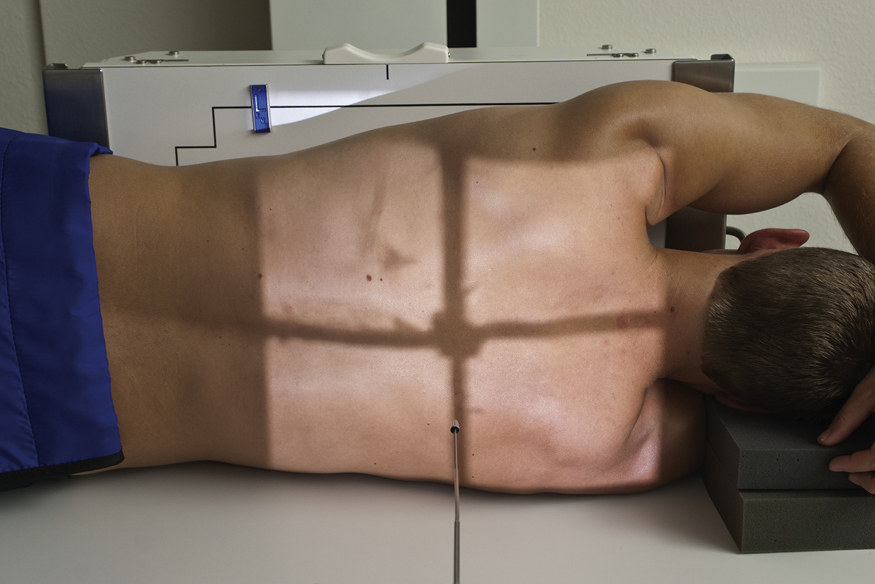
Dorsal Decubitus Position—Left or Right Lateral
In this position, the patient is lying on the dorsal (posterior) surface with the x-ray beam directed horizontally, exiting from the side closest to the IR (Fig. 1.69).
The position is named according to the surface on which the patient is lying (dorsal or ventral) and by the side closest to the IR (right or left).
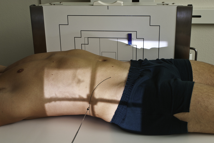
Ventral Decubitus Position—Right or Left Lateral
In this position, the patient is lying on the ventral (anterior) surface with the x-ray beam directed horizontally, exiting from the side closest to the IR (Fig. 1.70).

Additional Special-Use Projection Terms
Following are some additional terms commonly used to describe projections. These terms, as shown by their definitions, also refer to the path or projection of the CR and are projections rather than positions.
Axial Projection
Axial (ak′-se-al) refers to the long axis of a structure or part (around which a rotating body turns or is arranged).
Special application—AP or PA axial: In radiographic positioning, the term axial is used to describe any angle of the CR of 10° or more along the long axis of the body or body part.
7
However, in a true sense, an axial projection would be directed along, or parallel to, the long axis of the body or part. The term semiaxial, or “partly” axial, more accurately describes any angle along the axis that is not truly perpendicular or parallel to the long axis. However, for the sake of consistency with other references, the term
axial projection
is used throughout this text to describe both axial and semiaxial projections, as defined earlier and as illustrated in Figs. 1.71 through 1.73.
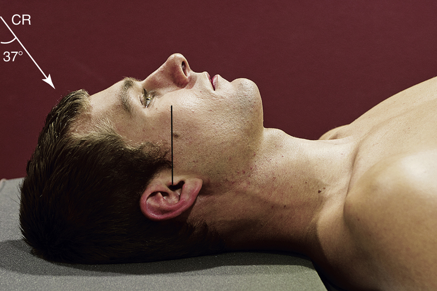
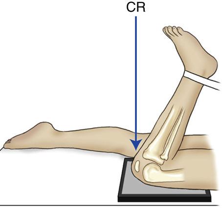
Inferosuperior and Superoinferior Axial Projections
Inferosuperior axial projections are frequently performed for the shoulder and hip, where the CR enters below or inferiorly and exits above or superiorly (see Fig. 1.73).
The opposite of this is the superoinferior axial projection, such as a special nasal bone projection (see Fig. 1.71).
Tangential Projection
Tangential (ta″-jen′-shal) means touching a curve or surface at only one point.
This is a special use of the term projection to describe the CR that skims a body part to project the anatomy into profile and free of superimposition of surrounding body structures.
Examples
Following are two examples or applications of the term tangential projection:
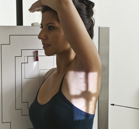
AP Axial Projection—Lordotic Position
This is a specific AP axial chest projection for demonstrating the apices of the lungs. It also is called the AP lordotic position. In this case, the long axis of the body rather than the CR is angled.
The term lordotic comes from lordosis, a term that denotes curvature of the cervical and lumbar spine (see Chapters 8 and 9). As the patient assumes this position (Fig. 1.76), the lumbar lordotic curvature is exaggerated, making this a descriptive term for this special chest projection.


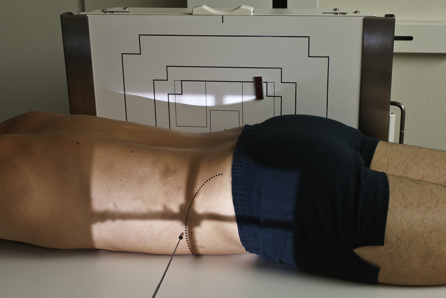
Transthoracic Lateral Projection (Right Lateral Position)
This is a lateral projection through the thorax. It requires a qualifying positioning term (right or left lateral position) to indicate which shoulder is closest to the IR and is being examined (Fig. 1.77).
NOTE: This is a special adaptation of the projection term, indicating that the CR passes through the thorax even though it does not include an entrance or exit site. In practice, this is a common lateral shoulder projection and is referred to as a right or left transthoracic lateral shoulder.
Dorsoplantar and Plantodorsal Projections
These are secondary terms for AP or PA projections of the foot.
Dorsoplantar (DP) describes the path of the CR from the dorsal (anterior) surface to the plantar (posterior) surface of the foot (Fig. 1.78).
A special plantodorsal projection of the heel bone (calcaneus) is called an axial plantodorsal projection (PD) because the angled CR enters the plantar surface of the foot and exits the dorsal surface (Fig. 1.79).
Parietoacanthial and Acanthioparietal Projections
The CR enters at the cranial parietal bone and exits at the acanthion (junction of nose and upper lip) for the parietoacanthial projection (Fig. 1.80).
The opposite CR direction would describe the acanthioparietal projection (Fig. 1.81).
These are also known as PA Waters and AP reverse Waters methods and are used to visualize the facial bones.
Submentovertical (SMV) and Verticosubmental (VSM) Projections
These projections are used for the skull and mandible.
The CR enters below the chin, or mentum, and exits at the vertex or top of the skull for the submentovertical (SMV) projection (Fig. 1.82).
The less common, opposite projection of this would be the verticosubmental (VSM) projection, entering at the top of the skull and exiting below the mandible (not shown).


Relationship Terms
Following are paired positioning or anatomic terms that are used to describe relationships to parts of the body with opposite meanings.
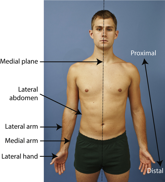
Medial Versus Lateral
Medial (me′-de-al) versus lateral refers to toward versus away from the center, or median plane.
In the anatomic position, the medial aspect of any body part is the “inside” part closest to the median plane, and the lateral part is away from the center, or away from the median plane or midline of the body (Fig. 1.83).
Examples
In the anatomic position, the thumb is on the lateral aspect of the hand. The lateral part of the abdomen and thorax is the part away from the median plane.
Proximal Versus Distal
Proximal (prok′-si-mal) is near the source or beginning, and distal (dis′-tal) is away from. In regard to the upper and lower limbs, proximal and distal would be the part closest to or away from the trunk, the source or beginning of that limb (see Fig. 1.83).
Examples
The elbow is proximal to the wrist. The finger joint closest to the palm of the hand is called the proximal interphalangeal (PIP) joint, and the joint near the distal end of the finger is the distal interphalangeal (DIP) joint (see Chapter 4).
Cephalad Versus Caudad
Cephalad (sef′-ah-lad) means toward the head end of the body; caudad (kaw′-dad) means away from the head end of the body.
A cephalad angle is any angle toward the head end of the body (Fig. 1.84; also see Fig. 1.86). (Cephalad, or cephalic, literally means “head” or “toward the head.”)
A caudad angle is any angle toward the feet or away from the head end (Fig. 1.85). (Caudad or caudal comes from cauda, literally meaning “tail.”)
In human anatomy, cephalad and caudad also can be described as superior (toward the head) or inferior (toward the feet).
Interior (Internal, Inside) Versus Exterior (External, Outer)
Interior is inside of something, nearer to the center, and exterior is situated on or near the outside.
Superficial Versus Deep
Superficial is nearer the skin surface; deep is farther away.
Example
The cross-sectional drawing in Fig. 1.87 shows that the humerus is deep compared with the skin of the arm, which is superficial.
Another example would be a superficial tumor or lesion, which is located near the surface, compared with a deep tumor or lesion, which is located deeper within the body or part.
Ipsilateral Versus Contralateral
Ipsilateral (ip″-si-lat′-er-al) is on the same side of the body or part; contralateral (kon″-trah-lat′-er-al) is on the opposite side.
Example
The right thumb and the right great toe are ipsilateral; the right knee and the left hand are contralateral.


Terms Related to Movement
The final group of positioning and related terms that every technologist should know relates to various movements. Most of these are listed as paired terms that describe movements in opposite directions.
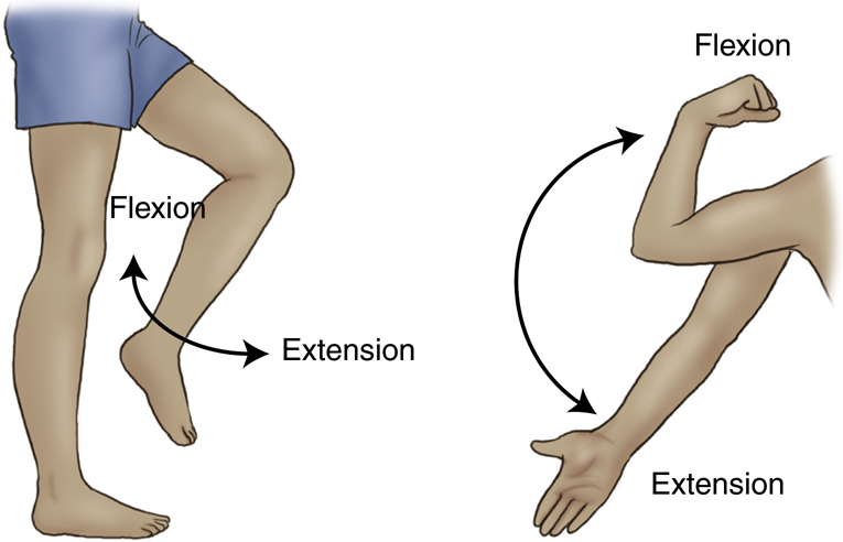
Flexion Versus Extension
When a joint is flexed or extended, the angle between parts is decreased or increased.
Flexion decreases the angle of the joint (see examples of knee, elbow, and wrist flexions in Fig. 1.88).
Extension increases the angle as the body part moves from a flexed to a straightened position. This is true for the knee, elbow, and wrist joints, as is shown in Fig. 1.88.
Hyperextension
Hyperextension is extending a joint beyond the straight or neutral position.
Abnormal Hyperextension
A hyperextended elbow or knee results when the joint is extended beyond the straightened or neutral position. This is not a natural movement for these two joints and results in injury or trauma.
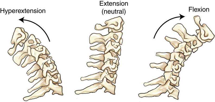
Normal Flexion and Hyperextension of the Spine
Flexion is bending forward, and extension is returning to the straight or neutral position. A backward bending beyond the neutral position is hyperextension. In practice, however, the terms flexion and extension are commonly used for these two extreme flexion and hyperextension projections of the spine (Fig. 1.89).
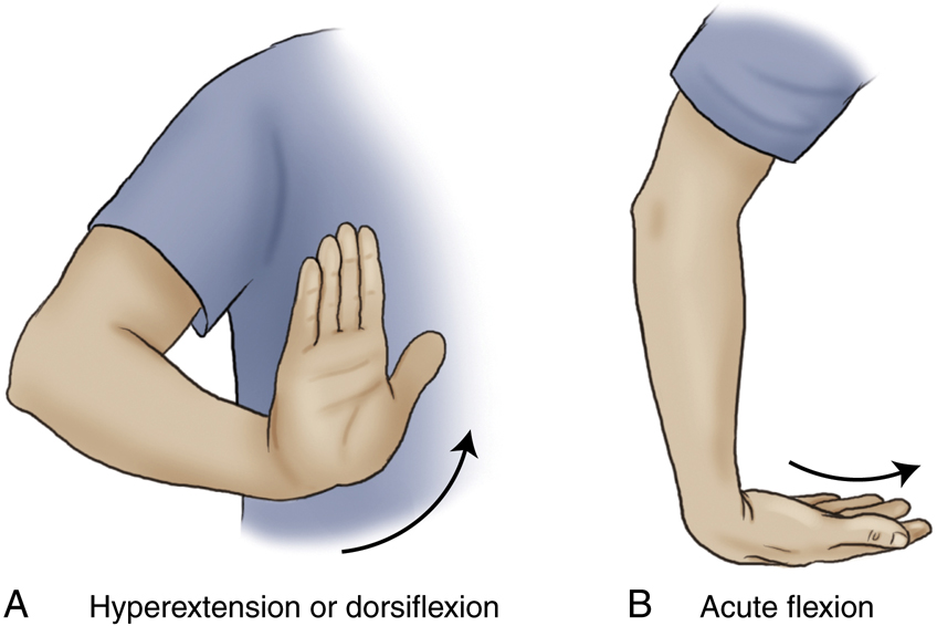
Normal Hyperextension of the Wrist
A second example of a special use of the term hyperextension concerns the wrist, where the carpal canal (tangential, inferosuperior) projection of the carpals is visualized by a special hyperextended wrist movement in which the wrist is extended beyond the neutral position. This specific wrist movement is also called dorsiflexion (backward or posterior flexion) (Fig. 1.90A).
Acute Flexion of the Wrist
An acute or full flexion of the wrist is required for a special tangential projection for a carpal bridge projection of the posterior aspect of the wrist (see Fig. 1.90B).
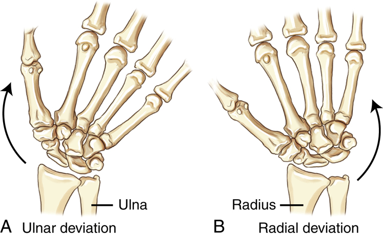
Ulnar Deviation Versus Radial Deviation of the Wrist
Deviation literally means “to turn aside” or “to turn away from the standard or course.”
8
Ulnar deviation (Fig. 1.91A) is to turn or bend the hand and wrist from the natural position toward the ulnar side, and radial deviation (Fig. 1.91B) is toward the radial side of the wrist.
NOTE: Earlier editions of this textbook and other positioning references have defined these wrist movements as ulnar and radial flexion movements because they describe specific flexion movements toward either the ulna or the radius.
9
However, because practitioners in the medical community, including orthopedic physicians, commonly use the terms ulnar and radial deviation for these wrist movements, this text also has changed this terminology to ulnar and radial deviation movements to prevent confusion and to ensure consistency with other medical references.
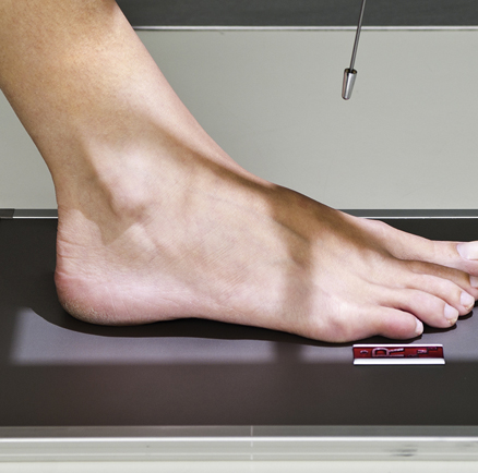
Dorsiflexion Versus Plantar Flexion of the Foot and Ankle
Dorsiflexion of the Foot
This term means to decrease the angle (flex) between the dorsum (top of foot) and the lower leg, moving foot and toes upward (Fig. 1.92A).
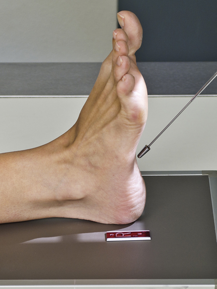
Plantar Flexion of the Foot
Extending the ankle joint, moving foot and toes downward from the normal position; flexing or decreasing the angle toward the plantar (posterior) surface of the foot (see Fig. 1.92B).
NOTE: See preceding page for dorsiflexion of the wrist (see Fig. 1.90A) compared with dorsiflexion of the foot (see Fig. 1.92A).
Eversion Versus Inversion
Inversion (in-ver′-zhun) is inward stress movement of the foot as applied to the foot without rotation of the leg (Fig. 1.94).
The plantar surface (sole) of the foot is turned or rotated away from the median plane of the body (the sole faces in a more lateral direction) for eversion and toward the median plane for inversion.
The leg does not rotate, and stress is applied to the medial and lateral aspects of the ankle joint for evaluation of possible widening of the joint space (ankle mortise).
Valgus Versus Varus 1
Valgus (val′-gus) describes an abnormal position in which a part or limb is forced outward from the midline of the body. Valgus sometimes is used to describe eversion stress of the ankle joint.
Varus (va′-rus) describes an abnormal position in which a part or limb is forced inward toward the midline of the body. The term varus stress sometimes is used to describe inversion stress applied at the ankle joint.
NOTE: The terms valgus and varus are also used to describe the loss of normal alignment of bones due to fracture (see Chapter 15).
Medial (Internal) Rotation Versus Lateral (External) Rotation
Medial rotation is a rotation or turning of a body part with movement of the anterior aspect of the part toward the inside, or median, plane (Fig. 1.95A).
Lateral rotation is a rotation of an anterior body part toward the outside, or away from the median plane (Fig. 1.95B).
NOTE: In radiographic positioning, these terms describe movement of the anterior aspect of the part that is being rotated. In the forearm movements (see Fig. 1.95A and B), the anterior aspect of the forearm moves medially or internally on medial rotation and laterally or externally on lateral rotation. Another example is the medial and lateral oblique projections of the knee, in which the anterior part of the knee is rotated medially and laterally in either the AP or PA projections (see Chapter 6).


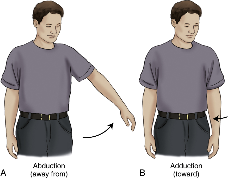
Abduction Versus Adduction
Abduction (ab-duk′-shun) is the lateral movement of the arm or leg away from the body.
Another application of this term is the abduction of the fingers or toes, which means spreading them apart (Fig. 1.96A).
Adduction (ah-duk′-shun) is a movement of the arm or leg toward the body, to draw toward a center or medial line (Fig. 1.96B).
Adduction of the fingers or toes means moving them together or toward each other.
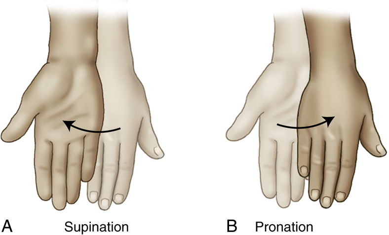
Supination Versus Pronation
Supination (su″-pi-na′-shun) is a rotational movement of the hand into the anatomic position (palm up in supine position or forward in erect position) (Fig. 1.97A). This movement rotates the radius of the forearm laterally along its long axis.
Pronation (pro-na′-shun) is a rotation of the hand into the opposite of the anatomic position (palm down or back) (Fig. 1.97B).
NOTE: To help remember these terms, relate them to the body positions of supine and prone. Supine or supination means face up or palm up, and prone or pronation means face down or palm down.
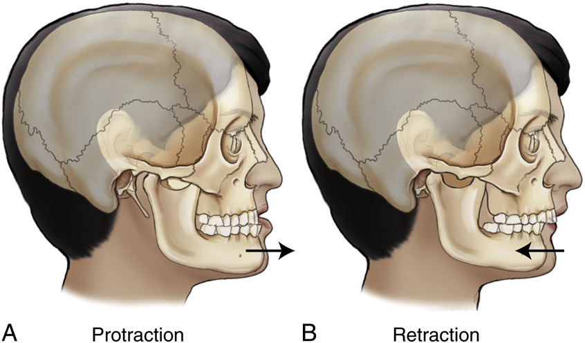
Protraction Versus Retraction
Retraction (re-trak′-shun) is a movement backward or the condition of being drawn back (Fig. 1.98B).
Example
Protraction is moving the jaw forward (sticking the chin out) or drawing the shoulders forward. Retraction is the opposite of this—that is, moving the jaw backward or squaring the shoulders, as in a military stance.
Elevation Versus Depression
Example
Shoulders are elevated when they are raised, as when shrugging the shoulders. Depressing the shoulders is lowering them.
Circumduction
Circumduction (ser″-kum-duk′-shun) means to move around in the form of a circle (Fig. 1.100). This term describes sequential movements of flexion, abduction, extension, and adduction, resulting in a cone-type movement at any joint where the four movements are possible (e.g., fingers, wrist, arm, leg).
Rotation Versus Tilt
Rotate is to turn or rotate a body part on its axis. In Fig. 1.101, the midsagittal plane of the entire body, including the head, is rotated.
Tilt is a slanting or tilting movement with respect to the long axis. Fig. 1.102 demonstrates no rotation of the head but a tilting (slanting) of the midsagittal plane of the head, which therefore is not parallel to the tabletop.
Understanding the difference between these two terms is important in cranial and facial bone positioning (see Chapter 11).
See Table 1.4 for a summary of positioning-related terminology.
Summary of Projections and Positions
The three terms position, projection, and view are sometimes confusing and may be used incorrectly in practice. These terms should be understood and used correctly (Table 1.5).
Position
Position is a term that is used to indicate the patient’s general physical position, such as supine, prone, recumbent, erect, or Trendelenburg.
TABLE 1.4
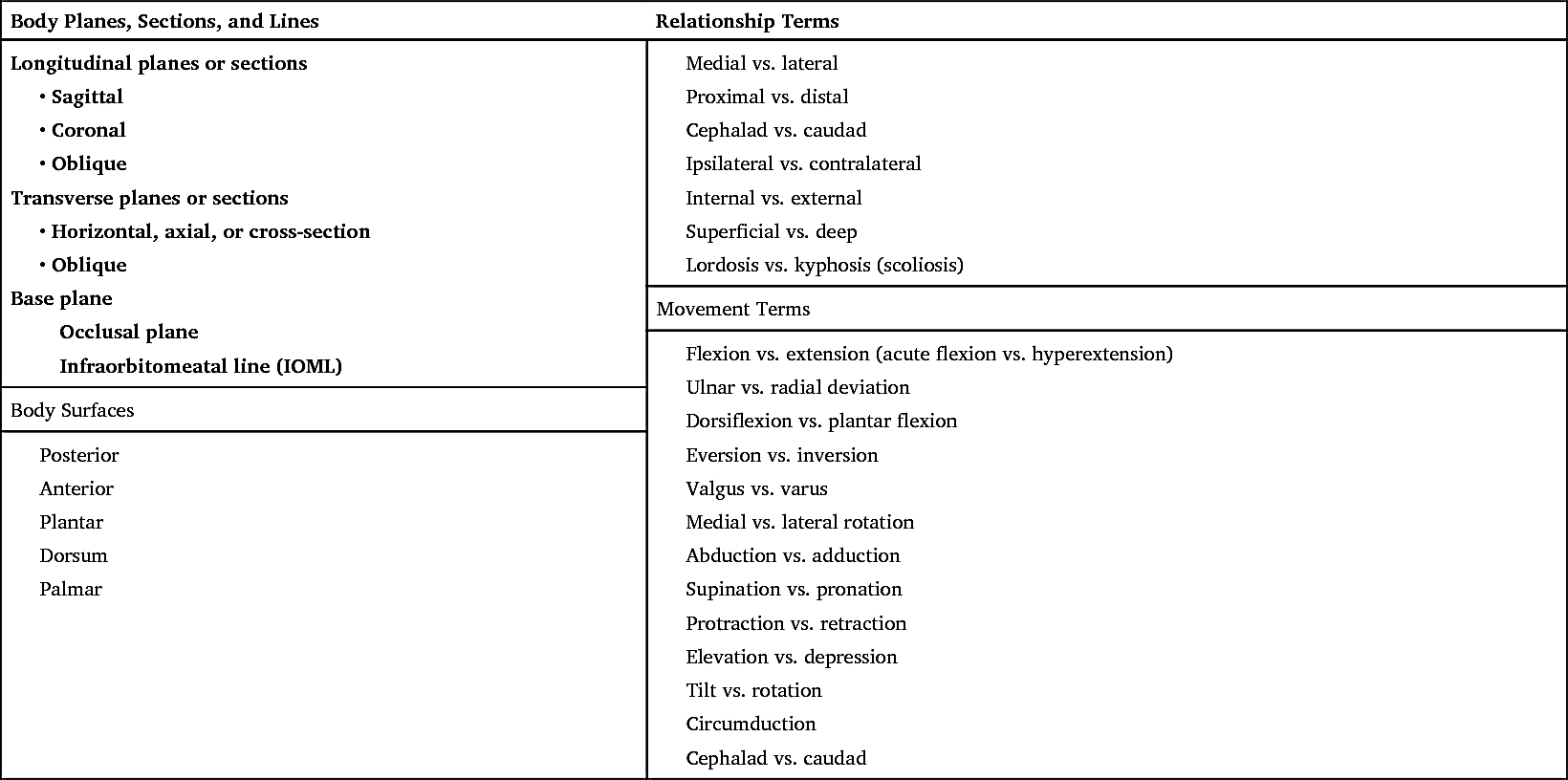
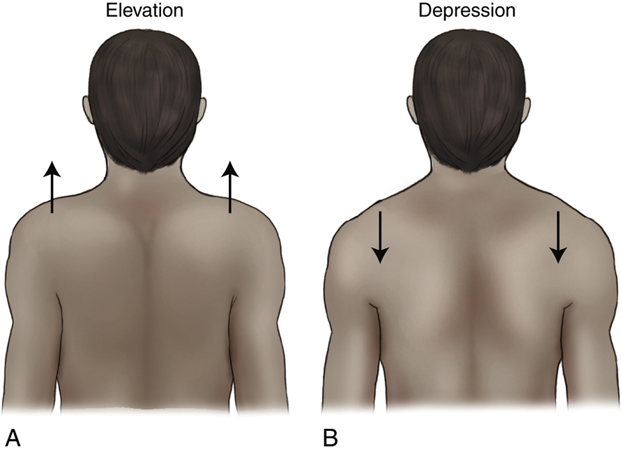
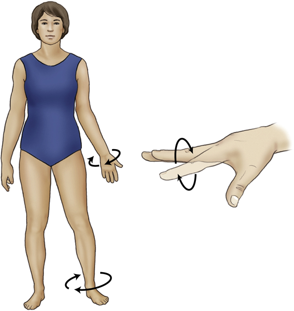


TABLE 1.5
Position also is used to describe specific body positions by the anatomy closest to the IR, such as lateral and oblique positions.
The term position should be “restricted to discussion of the patient’s physical position.”
11
Projection
Projection is a correct positioning term that describes or refers to the path or direction of the central ray, projecting an image onto an IR.
The term projection should be “restricted to discussion of the path of the central ray.”
10
View
View is not a correct positioning term in the United States.
View describes the anatomy or body part as seen by the IR or other recording medium, such as a fluoroscopic screen. In the United States, the term view should be “restricted to discussion of a radiograph or image.”
10