Nursing Care of Women with Complications During Labor and Birth
http://evolve.elsevier.com/Leifer.
- 1. Define each key term listed.
- 2. Describe each obstetric procedure discussed in this chapter.
- 3. Illustrate the nurse’s role in each obstetric procedure.
- 4. Analyze the nurse’s role in a cesarean birth.
- 5. Describe factors that contribute to an abnormal labor.
- 6. Explain each intrapartum complication discussed in this chapter.
- 7. Discuss the nurse’s role in caring for women with each intrapartum complication.
- 8. Review the nurse’s role in obstetric emergencies.
Key Terms
anaphylactoid syndrome (p. 206)
artificial rupture of membranes (AROM) (p. 185)
augmentation of labor (p. 183)
cephalopelvic disproportion (sĕf-ăh-lō-PĔL-vĭc dĭs-prŏ-PŎR-shŭn, p. 191)
chorioamnionitis (kō-rē-ō-ăm-nē-ō-NĪ-tĭs, p. 201)
complementary and alternative medicine (CAM) (p. 184)
fibronectin (fī-brō-NĔK-tĭn, p. 202)
hydramnios (hī-DRĂM-nē-ŏs, p. 194)
laminaria (lăm-ĭ-NĂ-rē-ăh, p. 185)
macrosomia (măk-rō-SŌM-ē-ă, p. 195)
oligohydramnios (ŏl-ĭ-gō-hī-DRĂM-nē-ŏs, p. 187)
shoulder dystocia (SHŌL-dŭr dĭs-TŌ-sē-ă, p. 195)
spontaneous rupture of membranes (SROM) (p. 185)
tocolytics (tō-kō-LĬT-ĭks, p. 185)
Childbirth is a normal, natural event in the lives of most women and their families. When the many factors that affect the birth process function in harmony, complications are unlikely. However, some women experience complications during childbirth that threaten their well-being or that of the infant.
Obstetric Procedures
Nurses assist with several obstetric procedures during birth; they also care for women after the procedures. Some procedures, such as amniotomy or amnioinfusion, are performed to prevent complications during birth. Other procedures are needed when the woman has a complication that necessitates an intervention to promote a positive outcome for the mother and fetus.
Induction or Augmentation of Labor
Induction of labor is the intentional initiation of labor before it begins naturally. Augmentation of labor is the stimulation of contractions after they have begun naturally.
Labor involves the complex interaction between fetus and mother. Before labor is induced, it is important that fetal maturity be confirmed, as induction is avoided before 39 weeks’ gestation. Fetal maturity can be assessed by ultrasound or amniotic fluid analysis (lecithin/sphingomyelin [L/S] ratio) (see Chapter 5). The Bishop score is used to assess the status of the cervix in determining its response to induction (Table 8.1). The presence of increased fetal fibronectin at the cervix and Bishop score above 6 determine cervical readiness for labor induction. Continuous monitoring of uterine activity and fetal heart rate during labor induction is essential.
Table 8.1
NOTE: High score is predictive of a successful labor induction because the cervix has ripened, or softened, in preparation for labor. The American College of Obstetricians and Gynecologists (ACOG) recommends a score of 6 or above before induction of labor.
Modified from Stables D, Rankin J: Physiology in childbearing: with anatomy and related biosciences, ed 2, Edinburgh, 2005, Elsevier; Gabbe M, et al, editors: Obstetrics: normal and problem pregnancies, ed 7, Philadelphia, 2017, Saunders.
Indications for Induction
Labor is induced if continuing the pregnancy is hazardous for the woman or the fetus. Following are some of the indications for labor induction:
- • Gestational hypertension (see Chapter 5)
- • Ruptured membranes without spontaneous onset of labor
- • Infection within the uterus
- • Medical problems in the woman that worsen during pregnancy, such as diabetes, kidney disease, or pulmonary disease
- • Fetal problems, such as slowed growth, prolonged pregnancy, or incompatibility between fetal and maternal blood types (see Chapter 5)
- • Placental insufficiency
- • Abnormal labor progress (defined as 6 cm of cervical dilatation with membranes ruptured and no cervical change after 4 to 6 hours of uterine contractions)
- • Fetal death
Convenience for the health care provider or the family is not an indication for inducing labor. However, a woman who has a history of rapid labors and lives a long distance from the birth facility may have her labor induced because she has a higher risk of giving birth en route if she awaits spontaneous labor.
Contraindications to Induction
Labor is not induced in the following conditions:
- • Placenta previa (see Chapter 5)
- • Umbilical cord prolapse
- • Abnormal fetal presentation
- • High station of the fetus (head not engaged), which can suggest a preterm fetus or a small maternal pelvis
- • Active herpes infection externally or in the birth canal, which the infant can acquire during birth
- • Abnormal size or structure of the mother’s pelvis
- • Previous classic (vertical) cesarean incision
The health care provider may attempt to induce labor in a preterm pregnancy if continuing the pregnancy is more harmful to the woman or the fetus than the hazards of prematurity would be to the infant.
Nonpharmacological Methods to Stimulate Contractions
Natural or Complementary Methods of Inducing Labor
Complementary and alternative medicine (CAM) offers “natural” methods of stimulating labor that have been practiced for centuries but often lack rigorous and controlled studies to prove effectiveness (see Chapter 34). Some CAM practices to stimulate labor follow.
Walking
Many women benefit from a change in activity if their labor slows. Walking stimulates contractions, eases the pressure of the fetus on the mother’s back, and adds gravity to the downward force of contractions. If the woman does not feel like walking, other upright positions often improve the effectiveness of each contraction. She can sit (in a chair, on the side of the bed, or in the bed), squat, kneel while facing the raised head of the bed for support, or maintain other upright positions.
Nipple Stimulation of Labor
Stimulating the nipples causes the woman’s posterior pituitary gland to se-crete oxytocin naturally. This improves the quality of contractions that have slowed or weakened, just as intravenous (IV) administration of synthetic oxytocin does. The woman can stimulate her nipples by doing the following:
- • Pulling or rolling them, one at a time
- • Gently brushing them with a dry washcloth
- • Using water in a whirlpool tub or a shower
- • Applying suction with a breast pump
- • Sexual intercourse: Orgasm that occurs during sexual intercourse stimulates uterine contractions, and the male ejaculate contains prostaglandins.
- • Acupuncture and acupressure have been used for centuries to stimulate labor when given by professionals. See Table 34.2 for common herbs contraindicated in pregnancy and lactation.
If contractions become too strong with these techniques, the woman simply stops stimulation.
Pharmacological and Mechanical Methods to Stimulate Contractions
Cervical Ripening
Cervical ripening is the physical softening of the cervix that leads to effacement and dilation. Induction of labor is more effective if the woman’s cervix is “ripe” (see Table 8.1). These prelabor cervical changes occur naturally in most women. Methods to hasten the changes, or “ripen” the cervix, ease labor induction, as oxytocic drugs have no effect on the cervix. Oxytoxin used to induce labor without a “ripe cervix” can result in the need for a cesarean section (Levine and Srinivas, 2020). Cervical ripening can be achieved by pharmacological or mechanical means.
Pharmacological Methods
The use of prostaglandins to ripen the cervix is contraindicated in women with a history of uterine myomectomy surgery or previous cesarean section because of the risk of uterine rupture.
Prostaglandin E2
Dinoprostone (Cervidil or Prepidil) vaginal insertion is recommended via a sustained-release vaginal insert.
 Safety Alert!
Prostaglandin E1
Misoprostol was designed for the treatment of peptic ulcer disease. Its use as a preinduction medication has been approved by the U.S. Food and Drug Administration (FDA) (Levine and Srinivas, 2020). It is stable at room temperature and can be administered orally (sublingual or buccal) or intravaginally. Prostaglandin E1 is more effective in achieving vaginal delivery within 24 hours, but it is associated with uterine tachysystole and fetal heart rate abnormalities.
The procedure should be explained to the woman and her family. A fetal heart rate baseline is recorded. An IV line with saline or heparin sodium (“hep-lock”) may be placed in case uterine tachysystole (increased uterine contractions) occurs, and IV tocolytics (drugs that reduce uterine contractions) may be needed. After insertion of the prostaglandin gel, the woman remains on bed rest for 1 to 2 hours and is monitored for uterine contractions. Vital signs and fetal heart rate are also recorded. Oxytocin induction can be started when the insert is removed—usually after 6 to 12 hours. Signs of uterine tachysystole include uterine contractions that last longer than 90 seconds or more than five contractions in 10 minutes.
The vaginal insert can be removed by pulling on the netted string that protrudes from the vaginal orifice. Some women who receive cervical ripening products begin labor without additional oxytocin stimulation.
Mechanical Methods
Stripping the Amniotic Membranes
Stripping the amniotic membranes involves separation of the chorioamniotic membranes from the wall of the lower uterine segment and cervix by insertion of the examiner’s gloved finger through the cervix and beyond the internal cervical os and rotating the finger along the lower uterine segment.
Hydroscopic Dilators
Laminaria and Lamicel are mech-anical dilators placed in the lower uterine segment that stimulate the release of prostaglandins from the fetal membranes and maternal decidua. They swell inside the cervix, resulting in mechanical cervical dilation.
Transcervical Balloon Dilators
A 16-Fr catheter with a 30-mL balloon can be inserted through the cervix and inflated. Mechanical pressure by gentle traction against the cervix dilates the cervix.
Amniotomy
Amniotomy is the artificial rupture of membranes (AROM) (amniotic sac) by using a sterile sharp instrument to puncture the amniotic sac and release the amniotic fluid for the purpose of inducing or augmenting labor. It may also be performed to permit internal fetal monitoring (see Chapter 6). A health care provider performs the procedure. The nurse assists the health care provider with the procedure and cares for the woman and fetus afterward. Confirmation of a vertex presentation and the station is essential to prevent umbilical cord prolapse. The amniotomy stimulates prostaglandin secretion, which stimulates labor, but the loss of amniotic fluid may result in umbilical cord compression.
Complications of Amniotomy
Three complications associated with amniotomy may also occur if a woman’s membranes rupture spontaneously (spontaneous rupture of membranes [SROM]). These complications are prolapse of the umbilical cord, infection, and abruptio placentae.
Prolapse of the Umbilical Cord
Prolapse may occur if the cord slips downward with the gush of amniotic fluid (see section Prolapsed Umbilical Cord).
Infection
Infection may occur because the membranes no longer block vaginal organisms from entering the uterus. Once performed, an amniotomy commits the woman to delivery within a certain time; the health care provider delays amniotomy until he or she is reasonably sure that birth will occur before the risk of infection markedly increases.
Abruptio Placentae
Abruptio placentae (separation of the placenta before birth) is more likely to occur if the uterus is overdistended with amniotic fluid (hydramnios) when the membranes rupture. The uterus becomes smaller with the discharge of amniotic fluid, but the placenta stays the same size and no longer fits its implantation site (see Chapter 5 for more information about abruptio placentae).
Nursing Care After Amniotomy
The nursing care after amniotomy is the same as that after spontaneous membrane rupture: observing for complications and promoting the woman’s comfort.
 Nursing Tip
Observe for wet underpads and linens after the membranes rupture. Change them as often as needed to keep the woman relatively dry and to reduce the risk for infection or skin breakdown.
Observing for Complications
The fetal heart rate is recorded for at least 1 minute after amniotomy. Rates outside the normal range of 110 to 160 beats/min for a term fetus suggest a prolapsed umbilical cord. A large quantity of fluid increases the risk for prolapsed cord, especially if the fetus is high in the pelvis.
The color, odor, amount, and character of amniotic fluid are recorded. The fluid should be clear, possibly with flecks of vernix (newborn skin coating) and lanugo, and should not have a bad odor. Cloudy, yellow, or malodorous fluid suggests infection. Green fluid means that the fetus passed the first stool (meconium) into the fluid before birth. Meconium-stained amniotic fluid is associated with fetal compromise during labor and infant respiratory distress after birth.
The woman’s temperature is taken every 2 to 4 hours after her membranes rupture according to facility policy. A maternal temperature of 38°C (100.4 °F) or higher suggests infection. An increase in the fetal heart rate, especially if more than 160 beats/min, may precede the woman’s temperature increase.
Promoting Comfort
When amniotomy is anticipated, several disposable underpads are placed under the woman’s hips to absorb the fluid that continues to leak from the woman’s vagina during labor. Disposable underpads are changed often enough to keep her reasonably dry and to reduce the moist, warm environment that favors the growth of microorganisms.
Oxytocin Induction or Augmentation of Labor
Initiation or stimulation of contractions with oxytocin (Pitocin) is the most common method of labor induction and augmentation in women with a favorable or “ripe” cervix (Levine and Srinivas, 2020). When oxytocin is administered to stimulate contractions, it is called induction of labor. When oxytocin is administered to stimulate contractions that have already begun, it is known as augmentation of labor. A registered nurse (RN), who has additional training in the induction of labor and electronic fetal monitoring, administers oxytocin. Augmentation of labor with oxytocin follows a similar procedure as other methods of induction.
Oxytocin for induction or augmentation of labor is diluted in an IV solution. The oxytocin solution is a secondary (piggyback) infusion that is inserted into the primary (nonmedicated) IV solution line so that it can be stopped quickly while an open IV line is maintained. Infusion of oxytocin solution is regulated with an infusion pump. Administration begins at a very low rate and is adjusted upward or downward according to how the fetus responds to labor and to the woman’s contractions. The dose is individualized for every woman. When contractions are well established, it is often possible to reduce the rate of oxytocin. Augmentation of labor usually requires less total oxytocin than induction of labor because the uterus is more sensitive to the drug when labor has already begun.
Continuous electronic monitoring is the usual method to assess and record fetal and maternal responses to oxytocin. Many health care providers prefer internal methods of monitoring when oxytocin is used because these techniques are more accurate, especially for contraction intensity. Oxytocin may be used in the fourth stage of labor to reduce uterine bleeding after the placenta has been delivered. Vital signs are monitored closely.
Complications of Augmentation of Labor
The most common complications related to overstimulation of contractions are fetal compromise and uterine rupture (see section Uterine Rupture). Fetal compromise can occur because blood flow to the placenta is reduced if contractions are excessive (tachysystole). Most placental exchange of oxygen, nutrients, and waste products occurs between contractions. This exchange is likely to be impaired if the contractions are too long, too frequent, or too intense.
Water intoxication sometimes occurs because oxytocin inhibits the excretion of urine and promotes fluid retention. Water intoxication is not likely with the small amounts of oxytocin and fluids given intravenously during labor, but it is more likely to occur if large doses of oxytocin and fluids are given intravenously after birth.
Oxytocin is discontinued or its rate is reduced if signs of fetal compromise or excessive uterine contractions occur. Fetal heart rates outside the normal range of 110 to 160 beats/min, late decelerations, and loss of variability (see Chapter 6) are the most common signs of fetal compromise.
Tachysystole is most often evidenced by contraction frequency greater than every 2 minutes; five or more contractions within 10 minutes, durations longer than 90 seconds; or resting intervals shorter than 60 seconds.
The resting tone of the uterus (muscle tension when it is not contracting) is often higher than normal. Internal uterine activity monitoring allows determination of peak uterine pressures and uterine resting tone.
In addition to stopping the oxytocin infusion, the RN chooses one or more of the following measures to correct adverse maternal or fetal reactions:
The health care provider is notified after corrective measures are taken. A tocolytic (drug that reduces uterine contractions such as magnesium sulfate or terbutaline) may be ordered if contractions do not quickly decrease after oxytocin is stopped.
IV oxytocin is considered to be a high-alert medication because it has an increased risk of causing a significant adverse reaction if used incorrectly.
The nurse must be aware of signs and symptoms of increased uterine activity and must monitor fetal heart rate every 15 minutes during active labor and every 5 minutes during the transitional phase. Safety interventions for oxytocin-induced uterine contractions or fetal heart rate abnormalities include the following:
- • Notifying the health care provider and the RN
- • Repositioning the woman to left or right lateral position
- • Decreasing the dose of oxytocin to half of the current rate or discontinuing oxytocin
- • Preparing an IV bolus of lactated Ringer’s solution
- • Administering oxygen at 10 L/min via a nonrebreather face mask
- • Preparing IV terbutaline for administration
- • Assessing uterine contractions and fetal heart rate every 5 minutes
Nursing Care During Induction or Augmentation
The American Academy of Pediatrics (AAP) and the American College of Obstetricians and Gynecologists (ACOG) recommend that an RN, with 1:1 or 1:2 ratio, care for patients undergoing oxytocin-induced labor. Fetal heart rate must be assessed and recorded every 15 minutes during active labor and every 5 minutes during transition. Baseline maternal vital signs are assessed, and a fetal monitor tracing is performed to identify contraindications to induction or augmentation before the procedure begins.
If abnormalities are noted in either fetal heart rate or maternal vital signs, the nurse stops the oxytocin and begins measures to reduce contractions and increase placental blood flow. The woman’s blood pressure, pulse rate, and respirations are measured every 30 to 60 minutes. Her temperature is taken every 2 to 4 hours. Recording her intake and output helps identify potential water intoxication.
Amnioinfusion
An amnioinfusion is the injection of warmed sterile saline or lactated Ringer’s solution into the uterus via an intrauterine pressure catheter during labor after the membranes have ruptured. Indications for this procedure include the following:
- • Oligohydramnios (lower-than-normal amount of amniotic fluid)
- • Umbilical cord compression resulting from lack of amniotic fluid
- • Goal of reducing recurrent variable decelerations in the fetal heart rate
- • Goal of diluting meconium-stained amniotic fluid to prevent meconium aspiration syndrome
Amnioinfusion replaces the “cushion” for the umbilical cord and relieves the variable decelerations of the fetal heart rate that may occur during contractions when decreased amniotic fluid is present. It can be administered as a one-time bolus for 1 hour or as a continuous infusion. Continuous monitoring of uterine activity and fetal heart rate is essential. The nurse should change the underpads on the bed as needed to maintain patient comfort and should document the color, amount, and any odor of the fluid expelled from the vagina.
Version
Version is a method of changing the fetal presentation, usually from breech or oblique to cephalic. There are two methods: external and internal. External version is the more common method. A successful version reduces the likelihood that the woman will need cesarean delivery.
Risks and Contraindications of Version
Few maternal and fetal risks are associated with version, especially external version. Version is not indicated if there is any maternal or fetal reason that vaginal birth should not occur because that is its goal. Examples of maternal or fetal conditions that are contraindications for version include the following:
- • Disproportion between the mother’s pelvis and fetal size
- • Abnormal uterine or pelvic size or shape
- • Abnormal placental placement
- • Previous cesarean birth with a vertical uterine incision
- • Active herpesvirus infection
- • Inadequate amniotic fluid
- • Poor placental function
- • Multifetal gestation
- • Malfunctioning placenta
Version may not be attempted in a woman who has a higher risk for uterine rupture, such as several previous cesarean births or high parity. Version is not usually attempted if the fetal presenting part is engaged in the pelvis. The main risk to the fetus is that it will become entangled in the umbilical cord, thus compressing the cord. This is more likely to happen if there is not adequate room to turn the fetus, such as in multifetal gestation (e.g., twins) or when the amount of amniotic fluid is minimal.
Technique
External version is done after 37 weeks’ gestation but before the onset of labor. The procedure begins with a non–stress test (NST) or biophysical profile (BPP) (see Table 5.1) to determine whether the fetus is in good condition and if there is adequate amniotic fluid to perform the version. The woman receives a tocolytic drug to relax her uterus during the version.
Using ultrasound to guide the procedure, the health care provider pushes the fetal buttocks upward out of the pelvis while pushing the fetal head downward toward the pelvis in either a clockwise or a counterclockwise turn. The fetus is monitored frequently during the procedure. The tocolytic drug is discontinued after the external version is completed (or the effort abandoned). The Rh-negative woman receives a dose of Rho (D) immune globulin (RhoGAM) to prevent development of RH-positive antibodies if the fetus is RH-positive.
Internal version is an emergency procedure. The health care provider usually performs internal version during a vaginal birth of twins to change the fetal presentation of the second twin.
Nursing Care During Version
Nursing care of the woman having external version includes assisting with the procedure and observing the mother and fetus afterward for 1 to 2 hours. Baseline maternal vital signs and a fetal monitor strip (part of the NST or BPP) are taken before the version. The mother’s vital signs and the fetal heart rate are observed to ensure return to normal levels after the version is complete.
Vaginal leaking of amniotic fluid suggests that manipulating the fetus caused a tear in the membranes, and this is reported. Uterine contractions usually decrease or stop shortly after the version. The health care provider is notified if they do not. The nurse reviews signs of labor with the woman because version is performed near term, when spontaneous labor is expected.
Episiotomy and Lacerations
Episiotomy is the surgical enlargement of the vaginal opening during birth. The health care provider performs and repairs an episiotomy. A laceration is an uncontrolled tear of the tissues that results in a jagged wound. Lacerations of the perineum and episiotomy incisions are treated similarly.
Perineal lacerations and often episiotomies are described by the amount of tissue involved, as follows:
- • First degree: Involves the superficial vaginal mucosa or perineal skin
- • Second degree: Involves the vaginal mucosa, perineal skin, and deeper tissues of the perineum
- • Third degree: Same as second degree, plus involves the anal sphincter
- • Fourth degree: Extends through the anal sphincter into the rectal mucosa
Women with third- and fourth-degree lacerations may have more discomfort postpartum if they are constipated after birth.
 Nutrition Considerations
Third- or Fourth-Degree Laceration
Pay special attention to a woman’s diet and fluids if she had a third- or fourth-degree laceration. A high-fiber diet and adequate fluids help prevent constipation that might result in a breakdown of the perineal area where the laceration was sutured.
Indications for Episiotomy
Maternal indications include the following:
Episiotomy is no longer routinely performed during vaginal delivery but is used with specific indications when problems occur during the expulsion stage of labor. Perineal massage and stretching exercises before labor are popular techniques to decrease the need for an episiotomy during birth.
Risks of Episiotomy or Laceration
As in other incisions, infection is the primary risk in an episiotomy or laceration. An additional risk is extension of the episiotomy with a laceration into or through the rectal sphincter (third or fourth degree), which can cause prolonged perineal discomfort and stress incontinence.
Technique
The episiotomy is performed with blunt-tipped scissors just before birth. One of the following two directions is chosen (Fig. 8.1):
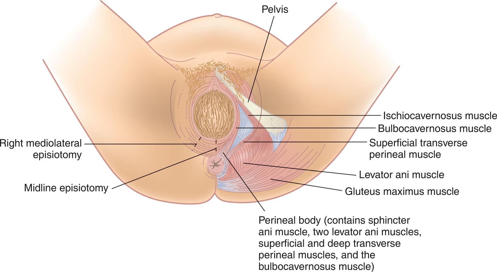
The two common types of episiotomies are midline (median) and mediolateral. (From Matteson PS: Women’s health during the childbearing years: a community-based approach, St Louis, 2001, Mosby.)
Illustration of genital area of female shows labels as follows: Pelvis, Ischiocavernosus muscle, bulbocavernosus muscle, superficial transverse perineal muscle, levator ani muscle, gluteus maximus muscle, perineal body (contains sphincter ani muscle, two levator ani muscles, superficial and deep transverse perineal muscles, and the bulbocavernosus muscle), midline episiotomy, and right mediolateral episiotomy.
A median episiotomy is easier to repair and heals neatly. The mediolateral incision provides more room, but greater scarring during healing may cause painful sexual intercourse. A laceration that extends a median episiotomy is more likely to involve the rectal sphincter than one that extends the mediolateral episiotomy.
Nursing Care for Episiotomy or Laceration
Nursing care for an episiotomy or laceration begins during the fourth stage of labor. Cold packs should be applied to the perineum for at least the first 12 hours to reduce pain, bruising, and edema. After 12 to 24 hours of cold applications, warmth in the form of heat packs or sitz baths increases blood circulation, enhancing comfort and healing. Mild oral analgesics are usually sufficient for pain management. See Chapter 9 for postpartum nursing care of the woman with an episiotomy or laceration.
Forceps And Vacuum Extraction Births
An obstetrician uses obstetric forceps and vacuum extractors to provide traction and rotation to the fetal head when the mother’s pushing efforts are insufficient to accomplish a safe delivery. Forceps are instruments with curved blades that fit around the fetal head without unduly compressing it (Fig. 8.2). Several different styles are available to assist the birth of the fetal head in a cephalic presentation or the after-coming head in a breech delivery. Forceps may also help the health care provider extract the fetal head through the incision during cesarean birth.
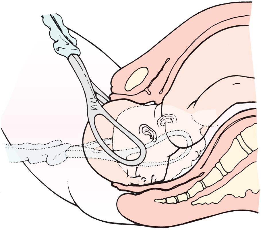
After applying the forceps to each side of the fetal head and locking the two blades, the physician pulls, following the pelvic curve.
A vacuum extractor uses suction applied to the fetal head so that the health care provider can assist the mother’s expulsive efforts (Fig. 8.3). The vacuum extractor is used only with an occiput presentation. One advantage of the vacuum extractor is that it does not take up room in the mother’s pelvis, as forceps do. Since 2001, the use of vacuum extractors has increased during delivery, whereas the use of forceps has decreased. However, since 2011, the use of cesarean sections has increased, whereas the use of both forceps and vacuum extractors has decreased (Foglia et al., 2020).
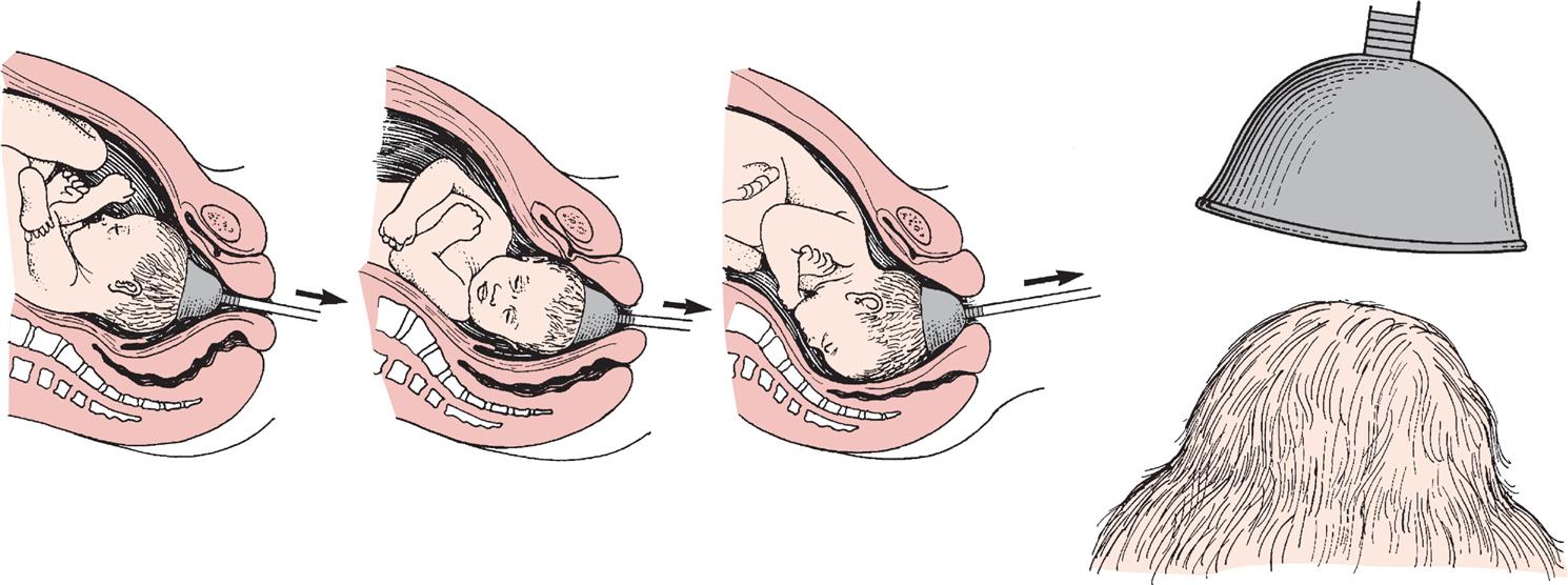
The arrows indicate the direction of traction on the vacuum cup. The vacuum cup is positioned on the midline, near the posterior fontanelle. (From Lowdermilk DL, Perry SE, Cashion KL, et al: Maternity and women’s health care, ed 12, St Louis, 2020, Elsevier.)
Indications for Forceps or Vacuum Extraction
Forceps or vacuum extraction may be used to end the second stage of labor if it is in the best interest of the mother or fetus. The mother may be exhausted, or she may be unable to push effectively. Women with cardiac or pulmonary disorders often have forceps or vacuum extraction births because prolonged pushing can worsen these conditions. Fetal indications include conditions in which there is evidence of an increased risk to the fetus near the end of labor. The cervix must be fully dilated, the membranes ruptured, the bladder empty, and the fetal head engaged and at +2 station for optimal outcome.
Contraindications for Forceps or Vacuum Extraction
Forceps or vacuum extraction cannot substitute for cesarean birth if the maternal or fetal condition requires a quicker delivery. Delivery by these techniques is not done if the delivery would be more traumatic than cesarean birth, such as when the fetus is high in the pelvis or too large for a vaginal delivery.
Risks Associated with Forceps or Vacuum Extraction
Trauma to maternal or fetal tissues is the main risk when forceps or vacuum extraction is used. The mother may have a laceration or hematoma (collection of blood in the tissues) in her vagina. The infant may have bruising, facial or scalp lacerations or abrasions, cephalhematoma (see Chapter 12), or intracranial hemorrhage. The vacuum extractor causes a harmless area of circular edema on the infant’s scalp (chignon) where it was applied.
Technique
The health care provider catheterizes the woman to prevent trauma to her bladder and to make more room in her pelvis. After the forceps are applied, the health care provider pulls in line with the pelvic curve. An episiotomy is sometimes done. After the fetal head is brought under the mother’s symphysis pubis, the rest of the birth occurs in the usual way.
Birth assisted with the vacuum extractor follows a similar sequence. The health care provider applies the cup over the posterior fontanelle of the fetal occiput, and suction is created with a machine to hold it there. Traction is applied by pulling on the handle of the extractor cup.
Nursing Care During Forceps or Vacuum Extraction Birth
If the use of forceps or vacuum extraction is anticipated, the nurse places the sterile equipment on the delivery instrument table. After birth, nursing care is similar to care for episiotomy and perineal lacerations. Ice is applied to the perineum to reduce bruising and edema. The health care provider is notified if the woman has signs of vaginal hematoma, which include severe and poorly relieved pelvic or rectal pain.
The infant’s head is examined for lacerations, abrasions, or bruising. Mild facial reddening and molding (alteration in shape) of the head are common and do not necessitate treatment. Cold treatments are not used on neonates because they would cause hypothermia.
Pressure from forceps may injure the infant’s facial nerve. This is evidenced by facial asymmetry (different appearance of right and left sides), which is most obvious when the infant cries. Facial nerve injury usually resolves without treatment. The scalp chignon from the vacuum extractor does not necessitate intervention and resolves quickly.
Cesarean Birth
Cesarean birth is the surgical delivery of the fetus through incisions in the mother’s abdomen and uterus. Cesarean section is currently the most common major surgical procedure in the United States. Cesarean delivery rates in the United States have been 31% to 33% for the last 10 years, mainly due to reduced operative vaginal deliveries, malpresentation (breech) deliveries, increase in maternal obesity and diabetes mellitus, failure of trial of labor after cesarean (TOLAC), and maternal request (Berghella et al., 2020). The goal of Healthy People 2030 (U.S. Department of Health and Human Services, 2018) is to reduce cesarean sections to 15%. This is the basis for some of the practices in the management of the second stage of labor, such as the following:
- • Position variation (upright or horizontal)
- • Epidural analgesia and subarachnoid analgesia that allows ambulation and delivery in squatting position
- • Oxytocin (Pitocin) augmentation of labor
- • Spontaneous open glottis pushing when fetus is at +1 station
- • Use of vacuum-assisted delivery replacing forceps delivery
- • Electronic fetal and uterine monitoring
Indications for Cesarean Birth
Several conditions may necessitate cesarean delivery, as follows:
- • Abnormal labor
- • Inability of the fetus to pass through the mother’s pelvis (cephalopelvic disproportion) (most breech presentations are delivered by cesarean section)
- • Maternal conditions such as gestational hypertension or diabetes mellitus
- • Active maternal herpesvirus infection, which may cause serious or fatal infant infection
- • Previous surgery on the uterus, including the classic type of cesarean incision
- • Fetal compromise, including prolapsed umbilical cord and abnormal presentations
- • Placenta previa or abruptio placentae
Contraindications for Cesarean Birth
There are few contraindications to cesarean birth, but it is not usually performed if the fetus is dead or too premature to survive or if the mother has abnormal blood clotting. A cesarean birth should not be planned for the convenience of the woman, as there are risks involved.
Risks of Cesarean Birth
Cesarean birth carries risks to both mother and fetus. Maternal risks are similar to those of other types of surgery and include the following:
- • Risks related to anesthesia (see Chapter 7)
- • Respiratory complications
- • Hemorrhage
- • Blood clots
- • Injury to the urinary tract
- • Delayed intestinal peristalsis (paralytic ileus)
- • Infection
Risks to the newborn may include the following:
To help prevent the unintentional birth of a preterm fetus, the physician often performs amniocentesis before a planned cesarean birth to determine whether the fetal lungs are mature (see Chapter 5).
Technique
Cesarean birth may occur under planned, unplanned, or emergency conditions. The preparation is similar for each and includes routine preoperative care such as obtaining informed consent. If the woman wears eyeglasses, those glasses should accompany her to the operating room because she is usually awake to bond with the infant after birth.
Preparations for Cesarean Birth
As with other surgery, several laboratory studies are performed to identify anemia or blood-clotting abnormalities. Complete blood count, coagulation studies, and blood typing and history screening are common, and appropriate consent is obtained. One or more units of blood may be typed and cross-matched if the woman is likely to need a transfusion. The baseline vital signs of the mother and the fetal heart rate are recorded. Administration of a clear oral antacid is routine for aspiration prophylaxis. Nothing by mouth (NPO) except clear fluids before the surgery is advised as aspiration of partially digested foods can cause serious complications. The woman is placed in a supine position with a wedge under the hip to prevent decreased blood flow to the fetus. A regional anesthetic is administered, and an IV medication to reduce gastric acidity and speed gastric emptying is provided. A prophylactic IV antibiotic may be administered before surgery. Shaving of the skin or hair removal may be necessary.
An indwelling Foley catheter is inserted to keep the bladder empty and to prevent trauma to the bladder. The catheter bag is placed near the head of the operating table so that the anesthesiologist can monitor urine output, an important indicator of the woman’s circulating blood volume. The circulating nurse scrubs the abdomen with chlorhexidine alcohol by using a circular motion that goes outward from the incisional area. The woman’s partner may don a hat, mask, and gown and provide support to the woman at the head of the table.
Types of Incisions
There are two incisions in cesarean birth: a skin incision and a uterine incision. The directions of these incisions are not always the same.
Skin Incisions
The skin incision is done in either a vertical or a transverse direction. A vertical incision allows more room if a large fetus is being delivered, and it is usually needed for an obese woman. In an emergency, the vertical incision can be accomplished more quickly. The transverse, or Pfannenstiel, incision is nearly invisible when healed but cannot always be used in an obese woman or in a woman with a large fetus.
Uterine Incisions
The more important of the two incisions is the one that cuts into the uterus. There are three types of uterine incisions (Fig. 8.4): low transverse, low vertical, and classic.
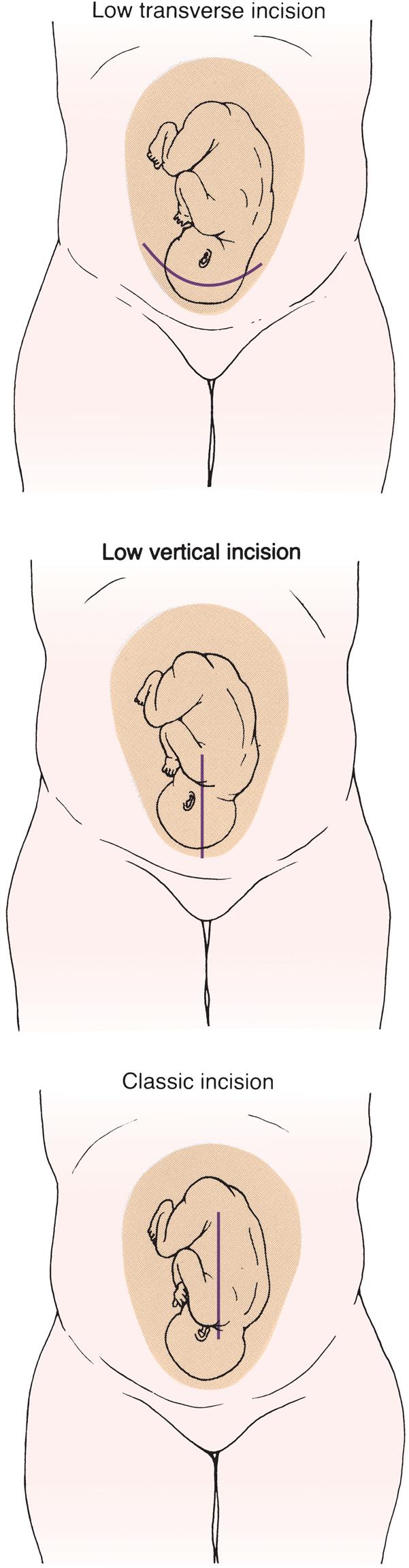
The low transverse uterine incision is preferred because it is not likely to rupture during a subsequent birth, allowing vaginal birth after a cesarean birth. The low vertical and classic incisions may occasionally be used. The skin incision and uterine incision do not always match.
Three illustrations show baby in the mother womb. The first illustration marked low transverse incision shows a curved line indicating cut at the lower part of uterus. The second illustration marked low vertical incision shows cut at the lower half of uterus. The third illustration marked classical incision shows vertical cut at the center of uterus.
Low Transverse Incision
A low transverse incision is preferred because it is not likely to rupture during another birth, causes less blood loss, and is easier to repair. It may not be an option if the fetus is large or if there is a placenta previa in the area where the incision would be made. This type of incision makes vaginal birth after cesarean (VBAC) possible for subsequent births.
Low Vertical Incision
A low vertical incision produces minimal blood loss and allows delivery of a larger fetus. However, it is more likely to rupture during another birth, although less so than the classic incision.
Classic Incision
The classic incision is rarely used because it involves more blood loss, and it is the most likely of the three types to rupture during another pregnancy. However, it may be the only choice if the fetus is in a transverse lie or if there is scarring or a placenta previa in the lower anterior uterus.
Sequence of Events
After the woman has received a spinal anesthetic and has been scrubbed and draped, the obstetrician makes the skin incision. After making the uterine incision, the physician ruptures the membranes (unless they are already ruptured) with a sharp instrument. The amniotic fluid is suctioned from the operative area, and its amount, color, and odor are noted.
The physician reaches into the uterus to lift out the fetal head or buttocks. Forceps or vacuum extraction may be used to assist birth of the head. The infant’s mouth and nose are quickly suctioned to remove secretions, and the cord is clamped. Delayed cord clamping of 30 to 60 seconds is recommended to increase fetal blood volume and the infant should be monitored for jaundice (Berghella et al., 2020). The physician hands the infant to the nurse, who receives the infant into sterile blankets and places the infant into a radiant warmer. A pediatrician is usually available for resuscitation.
After the birth of the infant, the physician scoops out the placenta and examines it for intactness. The uterine cavity is sponged to remove blood clots and other debris. The uterine and skin incisions are then closed and secured in layers with sutures, staples, or Dermabond (Fig. 8.5).
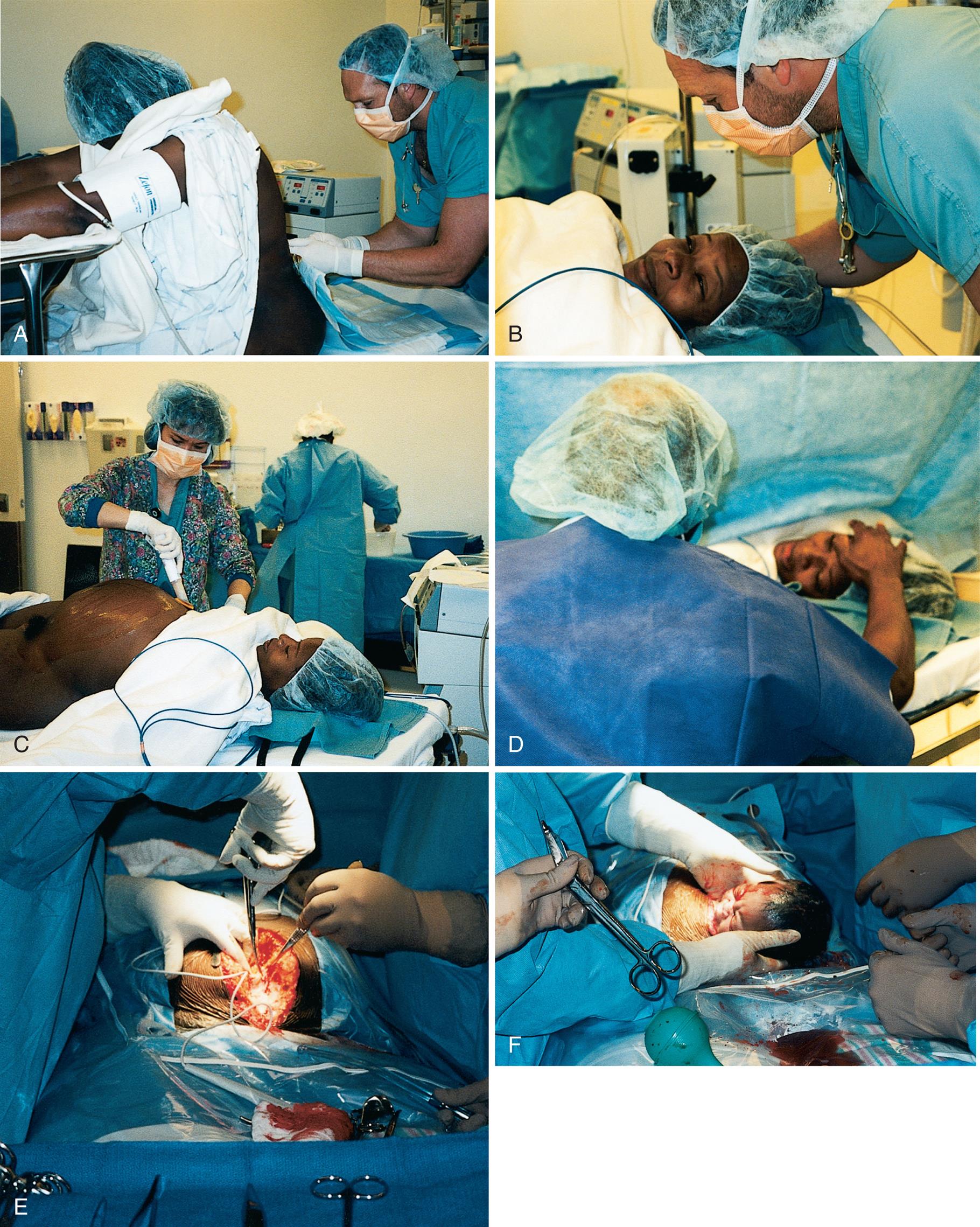
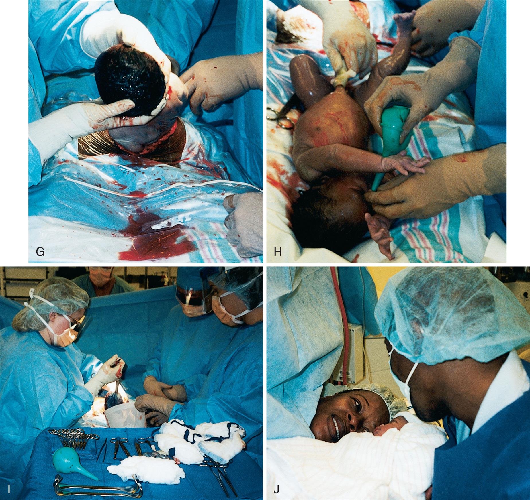
(A) Spinal anesthetic is given. (B) The anesthesiologist reassures the woman. (C) The nurse prepares the abdomen. (D) The partner encourages the woman. (E) A vertical incision is made. (F) The head of the infant is lifted out of the uterus. (G) The body of the infant is lifted out of the uterus. (H) The infant is placed on the mother’s abdomen, and the infant’s secretions are suctioned with a bulb syringe. Note the active muscle tone of the newborn. (I) The placenta is delivered from the uterus. (J) The parents and the newborn bond. (Courtesy Pat Spier, RN-C.)
Six close-up views marked A through F shows step involved in delivering a baby through Cesarean section. A) Side view of a nurse with gloved hands inserting a needle at the lower back of woman. B) The woman is lying supine with frightened expressions. A nurse is talking to the woman. C) A nurse starts a cut on the abdomen of woman. D) Nurse places hand on the forehead of woman. E) Nurses with gloved hands cut the abdomen of woman. F) Head of the baby is outside the abdomen of woman." "Four close-up views marked G through J shows step involved in delivering a baby through Cesarean section. G) Nurses with gloved hands gently pull the baby from uterus. H) Baby is outside the uterus. Tip of bulb syringe is inserted into the nostril of baby by a gloved hand. I) Assortment of various equipments used during and after the delivery along with cotton. J) Nurse brings the newborn near mother.
Nursing Care During and After Cesarean Birth
The RN assumes most of the preoperative and postoperative care of the woman. This includes obtaining the required laboratory studies, administering medications, performing preoperative teaching, and preparing for surgery. Women who have cesarean births usually need greater emotional support than women having vaginal births. They are usually happy and excited about the newborn, but they may also feel grief, guilt, or anger because the expected course of birth did not occur. These feelings may linger and resurface during another pregnancy. Emotional care of the partner and family is essential; they are included in explanations of the surgery as much as the woman wishes. The partner may be frightened when an emergency cesarean is needed but may not express these feelings because the woman needs so much support. The nurse informs the partner when he or she may enter the operating room, as 30 minutes or longer may be needed to administer a regional anesthetic and for surgical preparations if there is no emergency. The partner dons surgical attire during this time.
The partner may be almost as exhausted as the woman if a cesarean birth is performed after hours of labor. The thoughtful nurse includes the partner and promotes his or her emotional and physical well-being. The mother, neonate, and partner are kept together as much as possible after birth, just as for a vaginal birth. The woman and her partner are encouraged to talk about the cesarean birth so that they can integrate the experience. The nurse answers questions about events surrounding the birth. The focus is on the birth, rather than on the surgical aspects of cesarean delivery.
Nursing assessments after cesarean birth are similar to assessments after vaginal birth, including assessment of the uterine fundus. Assessments are done every 15 minutes for the first 1 or 2 hours and then every 30 minutes for 1 hour according to hospital policy. Recovery-room assessments after cesarean birth include the following:
- • Vital signs to identify hemorrhage or shock; a pulse oximeter is used to better identify depressed respiratory function
- • IV site and rate of solution flow
- • Fundus for firmness, height, and midline position
- • Dressing for drainage
- • Lochia for quantity, color, and presence of clots
- • Urine output from indwelling catheter
- • Return of sensation to the lower body
The fundus is checked as gently as possible. The woman flexes her knees slightly and takes slow, deep breaths to minimize the discomfort of fundal assessments. While supporting the lower uterus with one hand, the fingers of the other hand are gently “walked” from the side of the uterus toward the midline. Massage is not needed if the fundus is already firm.
Although assessing the uterus after cesarean birth causes discomfort, it is important to do so regularly because the woman may have a relaxed uterus that causes excessive blood loss.
The woman is told to take deep breaths at each assessment and to cough to move secretions from her airways. A small pillow or folded blanket supports her incision when she coughs or moves, which reduces pain. Changing her position every 1 or 2 hours helps expand her lungs and also makes her more comfortable. Early oral intake also enhances return of bowel function and early ambulation enhances rapid recovery (Berghella et al., 2020).
Pain relief after cesarean birth may be accomplished by a patient-controlled analgesia (PCA) pump or by intermittent injections of narcotic analgesics. Epidural narcotics provide long-lasting pain relief but are associated with delayed respiratory depression and itching (see Chapter 7), which vary with the drug injected. The woman is changed to oral analgesics such as nonsteroidal antiinflamatory drugs (NSAIDS) or acetaminophen after about the first 12 to 24 hours. Nursing Care Plan 8.1 details interventions for selected nursing diagnoses that pertain to the woman with an unplanned cesarean birth.
 Nursing Care Plan 8.1
The Woman with an Unplanned Cesarean Birth
Patient Data
A woman, para 0, gravida 1, has been using breathing, relaxation, and imagery techniques during the first stage of labor, and her husband has been helpful and supportive. However, the labor is not progressing, and there are signs of fetal distress. The health care provider orders that the patient be prepared for an emergency cesarean section.
Selected Nursing Diagnosis:
Anxiety related to development of complications
| Goals | Nursing Interventions | Rationales |
|---|---|---|
| The woman and her partner will express decreased anxiety after explanations about the planned surgery. | Determine stress level and learning needs. | Provides a database to build on to provide information that will decrease anxiety. |
| Reinforce all explanations given by health care provider, expressing them in simpler terms if needed. | Anxiety tends to narrow attention; although health care provider may have explained the need for surgery, the woman and her partner may not have comprehended everything they were told. | |
| Encourage the woman to continue using the breathing and relaxation techniques she learned in prepared childbirth classes as long as contractions continue. | Learned pain management techniques increase the woman’s sense of control. Control over a situation reduces feelings of helplessness and decreases anxiety. | |
| Tell the woman what the operating room looks like and who will be present. Explain basic equipment such as catheter, narrow table, monitors for her heart rate and blood pressure, anesthesia machine, and large overhead lights. Explain that personnel will wear protective equipment such as masks, eye protection, gowns, gloves, hats, and shoe covers. | Commonplace equipment and attire in an operating room can be intimidating for someone who has not seen them before. Unfamiliarity increases anxiety; preparation reduces anxiety and fear of the unknown. | |
| Describe the usual postoperative care: assessment of vital signs, fundus, vaginal bleeding, dressing, and catheter. Tell her she will be asked to take deep breaths and change position regularly. | If the woman understands common postoperative care, she is more likely to cooperate with it, even if assessments are uncomfortable. | |
| Encourage her partner to be with her during surgery, and do not separate family afterward, if possible. | Companionship of familiar persons helps to reduce anxiety; keeping new family together promotes attachment to the newborn. | |
| Stay with the woman. Encourage verbalization and support her coping mechanisms. | The presence of a professional person reduces anxiety. |
Selected Nursing Diagnosis:
Impaired comfort related to decreased coping ability
| Goals | Nursing Interventions | Rationales |
|---|---|---|
| The woman will verbalize reduced discomfort or will be able to use effective techniques to decrease perception of pain. | Determine the nature, duration, and location of pain. | Never assume that the pain is related to a contraction. Locating the site of pain helps identify complications that may be occurring (e.g., embolism). Assessing pain and contractions can help identify a prolonged contraction that can cause fetal hypoxia. |
| Encourage the woman to continue to use coping mechanisms learned during prenatal classes. Use therapeutic touch to increase comfort. | A feeling of loss of control can increase the perception of pain. Reduction of tension can promote comfort. | |
| Maintain a calm manner and environment. | A calm manner calms the parents and reduces anxieties and tensions that elevate pain perception. |
Critical Thinking Question
Abnormal Labor
A normal labor evidences a regular progression in cervical effacement, dilation, and descent of the fetus. Abnormal labor, called dysfunctional labor, does not progress. Dystocia is a term used to describe a difficult labor.
The “four Ps” of labor (see Chapter 6) interact constantly throughout the birth. Abnormalities in the powers, passengers, passage, or psyche may result in a dysfunctional labor. In addition, the length of labor may be unusually short or long. Labor abnormalities may necessitate use of forceps or cesarean delivery, and they are more likely to result in injury to the mother or fetus.
It is essential for nurses to understand the normal birth process so that deviations from normal can be recognized and prompt interventions can be implemented. Effective support for the woman and her family is part of competent and compassionate care. Risk factors for dysfunctional labor include the following:
- • Advanced maternal age
- • Obesity
- • Overdistention of uterus (hydramnios or multifetal pregnancy)
- • Abnormal presentation
- • Cephalopelvic disproportion (CPD)
- • Overstimulation of the uterus
- • Maternal fatigue, dehydration, fear
- • Lack of analgesic assistance
Problems with the Powers of Labor
Increased Uterine Muscle Tone
Increased uterine muscle tone usually occurs during the latent phase of labor (before 4 cm of cervical dilation) and is characterized by contractions that are frequent, cramplike, and poorly coordinated. These contractions are painful but nonproductive. Even between contractions the uterus is tense, which reduces blood flow to the placenta. Hypertonic labor dysfunction is less common than hypotonic dysfunction. Table 8.2 summarizes the differences between hypertonic and hypotonic labor dysfunction.
Table 8.2
Medical Treatment
Medical treatment may include mild sedation to allow the woman to rest. Tocolytic drugs (see section Tocolytic Therapy) such as terbutaline (Brethine) may be ordered.
Nursing Care
Women with increased uterine muscle tone are uncomfortable and frustrated. Anxiety about the lack of progress and fatigue impair their ability to tolerate pain. They may lose confidence in their ability to give birth. The nurse should accept the woman’s frustration and that of her partner. Both may be exhausted from the near-constant discomfort. Warm showers or baths may help promote relaxation. It is important not to equate the amount of pain a woman reports with how much she “should” feel at that point in labor. The nurse provides general comfort measures that promote rest and relaxation.
Decreased Uterine Muscle Tone
A woman who has decreased uterine muscle tone has contractions that are too weak to be effective during active labor. The woman begins labor normally, but contractions diminish (hypotonic labor dysfunction) during the active phase (after 4 cm of cervical dilation), when the pace of labor is expected to accelerate. This is more likely to occur if the uterus is overdistended, such as with twins, a large fetus, or excess amniotic fluid (hydramnios). Uterine overdistention stretches the muscle fibers and thus reduces their ability to contract effectively.
Medical Treatment
The physician usually performs an amniotomy if the membranes are intact. Augmentation of labor with oxytocin or by nipple stimulation increases the strength of contractions. IV or oral fluids may improve the quality of contractions if the woman is dehydrated.
Nursing Care
The woman is reasonably comfortable but frustrated because her labor is not progressing. In addition to providing care related to amniotomy and labor augmentation, the nurse provides emotional support to the woman and her partner. The woman is allowed to express her frustrations. The nurse tells the woman when she is making progress to encourage continuation of her efforts.
Position changes may help relieve discomfort and enhance progress. Contractions are usually stronger and more effective when the woman assumes an upright position or lies on her side, although they may be less frequent. Walking or nipple stimulation may intensify contractions (see Nursing Care Plan 8.2).
Nursing Care Plan 8.2
The Woman with Hypotonic Labor Dysfunction
Patient Data
A woman, para 0, gravida 1, is admitted at 7 p.m. because of premature rupture of the membranes. Contractions remain irregular at 7 a.m. the next morning. The woman appears anxious and fearful concerning her lack of progress.
Selected Nursing Diagnosis:
Risk for infection related to loss of barrier (ruptured membranes)
| Goals | Nursing Interventions | Rationales |
|---|---|---|
| The woman’s temperature will remain under 38°C (100.4 °F), and the amniotic fluid will remain clear with a mild odor. | Take the woman’s temperature every 2–4 hours, or more often if elevated. At the same time, assess the amniotic fluid drainage for color, clarity, and odor. | Elevated temperature is a sign of infection; cloudy, yellow, or foul-smelling fluid suggests infection; and meconium (green) staining suggests fetal compromise but is also seen with prolonged pregnancy. |
| Observe fetal heart rates (see Chapter 6). | Fetal tachycardia (rate >160 beats/min) may be the first sign of infection. Poor fetal oxygenation may also occur, especially with abnormal labor. | |
| Assist the woman to maintain good perineal hygiene (wiping front to back). Keep underpads clean and dry. | Good hygiene reduces the possibility of introducing bacteria into the birth canal. | |
| Monitor intravenous (IV) line, electrode sites, and incision sites for signs of redness, edema, pain, and drainage. | These are the primary sites where infection can occur. | |
| After birth, continue to assess the woman’s temperature at least every 4 hours. Assess the lochia (postbirth vaginal drainage) for a foul odor or brown color. | The woman may not show these signs of infection until after birth. | |
| Observe the neonate for a temperature below 36.2°C (97 °F) or above 38°C (100.4°F). Observe for poor feeding, lethargy, irritability, or “not looking right.” | The neonate may become infected in utero and display these signs of infection after birth. Neonatal sepsis may occur with prolonged rupture of membranes and is a potentially fatal infection. |
Selected Nursing Diagnosis:
Ineffective coping related to frustration with slow labor and delayed birth
Critical Thinking Question
- 1. A woman, para 0, gravida 1, has been admitted with ruptured membranes. Contractions are irregular and ineffective, and progress in dilation and effacement of the cervix is very slow. An oxytocin IV infusion is started after 15 hours. What could happen if the health care provider decided not to augment labor?
Ineffective Maternal Pushing
The woman may not push effectively during the second stage of labor because she does not understand which techniques to use or fears tearing her perineal tissues. Epidural or subarachnoid blocks (see Chapter 7) may depress or eliminate the natural urge to push. An exhausted woman may be unable to gather her resources to push appropriately.
Nursing Care
Nursing care focuses on coaching the woman about the most effective techniques for pushing. If she cannot feel her contractions because of a regional block, the nurse tells her when to push as each contraction reaches its peak.
The exhausted woman may benefit from pushing only when she feels a strong urge. The fearful woman may benefit from explanations that sensations of tearing or splitting often accompany fetal descent but that her body is designed to accommodate the fetus. Promoting relaxation, relieving fatigue, changing position, and increasing hydration can help the woman sustain the energy level needed for effective pushing.
Problems with the Fetus
Fetal Size
A large fetus (macrosomia) is generally considered to be one that weighs more than 4000 g (8.8 lb) at birth. The large fetus may not fit through the woman’s pelvis. A very large fetus also distends the uterus and can contribute to hypotonic labor dysfunction.
Sometimes a single part of the fetus is too large. For example, the fetus may have hydrocephalus (an abnormal amount of fluid in the brain) (see Chapter 14). In that case, the fetal body size and weight may be normal, but the head is too large to fit through the pelvis. These infants are often in abnormal presentations as well.
Shoulder dystocia may occur, usually when the fetus is large. The fetal head is born, but the shoulders become impacted above the mother’s symphysis pubis. A shoulder dystocia is an emergency because the fetus needs to breathe. The head is out, but the chest cannot expand. The cord is compressed between the fetus and the mother’s pelvis. The health care provider may request that the nurse apply firm downward pressure just above the symphysis pubis (suprapubic pressure) to push the shoulders toward the pelvic canal. Squatting or sharp flexion of the thighs against the abdomen may also loosen the shoulders.
Nursing Care
If the woman successfully delivers a large infant, both mother and child should be observed for injuries after birth. The woman may have a large episiotomy or laceration. The large infant is more likely to have a fracture of one or both clavicles (collarbones). The infant’s clavicles are felt for crepitus (crackling sensation) or deformity of the bones, and the arms are observed for equal movement (unilateral Moro reflex). The woman is more at risk for uterine atony and postpartum hemorrhage because her uterus does not contract well after birth to control bleeding at the placental site.
Abnormal Fetal Presentation or Position
Labor is most efficient if the fetus is in a flexed, cephalic presentation and in one of the occiput anterior positions (see Chapter 6). Abnormalities of fetal presentation and position prevent the smallest diameter of the fetal head from passing through the smallest diameter of the pelvis for the effective progress of labor.
Abnormal Presentations
The fetus in an abnormal presentation such as the breech or face presentation does not pass easily through the woman’s pelvis and interferes with the most efficient mechanisms of labor (see Chapter 6).
In the United States most fetuses in the breech presentation are born by cesarean delivery. During vaginal birth in this presentation, the trunk and extremities are born before the head. After the fetal body delivers, the umbilical cord can be compressed between the fetal head and the mother’s pelvis. The head, which is the single largest part of the fetus, must be quickly delivered to avoid fetal hypoxia. Fig. 8.6 illustrates the sequence of delivery for a vaginal breech birth.
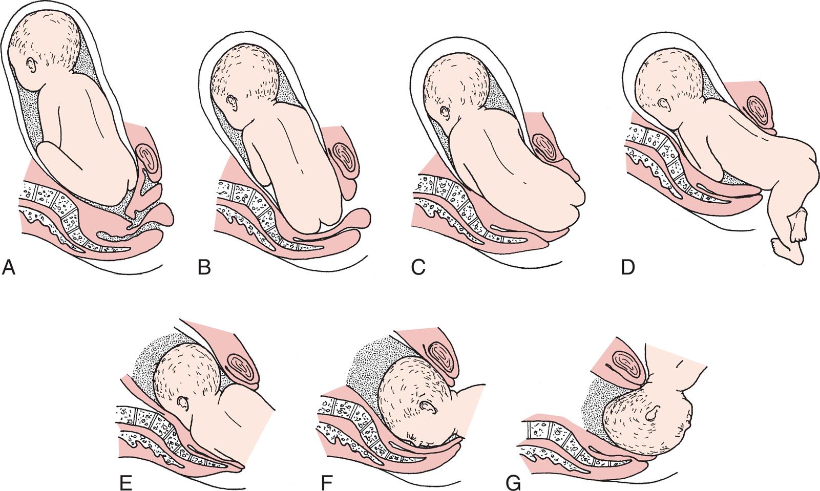
(A) Breech before onset of labor. (B) Engagement and internal rotation of buttocks. (C) Lateral flexion. (D) External rotation and restitution of buttocks. (E) Internal rotation of shoulders and head. (F) Face rotates to sacrum. Note that there is no flexion of the head so that the smallest diameter of the fetal head is not passing through the pelvis. The umbilical cord is compressed between the fetal head and the bony pelvis. (G) The head is born as the fetal body is elevated. (From Lowdermilk DL, Perry SE, Cashion KL, et al: Maternity and women’s health care, ed 12, St Louis, 2020, Elsevier.)
A series of illustrations labelled A through G shows the mechanism of labor in breech. Part A shows the mature fetus in the womb with knees bent and hips in contact with the vaginal opening. Parts B and C show fetus’s hips moving out of the expanded vagina. Part D shows the lower half of the body presented outside the vaginal opening. Part E shows the chest region moving out. Parts F and G show face and head moving out of the vaginal opening, respectively.
Intrapartum nurses must be prepared to assist with a breech birth because a woman sometimes arrives at the birth facility in advanced labor with her fetus in a breech presentation. External version is sometimes used to avoid the need for cesarean delivery in the case of a breech presentation; however, external version is not always successful, and the fetus sometimes returns to the abnormal presentation.
Abnormal Positions
A common cause of abnormal labor is a fetus that remains in a persistent occiput posterior position (left occiput posterior [LOP] or right occiput posterior [ROP]). The fetal occiput occupies either the left or the right posterior quadrant of the mother’s pelvis. In most women, the fetal head rotates in a clockwise or counterclockwise direction until the occiput is in one of the anterior quadrants of the pelvis (left occiput anterior [LOA] or right occiput anterior [ROA]).
Labor is likely to be longer when rotation does not occur. Intense and poorly relieved back and leg pain characterize labor when the fetus is in the occiput posterior position. Women with a small or average-sized pelvis may have difficulty delivering infants who remain in an occiput posterior position. The physician may use forceps to rotate the fetal head into an occiput anterior position.
Nursing Care
During labor, the nurse should encourage the woman to assume positions that favor fetal rotation and descent. These positions also reduce some of the back pain. Good positions for back labor include the following:
- • Sitting, kneeling, or standing while leaning forward
- • Rocking the pelvis back and forth while on hands and knees (Fig. 8.7) to encourage rotation
- • Side-lying (on the left side for an ROP position, on the right side for an LOP position)
- • Squatting (for second-stage labor)
- • Lunging by placing one foot in a chair with the foot and knee pointed to that side; lunging sideways repeatedly during a contraction for 5 seconds at a time

After birth, the mother and infant are observed for signs of birth trauma. The mother is more likely to have a hematoma of her vaginal wall if the fetus remained in the occiput posterior position for a long time. The infant may have excessive molding (alteration in shape) of the head, caput succedaneum (scalp edema) (see Chapter 12), and possibly injury from forceps or the vacuum extractor.
Multifetal Pregnancy
If the woman has more than one fetus, several factors can make dysfunctional labor likely, as follows:
Because of the difficulties inherent in multifetal deliveries, cesarean birth is common. Birth is almost always cesarean if three or more fetuses are involved.
Nursing Care
When the woman has a multifetal pregnancy, each fetus is monitored separately during labor. An upright or side-lying position with the head slightly elevated aids breathing and is usually most comfortable. Labor care is similar to that for single pregnancies, with observations for hypotonic labor.
The nursery and intrapartum staffs prepare equipment and medications for every infant expected. An anesthesiologist and a pediatrician are often present at birth because of the potential for maternal or neonatal problems. One nurse is available for each infant. Another nurse focuses on the mother’s needs.
Problems with the Pelvis and Soft Tissues
Bony Pelvis
Some women have a small or abnormally shaped pelvis that impedes the normal mechanisms of labor. The gynecoid pelvis is the most favorable for vaginal birth. Absolute pelvic measurements are rarely helpful to determine whether a woman’s pelvis is adequate for birth. A woman with a “small” pelvis may still deliver vaginally if other factors are favorable. She often delivers vaginally if her fetus is not too large, the head is well flexed, contractions are good, and her soft tissues yield easily to the forces of labor.
In contrast, a woman may vaginally deliver several infants much larger than 4082 g (9 lb) yet cannot deliver an infant weighing 4536 g (10 lb). Obviously, the woman’s pelvis was “adequate,” or even “large,” according to standard measurements; however, the pelvis was not large enough for her largest infant. The ultimate test of a woman’s pelvic size is whether her child fits through it at birth. A trial of labor may be indicated, and a cesarean delivery is done if necessary.
Soft Tissue Obstructions
The most common soft tissue obstruction during labor is a full bladder. The woman is encouraged to urinate every 1 or 2 hours. Catheterization may be needed if she cannot urinate, especially if a regional anesthetic or large quantities of IV fluids were given, which fill her bladder quickly yet reduce her sensation to void.
Soft tissue obstructions that are less common include pelvic tumors such as benign (noncancerous) fibroids. Some women have a cervix that is scarred from previous infections or surgery. The scar tissue may not readily yield to forces of labor to efface and dilate.
Problems with the Psyche
Labor is stressful, but women who have had prenatal care and have adequate social and professional support usually adapt to this stress and can labor and deliver normally. The most common factors that can increase stress and cause dystocia include lack of analgesic control of excessive pain, absence of a support person or coach to assist with nonpharmacological pain-relief measures, immobility and restriction to bed, and a lack of the ability to carry out cultural traditions.
Increased anxiety releases hormones such as epinephrine, cortisol, and adrenocorticotropic hormone that reduce contractility of the smooth muscle of the uterus. The body reacts to stress with the fight-or-flight response, which impedes normal labor by the following mechanisms:
Nursing Care
Promoting relaxation and helping the woman conserve her resources for the work of childbirth are the principal nursing goals. The nurse uses every opportunity to spare the woman’s energy and promote her comfort (see Chapter 7).
Abnormal Duration of Labor
Prolonged Labor
Any of the previously discussed factors may be associated with a long or difficult labor (dystocia). The average rate of cervical dilation during the active phase of labor is about 1.2 cm/hour for the woman having her first child and about 1.5 cm/hour if she has had a child previously. Descent is expected to occur at a rate of at least 1.0 cm/hour in a first-time mother and 2.0 cm/hour in a woman who has had a child before.
A Friedman curve is often used to graph the progress of cervical dilation and fetal descent. The Friedman curve is used as a guide to assess and manage the normal progress of labor rather than using a rigid determination of “normal length of labor.” Nursing interventions such as encouraging a delay in pushing during the second stage of labor until after full cervical dilation has occurred, alternative positioning of the patient during the first and second stages of labor, and electronic fetal monitoring along with the use of epidural anesthesia have had an impact on the length of the first and second stages of labor and on the positive outcome for mother and newborn. Therefore the Friedman curve remains a management guide in assessing cervical dilation in relation to the descent of the fetal head along with other factors and assessments. It may be referred to when determining the need for a cesarean section.
Prolonged labor can result in several problems, including the following:
- • Maternal or newborn infection, especially if the membranes have been ruptured for a long time (usually about 24 hours)
- • Maternal exhaustion
- • Postpartum hemorrhage (see Chapter 10)
- • Greater anxiety and fear in an ensuing pregnancy
In addition, mothers who have difficult and long labors are more likely to be anxious and fearful about their next labor.
Nursing Care
Nursing care focuses on helping the woman conserve her strength and encouraging her as she copes with the long labor. The nurse should observe for signs of infection during and after birth in both the mother (see Chapter 10) and the newborn (see Chapter 12).
Precipitate Birth
A precipitate birth is completed in less than 3 hours, and there may be no health care provider present. Labor often begins abruptly and intensifies quickly, rather than having a more subtle onset and gradual progression. Contractions may be frequent and intense, often from the onset. If the woman’s tissues do not yield easily to the powerful contractions, she may have uterine rupture, cervical lacerations, or hematoma.
Fetal oxygenation can be compromised by intense contractions because normally the placenta is resupplied with oxygenated blood between contractions. In precipitate labor, this interval may be very short. Birth injury from rapid passage through the birth canal may become evident in the infant after birth. These injuries can include intracranial hemorrhage or nerve damage.
Nursing Care
Women who experience precipitate birth may have panic responses about the possibility of not getting to the hospital in time or not having their health care provider present. Although they are relieved after birth, they require continued support and reassurance concerning the deviation from their expected experience. After birth, the nurse observes the mother and the infant for signs of injury. Excessive pain or bruising of the woman’s vulva is reported. Cold applications limit pain, bruising, and edema. Abnormal findings on the newborn’s assessment (see Chapter 12) are reported to the health care provider.
Premature Rupture of Membranes
Premature rupture of membranes (PROM) is spontaneous rupture of the membranes at term (38 or more weeks of gestation) more than 1 hour before labor contractions begin. A related term, preterm premature rupture of membranes (PPROM), refers to rupture of the membranes before term (before 37 weeks of gestation) with or without uterine contractions. Vaginal or cervical infection may cause prematurely ruptured membranes.
Diagnosis is confirmed by testing the fluid with nitrazine paper, which turns blue in the presence of amniotic fluid. A sample of vaginal fluid placed on a slide and sent to the laboratory will show a ferning pattern under the microscope, confirming that it is amniotic fluid (see Chapter 6). Treatment is based on weighing the risks of early delivery of the fetus against the risks of infection in the mother (chorioamnionitis, or inflammation of the fetal membranes) and sepsis in the newborn. An ultrasound determines gestational age, and oligohydramnios is confirmed if the amniotic fluid index (AFI) is less than 5 cm. Oligohydramnios in a gestation of less than 24 weeks can lead to fetal pulmonary and skeletal defects. If PROM occurs at 36 weeks of gestation or later, labor is induced within 24 hours. Because the cushion of amniotic fluid is lost, the risk for umbilical cord compression is great.
Nursing Care
The nurse should observe, document, and report maternal temperature above 38°C (100.4 °F), fetal tachycardia, and tenderness over the uterine area. Antibiotic and steroid therapy may be anticipated, cultures may be ordered, and labor may be induced or a cesarean section may be indicated. Nursing care for the woman who is not having labor induced immediately primarily involves monitoring and teaching the woman. Teaching combines information about infection and preterm labor and includes the following:
- • Report a temperature that is above 38°C (100.4 °F).
- • Avoid sexual intercourse or insertion of anything in the vagina, which can increase the risk for infection.
- • Avoid orgasm, which can stimulate contractions.
- • Avoid breast stimulation, which can stimulate contractions because of natural oxytocin release.
- • Maintain any activity restrictions prescribed.
- • Note any uterine contractions, reduced fetal activity, or other signs of infection (see section Amniotomy).
- • Record fetal kick counts daily, and report fewer than 10 kicks in a 12-hour period.
Preterm Labor
In the United States the preterm birth rate rose for a fifth year in a row to 10.23% of live births in 2019 (Hamilton et al., 2020). Preterm labor occurs after 20 weeks and before 37 weeks of gestation. The main risks are the problems of immaturity in the newborn. One goal of Healthy People 2030 is that 90% of all women will receive prenatal care starting in the first trimester. Preterm delivery is a major cause of perinatal morbidity and mortality, has a major medical and economic impact, and is a factor in the rising costs of health care. Early prenatal care can prevent premature labor or identify women at risk (Box 8.1). Environmental, biological, and socioeconomic factors affect the occurrence of preterm labor. Some biological causes include advanced maternal age, short pregnancy interval, fetal anomalies, and coexisting health disorders. Socioeconomic causes may include lack of access to care, inadequate nutrition, or intimate partner violence. Environmental causes may include smoking, drug use, long work hours, and long periods of standing (Griggs et al., 2020).
Early prenatal care allows women to be educated about the signs of preterm labor so that interventions can occur early. Home uterine activity monitoring can be initiated for women at risk for preterm labor.
Signs of Impending Preterm Labor
A transvaginal ultrasound showing a shortened cervix at 20 weeks of gestation may be predictive of impending preterm labor. Ultrasound may be advised for high-risk women. A cervicovaginal test for fetal fibronectin is also used to predict preterm labor. Fibronectin is a protein produced by the fetal membranes that can leak into vaginal secretions if uterine activity, infection, or cervical dilation of 2 cm or more occurs. The presence of increased fibronectin in vaginal secretions between 22 and 24 weeks’ gestation is predictive of preterm labor. Diagnosis of preterm labor is based on cervical effacement and dilation of more than 2 cm.
Maternal symptoms of preterm labor that cause women to seek medical care include the following:
- • Contractions that may be either uncomfortable or painless
- • Feeling that the fetus is “balling up” frequently
- • Menstrual-like cramps
- • Constant low backache
- • Pelvic pressure or a feeling that the fetus is pushing down
- • A change in vaginal discharge
- • Abdominal cramps with or without diarrhea
- • Pain or discomfort in the vulva or thighs
- • “Just feeling bad” or “coming down with something”
An ultrasound of the fetus to determine maturity, position, and other problems that may exist may be ordered. Treatment of preterm labor is more aggressive at 28 weeks’ gestation than at 34 weeks’ gestation.
Standardized Assessment of Preterm Labor
A preterm labor assessment toolkit (PLAT) is a toolkit designed to standardize care and assessment of women at risk for preterm labor; it was designed by the March of Dimes in accordance with ACOG guidelines (Wattie et al., 2017). The standardized approach reduces unnecessary antepartum admissions as well as unnecessary interventions, such as administration of tocolytic drugs or steroid medications. Using the PLAT patient triage protocol, patients in triage undergo a standardized assessment. The algorithm enables optimum delivery of steroid medication within 7 days of delivery if the findings are positive for likelihood of premature labor. The assessment guidelines can be completed within 2 to 4 hours to determine whether preterm labor is present. Contractions alone are not a reliable indicator of labor. The assessment includes history, physical examination, transvaginal ultrasound to determine cervical length (less than 20 mm), and fetal fibronectin in vaginal secretions. Fetal fibronectin is undetectable in vaginal secretions between gestational weeks 22 and 35. The presence of fetal fibronectin may indicate delivery is likely to occur within 14 days. Details of the algorithm can be accessed at https://www.marchofdimes.org/professionals/preterm-labor-assessment-toolkit.aspx.
Tocolytic Therapy
Tocolysis is the inhibition of myometrial uterine contractions. The goal of tocolytic therapy is to stop uterine contractions and keep the fetus in utero until the lungs are mature enough to adapt to extrauterine life, usually 2 to 7 days.
Magnesium sulfate is the drug of choice. It is not a very effective tocolytic, but it is used to protect the fetus from developing cerebral palsy (Simham et al., 2020). A continuous IV infusion is administered, and therapeutic levels are monitored. The woman should be informed that a warm flush may be perceived during the initiation of therapy. Overdose can affect the cardiorespiratory system, and vital signs are recorded every hour. If the fetus is born during magnesium therapy, drowsiness may be present, and resuscitation may be required. The nursery staff should be notified if magnesium sulfate therapy was used within 2 hours before delivery. Calcium gluconate should be on hand to treat adverse effects in the newborn. The FDA recommends limiting the use of magnesium sulfate to fewer than 5 to 7 days because the fetus can develop low blood calcium, bone problems, and respiratory depression with prolonged use (FDA, 2013). When magnesium sulfate is used, the nurse should monitor the patient for respiratory rate and lung sounds and signs of fluid overload, urine output, deep tendon reflexes, and bowel sounds because the intestinal muscles also relax in response to the drug.
β-Adrenergic drugs such as terbutaline (Brethine) are administered subcutaneously to stop uterine contractions within minutes (Simham et al., 2020). Cardiac side effects such as increased pulse rate and blood pressure can occur. Nasal stuffiness and hyperglycemia can occur, and the drug should be discontinued 2 hours before delivery to avoid side effects in the newborn. These drugs are given with caution to women with poorly controlled diabetes mellitus.
Prostaglandin synthesis inhibitors such as indomethacin are another type of drug that can be used to stop labor contractions. This type of drug causes a reduction in amniotic fluid, which is helpful when polyhydramnios is a problem. However, this drug is not commonly used because it can stimulate the ductus arteriosus to close prematurely, causing fetal death. Close fetal monitoring is essential.
Calcium channel blockers such as nifedipine (Procardia) are most commonly used to stop labor contractions. Because the drug causes vasodilation, maternal flushing and hypotension could be side effects, and blood pressure and pulse should be closely monitored. Magnesium sulfate should not be used when nifedipine is used or when intrauterine infection is suspected. Nifedipine also should be given with caution to women with hypotension (Simham et al., 2020).
Antimicrobial therapy is often initiated in women with preterm labor because studies have shown that subclinical chorioamnionitis is often present and for prevention of group B streptococcus infection.
Contraindications
Tocolytics should not be used in women with preeclampsia, placenta previa, abruptio placentae, gestational age greater than 37 weeks, chorioamnionitis, or fetal demise. In these cases, it would not improve the obstetric outcome to delay birth of the fetus.
Speeding Fetal Lung Maturation
If it appears that preterm birth is inevitable, the physician may give the woman steroid drugs (glucocorticoids) to increase fetal lung maturity if the gestation is between 24 and 34 weeks. Glucocorticoids are often used together with tocolytics. Betamethasone may be administered for this purpose in two intramuscular injections 24 hours apart.
Activity Restrictions
Bed rest was often prescribed for women at risk for preterm birth. However, the benefits of bed rest are not clear, and many adverse maternal effects can occur. Therefore total bed rest is prescribed less frequently than it was in the past. Activity restrictions are often more moderate, such as resting in a semi-Fowler’s position or partial bed rest.
Nursing Care
Nurses should be aware of the symptoms of preterm labor because they may occur in any pregnant woman, with or without risk factors. Symptoms are taught and regularly reinforced for women who have increased risk factors. Nursing care includes positioning the woman on her side for better placental blood flow, assessing vital signs frequently, and notifying the health care provider if tachycardia occurs. Signs of pulmonary edema (chest pains, cough, crackles, or rhonchi) and intake and output should be closely monitored. If the woman is monitored at home, appropriate activities and restrictions are identified, and arrangements for household responsibilities such as child care should be made with family or with the help of social services. If delivery occurs, monitoring the fetal heart rate is essential, and preparation for admission to the neonatal intensive care unit is initiated. Full emotional support of the parents is offered because they may be grieving the loss of the normal birth process.
Prolonged Pregnancy
A late-term pregnancy lasts between 41 weeks and 41 weeks and 6 days. A postterm pregnancy lasts 42 weeks. The term postmature most accurately describes the infant whose characteristics are consistent with a prolonged gestation (see Chapter 13).
Risks
The greatest risks of prolonged pregnancy are to the fetus. As the placenta ages, it delivers oxygen and nutrients to the fetus less efficiently. The fetus may lose weight, and the skin may begin to peel; these are the typical characteristics of postmaturity. Meconium may be expelled into the amniotic fluid, which can cause severe respiratory problems at birth. Low blood glucose levels are a likely complication after birth.
The fetus with placental insufficiency does not tolerate labor well. Because the fetus has less reserve than needed, the normal interruption in blood flow during contractions may cause excessive stress on the infant. If the placenta continues to function well, the fetus continues growing. This can lead to a large fetus and the problems accompanying macrosomia.
If placental function remains normal, there is little physical risk to the mother other than laboring with a large fetus. Psychologically, however, she often feels that pregnancy will never end. She becomes more anxious about when labor will begin and when her health care provider will “do something.”
Medical Treatment
The health care provider will evaluate whether the pregnancy is truly prolonged or if the gestation has been miscalculated. If the woman had early and regular prenatal care, ultrasound examinations have usually clarified her true gestation. Any pregnancy that lasts longer than 41 weeks must be monitored closely with NST, AFI, and BPP twice weekly and daily kick counts (see Chapter 5). Oligohydramnios (decreased amniotic fluid) in a postterm pregnancy is an indication for labor induction. If the woman’s pregnancy has definitely reached 41 weeks and 6 days, labor is usually induced by oxytocin. Prostaglandin application to ripen the cervix before oxytocin administration increases the probability of successful induction.
Nursing Care
Nursing care involves careful observation of the fetus during labor to identify signs associated with poor placental blood flow, such as late decelerations (see Chapter 6). After birth, the newborn is observed for respiratory difficulties and hypoglycemia.
Emergencies During Childbirth
Several intrapartum conditions can endanger the life or well-being of the woman or fetus. These conditions necessitate prompt nursing and medical action to reduce the likelihood of damage. Nursing and medical management often overlap in emergencies.
Prolapsed Umbilical Cord
The umbilical cord prolapses if it slips downward in the pelvis after the membranes rupture. In this position, it can be compressed between the fetal head and the woman’s pelvis, interrupting blood supply to and from the placenta. It may slip down immediately after the membranes rupture, or the prolapse may occur later. A prolapsed cord (Fig. 8.8) can be classified in the following ways:
The risk for prolapsed cord is increased if the membranes rupture before the fetal presenting part is completely engaged in the pelvis. Documenting the fetal heart rate after the membranes rupture is an essential nursing responsibility.
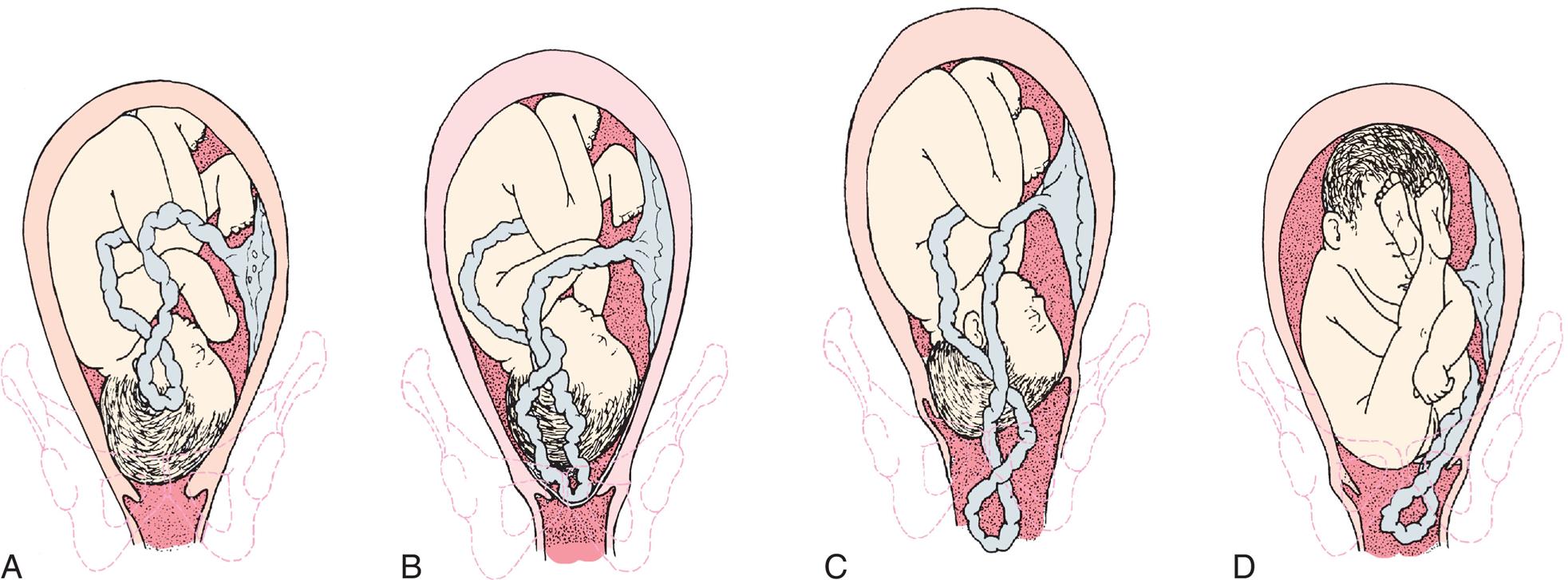
Note the pressure of the presenting part on the umbilical cord, which will interfere with oxygenation of the fetus. (A) Occult (hidden) prolapse of cord. The cord will be compressed between the fetal head and the mother’s bony pelvis. (B) Complete prolapsed cord. Note that the membranes are intact. (C) Cord is presenting in front of the fetal head and may be seen in the vagina. (D) Frank breech presentation with prolapsed cord. (From Lowdermilk DL, Perry SE, Cashion KL, et al: Maternity and women’s health care, ed 12, St Louis, 2020, Elsevier.)
Four illustrations labelled A through D, show anterior view of a pregnant womb highlighting the fully matured fetus attached to the umbilical cord. Parts A through C show the fetus in curved C-position with the head toward the vaginal opening. Part A shows the umbilical cord on the fetus side, curved near the head. Part B shows the umbilical cord near the vaginal opening. Part C shows the umbilical cord entered into the vagina. Part D shows the fetus in V-shape position with feet near the head and the hips toward the vaginal opening. The umbilical cord has entered the vagina.
Risk Factors
Prolapse of the umbilical cord is more likely if the fetus does not completely fill the space in the pelvis or if fluid pressure is great when the membranes rupture. These conditions are more likely to occur in the following situations:
Medical Treatment
The experienced physician may push the fetus upward from the vagina. Oxygen and a tocolytic drug such as terbutaline may be administered. The primary focus is to deliver the fetus by the quickest means possible, usually cesarean delivery.
Nursing Care
The main risk of a prolapsed cord is to the fetus. When a prolapsed cord occurs, the first action is to displace the fetus upward to stop compression against the pelvis. Maternal positions such as the knee-chest or Trendelenburg (head down) can accomplish this displacement (Fig. 8.9). Placing the mother in a side-lying position with her hips elevated on pillows also reduces cord pressure.
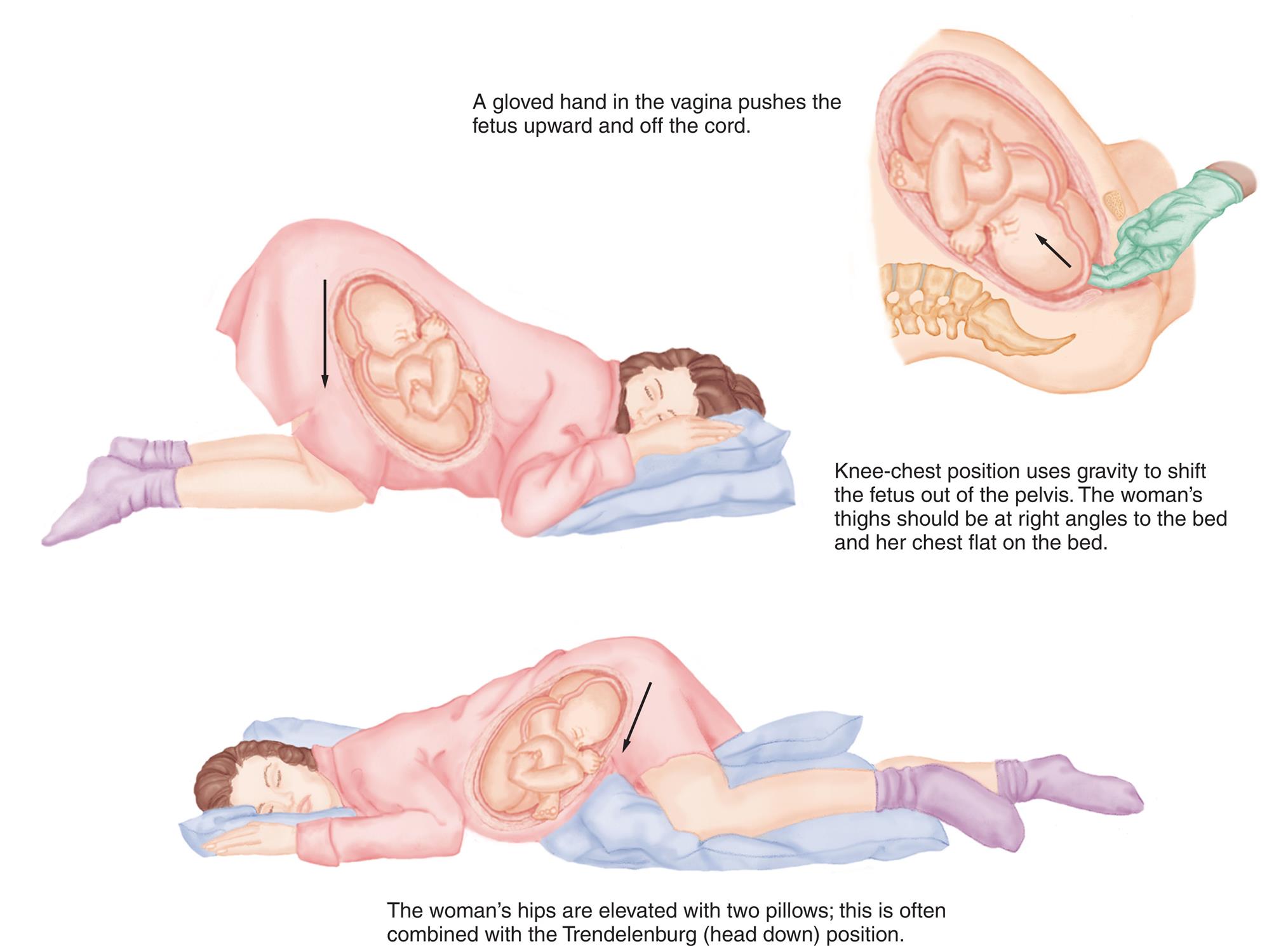
These positions can be used to relieve pressure on the prolapsed umbilical cord until delivery can take place. (From Murray SS, McKinney ES: Foundations of maternal-newborn and women’s health nursing, ed 6, St Louis, 2014, Saunders.)
Three illustration show different positions of pregnant woman. In the first illustration, a gloved hand in the vagina pushes the fetus upward and off the cord, indicated by a slant, upward arrow. In the second illustration, knee-chest position uses gravity to shift the fetus out of the pelvis. The woman’s thighs should be at right angles to the bed and her chest flat on the bed, indicated by a downward arrow. In the third illustration, the woman’s hips are elevated with two pillows; this is often combined with the Trendelenburg (head down) position, indicated by a slant, downward arrow.
In addition to prompt corrective actions and assisting with emergency procedures, the nurse should remain calm to avoid increasing the woman’s anxiety. Prolapsed cord is a sudden development; anxiety and fear are inevitable reactions in the woman and her partner. Calm, quick actions on the part of nurses help the woman and her family to feel that she is in competent hands. After birth, the nurse helps the woman to understand the experience. She may need several explanations of what happened and why.
Placenta Accreta
An abnormal attachment of the placenta to the uterine wall occurs in 3 out of 1000 deliveries (Francois and Foley, 2018). It is common in women with a previous cesarean section delivery, fibroids, increased maternal age, or endometrial defects. Symptoms include profuse bleeding at attempts to manually deliver the placenta after the fetus is delivered. The condition can be diagnosed before delivery via ultrasound and interventions used to minimize postpartum blood loss, but a hysterectomy often is required. The nurse should give care and support to the woman, who may not have the opportunity to carry another pregnancy. Nursing responsibilities include monitoring and documenting vital signs, IV therapy, providing pain relief, and observing the principles of blood transfusion therapy with similar interventions as other bleeding disorders of pregnancy.
Uterine Rupture
A tear in the uterine wall occurs if the muscle cannot withstand the pressure inside the organ. There are three variations of uterine rupture:
- 1. Complete rupture: There is a hole through the uterine wall, from the uterine cavity to the abdominal cavity.
- 2. Incomplete rupture: The uterus tears into a nearby structure, such as a ligament, but not all the way into the abdominal cavity.
- 3. Dehiscence: An old uterine scar, usually from a previous cesarean birth, separates.
Risk Factors
Uterine rupture is more likely to occur if the woman has had previous surgery on her uterus such as a previous cesarean delivery. The low transverse uterine incision (goes side to side) (see Fig. 8.5) is least likely to rupture. Because the classic uterine incision (goes up and down) is prone to rupture, a vaginal birth after this type of incision is not recommended. The woman who has had a previous cesarean section who wants to try to deliver vaginally (VBAC) should undergo a trial of labor (TOLAC), but a surgical team must be available to prevent or treat uterine rupture during labor in case an emergency cesarean section is indicated. Surgical intervention must be available within 30 minutes.
Uterine rupture may also occur if tachysystole develops as a result of labor induction with oxytocin or the woman has sustained blunt abdominal trauma, such as from a motor vehicle accident or from battering.
Characteristics
The woman may have no symptoms, or she may have sudden onset of severe signs and symptoms, such as the following:
- • Shock caused by bleeding into the abdomen (vaginal bleeding may be minimal)
- • Abdominal pain
- • Pain in the chest, between the scapulae (shoulder blades), or with inspiration
- • Cessation of contractions
- • Abnormal or absent fetal heart tones
- • Palpation of the fetus outside the uterus because the fetus has pushed through the torn area
Medical Treatment
If the fetus is living when the rupture is detected or if blood loss is excessive, the obstetrician performs surgery to deliver the fetus and to stop the bleeding. Hysterectomy (removal of the uterus) is likely to be required for an extensive tear. Smaller tears may be surgically repaired.
Nursing Care
The nurse should be aware of women who are at high risk for uterine rupture, and close monitoring during labor is essential. When uterine rupture occurs, the woman is prepared for immediate cesarean section. Measures to alleviate anxiety in the woman and her partner are necessary as emergency measures are being initiated.
Uterine rupture is sometimes not discovered until after birth. In these cases, the woman does not have dramatic symptoms of blood loss. However, she may have continuous bleeding that is brighter red than the normal postbirth bleeding. A rising pulse rate and falling blood pressure reading are signs of hypovolemic shock, which may occur if blood loss is excessive.
Amniotic Fluid Embolism
Amniotic fluid embolism, also known as anaphylactoid syndrome, occurs when amniotic fluid, with its particles such as vernix, fetal hair, and sometimes meconium, enters the woman’s circulation and obstructs small blood vessels in her lungs. It is more likely to occur during a very strong labor because the fluid is “pushed” into small blood vessels that rupture as the cervix dilates. Amniotic fluid embolism is characterized by abrupt onset of hypotension, respiratory distress, and coagulation abnormalities triggered by the thromboplastin contained in the amniotic fluid.
Treatment includes providing respiratory support with intubation and mechanical ventilation as necessary, treating shock with electrolytes and volume expanders, and replacing the coagulation factors such as platelets and fibrinogen. Packed red blood cells are sometimes given intravenously.
The woman’s intake and output are monitored closely. A pulse oximeter monitors oxygen saturation. The woman may be transferred to the intensive care unit for closer monitoring and nursing care.

Tess and Luis were introduced to the reader in Chapter 4, and Tess’ pregnancy experience has unfolded in each chapter. Refer to earlier chapters for her history and progress.
Tess was admitted to the labor unit and has been in active labor. However, after many hours, her labor contractions stop. The health care provider determines that her Bishop score is 7 and decides to augment her labor.
Questions
- 1. What is a Bishop score?
- 2. What is the difference between induction of labor and augmentation of labor?
- 3. When is induction or augmentation of labor contraindicated?
- 4. How can the nurse help Tess stimulate labor contractions using nonpharmacological methods? How can her husband Luis help?
- 5. What nursing responsibilities are involved during the administration of oxytocin during labor?
Get Ready for the Next-Generation NCLEX® Examination!
Key Points
- • An amnioinfusion is the insertion of fluid directly into the uterus to provide a cushion for the umbilical cord after amniotic fluid is lost.
- • Induction of labor is the intentional initiation of labor before it occurs naturally.
- • Augmentation of labor is the stimulation of contractions after they started naturally.
- • A Bishop score greater than 6 may predict successful labor induction because of a “ripe cervix.”
- • The nurse observes the character of the amniotic fluid and fetal heart rate when the membranes are ruptured. Fluid should be clear, but it may contain bits of lanugo and have a mild odor; the fetal heart rate should remain near its baseline level and between 110 and 160 beats/minute at term.
- • The nurse observes the fetal condition and character of contractions if any methods to stimulate labor are used.
- • Dystocia is a term used to describe a difficult labor, which can be caused by an abnormality of the power, passenger, passage, or psyche of the woman.
- • Nursing measures such as encouraging position changes, aiding relaxation, and reminding the woman to empty her bladder can promote a more normal labor.
- • Pharmacological, nonpharmacological, or mechanical methods can be used to stimulate labor.
- • After version, the nurse observes for leaking amniotic fluid and for a pattern of contractions that may indicate labor has begun. Before discharge, signs of labor are reviewed with the woman so she will know when to return to the birth center.
- • Nursing care after episiotomy or perineal lacerations includes comfort measures such as cold applications, analgesics, and wound assessment.
- • Nursing care after cesarean birth is similar to care after vaginal birth with the addition of assessing the wound, indwelling catheter patency, and IV flow. The woman and her partner may need extra emotional support after cesarean birth.
- • Nursing care after births involving instruments (forceps or vacuum extraction) and after abnormal labor and birth include observations for maternal and newborn injuries or infections. Infection is the most common hazard after membranes rupture prematurely, especially if there is a long interval before delivery.
- • When oxytocin is given to the woman in labor, the nurse must be aware of signs and symptoms of increased uterine activity and monitor the fetal heart rate every 15 minutes during active labor and every 5 minutes during the transition phase.
- • Induction of labor is more effective if cervical ripening is achieved before oxytocin is administered to stimulate contractions.
- • The nurse should be aware of the subtle symptoms a woman may experience at the beginning of preterm labor and should encourage her to seek care at the hospital promptly.
- • The nurse must be aware of the side effects of tocolytic drugs and monitor the mother and fetus closely.
- • Anaphylactoid syndrome occurs when amniotic fluid enters the woman’s circulation.
- • After any type of emergency, the woman and her family need emotional support, explanations of what happened, and patience with their repeated questions.
Additional Learning Resources
 Go to your Study Guide for additional learning activities to help you master this chapter content.
Go to your Evolve website (http://evolve.elsevier.com/Leifer) for the following learning resources:
- • Animations
- • Answer Guidelines for Critical Thinking Questions
- • Answers and Rationales for Review Questions for the NCLEX® Examination
- • Glossary with English and Spanish pronunciations
- • Interactive Review Questions for the NCLEX® Examination
- • Patient Teaching Plans in English and Spanish
- • Skills Performance Checklists
- • Video clips and more!
 Online Resources
- • Healthy People 2030: https://www.health.gov/healthypeople
- • National Center for Health Statistics: https://www.cdc.gov/nchs/fastats
Clinical Judgment and Next-Generation NCLEX® Examination-Style Questions
- 1. A nurse is caring for a patient receiving oxytocin for induction of labor. Which of the following observations require immediate report to the health care provider?
- 2. Place an X in the appropriate column to indicate whether the following observations are indications, contraindications, or not related to the plan to induce labor for a patient who is P0G1.
Observation Indica-tion Contraindi-cation Not Related Active herpes infection in the birth canal Fetal heart rate of 140 bpm 39 weeks’ gestation in a transverse lie 40 weeks’ gestation with gestational hypertension 40 weeks’ gestation with fetal macrosomia Infection within the uterus Expected due date approaching Rupture of membranes without spontaneous onset of labor Previous vertical cesarean incision Contractions at 5- minute interval with 50-second duration 40 weeks’ gestation with a prolapsed umbilical cord - 3. A woman has an emergency cesarean delivery after the umbilical cord was found to be prolapsed. She repeatedly asks similar questions about what happened at birth. The nurse’s interpretation of the woman’s behavior is that she:
- 1. cannot accept that she did not have the type of delivery she planned.
- 2. is trying to understand her experience and move on with postpartum adaptation.
- 3. thinks the staff is not telling her the truth about what happened at birth.
- 4. is confused about events because the effects of the general anesthetic are persisting.
- 4. A woman is being observed in the hospital because her membranes ruptured at 30 weeks’ gestation. While providing morning care, the nursing student notices that the draining fluid has a strong odor. The priority nursing action is to:
- 5. Following a vacuum extraction delivery, the nurse notices the newborn’s head is not symmetrical, with a chignon over the posterior fontanelle. Place an X in the appropriate column to indicate which of the following nursing actions as either indicated, contraindicated, or unnecessary.
| Nursing action | Indicat-ed | Contraindi-cated | Unnecess-ary |
|---|---|---|---|
| Place infant in Trend-elenberg position | |||
| Apply cold compress to the swollen area | |||
| Move infant to acute care nursery | |||
| Explain to the parents that the swelling will resolve without treatment | |||
| Document and continue routine observation | |||
| Immediately notify the health care provider |
- 6. A 32-year-old has been in active labor for nearly 15 hours without appreciable advancement of the fetus. The patient, her partner, and her obstetrician have discussed the options and have decided on a cesarean section. The obstetrical nurse begins implementation of interventions with the presurgical preparation of the patient.
| Nursing action | Indicat-ed | Contraindi-cated | Noness-ential |
|---|---|---|---|
| Request a bedside electrocardiogram | |||
| Send urine sample to lab | |||
| Administer IV antibiotic | |||
| Request a coagulation study | |||
| Provide preoperative teaching | |||
| Assure patency of intravenous line | |||
| Assess and record fetal heart rate | |||
| Request a complete blood count | |||
| Assess and record maternal vital signs | |||
| Shave both abdominal and pubic area | |||
| Request a blood typing and cross match | |||
| Insert indwelling urinary catheter | |||
| Administer IV proton pump inhibitor | |||
| Secure completion and review of consent form | |||
| Request 1 unit of appropriate blood be placed on reserve | |||
| Position client in a supine position with hip wedge in place |