18: The Patient With an Ostomy
Indications and Preparation for Ostomy Surgery
Nursing Care of the Patient Having Ostomy Surgery
Focused Assessment
| The patient problems and goals that require particular attention are as follows. |
| Patient Problems | Goals and Outcome Criteria |
|---|---|
| Anxiety and grieving related to perceived threat to self-image, anticipated changes in body appearance and function |
Reduced anxiety: patient states anxiety is reduced; relaxed manner
Grieving: verbalizes reality of impending loss
|
| Inadequate knowledge of what to expect after surgery in relation to the stoma related to lack of exposure to information | Patient understands postoperative routines and procedures in relation to ostomy surgery: patient correctly describes what to expect before and after surgery |
Interventions
Anxiety and Grieving
Inadequate Knowledge
Fecal Diversion

From Lewis SM, Bucher L, Heitkemper MM, Harding MM: Medical-surgical nursing: assessment and management of clinical problems, ed 10, St. Louis, 2017, Elsevier.

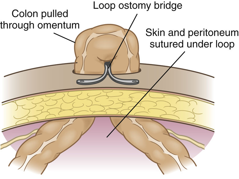
Three diagrams show the location of the stoma in the large intestine. The first diagram, labeled single-barrel, shows a circular structure in the mid-part of the descending colon, and it is not followed by a rectum further. The second diagram, labeled double-barrel, shows two circular structures in the mid part of the transverse colon. The third diagram, labeled loop, shows a structure with two extensions on both ends placed in the mid-part of the transverse colon.

A diagram shows the sigmoid colostomy. It shows a large intestine with a descending colon, with the end region separated from the rectum. The colon ends in a circular shaped structure representing sigmoid colostomy, and the end of the rectum facing the colon is labeled Hartman’s pouch.
Ileostomy
Procedure

Two diagrams, labeled A and B, show a continent ileostomy. (A) shows a diagram of the ileum with its lumens sutured. It shows labels for fascia above the ileum, distal ileum formed into a nipple valve, fold sutured to hold the valve in place, intussusception of the ileum to form a nipple valve, loop opened, lumens of terminal ileum that are sutured together, loop of terminal ileum sutured together, and suture needle and thread on the loop. (B) shows a diagram of the sutured ileum forming a pouch. It shows labels for nipple valve or stoma sutured to the skin layers, stoma sutured flush with skin, pouch sutured to the abdominal wall, and knock pouch in which edges are joined to form a pouch. The fascia is above the ileum.

Two diagrams depict the creation of an ileoanal reservoir in two stages. Stage 1: After removal of the colon, a temporary loop ileostomy is created and an ileo anal reservoir is formed. The reservoir is created in an S-shaped reservoir (using three loops of ileum) or a J-shaped reservoir (suturing a portion of ileum to the rectal cuff, with an upward loop). The diagram shows the label for the loop ileostomy and the S-shaped reservoir. The enlarged view of the diagram shows a J-shaped reservoir. Stage 2: After the reservoir has had time to heal-usually several months- the temporary loop ileostomy is reversed and stool is allowed to drain into the reservoir. The diagram shows the label for loop ileostomy reversed.
 Pharmacology Capsule
Postoperative Nursing Care of the Patient With an Ileostomy
Focused Assessment
Health History
Physical examination
| The patient problems and goals common to most postoperative patients (pain, potential for airway obstruction, potential for infection, inadequate circulation, and urinary retention) are discussed in Chapter 17. Additional diagnoses specific to the ileostomy patient in the immediate postoperative phase are as follows. |
| Patient Problems | Goals and Outcome Criteria |
|---|---|
| Potential for fluid volume deficit (dehydration) related to nothing by mouth (NPO) status, nasogastric suction, passage of liquid stool | Normal fluid balance: pulse and blood pressure consistent with patient norms, moist mucous membranes, urine output approximately equal to fluid intake, absence of neuromuscular symptoms. |
| Potential for inadequate tissue perfusion of stoma | Effective stoma perfusion: stoma is beefy red. |
| Potential for disrupted skin tissue integrity related to adhesive, fecal drainage, leaking appliance | Normal skin integrity: skin intact with minimal redness around stoma. |
| Altered body image related to presence of stoma, altered body function, loss of fecal continence | Adjustment to change in body image: patient makes positive statements about ability to adapt, learns and takes over ostomy care. |
| Potential for impaired sexual function related to altered body structure and function or reactions to those changes | Fulfilling sexual expression: patient reports making adaptations as necessary for satisfying sexual expression. |
| Inability to manage self-care related to lack of knowledge of ostomy management, failure to accept ostomy, lack of resources for proper care | Patient effectively manages ostomy: patient accepts responsibility for self-care, demonstrates proper care of ostomy, obtains necessary supplies. |
| Potential for injury related to obstruction of the ileum | Absence of injury: ostomy remains patent, drainage is continuous or frequent. |
Interventions
Potential for fluid volume deficit
Potential for inadequate tissue perfusion of stoma
Potential for disrupted skin tissue integrity
 inch larger than the stoma because a larger opening permits more fecal matter to come in contact with the skin. If a paste, barrier ring, or strip is used, apply it around the stoma or on the cut edge of the wafer. Place the wafer over the stoma and press down. Some pouching systems have the pouch preattached to the wafer (one piece); other systems consist of a wafer that is placed around the stoma and a separate pouch that attaches to the wafer (two-piece). Some patients have an uneven skin surface around the stoma, making it difficult to create a seal. In that case, a caulking material such as barrier strips or rings can be applied around the stoma to create a smooth surface. Clamp or close the pouch tail after it is securely placed. Initially, the pouch opening will probably need to be custom cut. As the stoma heals and shrinks, precut appliances may work well. Custom-cut pouches can be ordered for stomas of unusual size or shape. Patients whose barriers quickly erode and those with very flush or retracted stomas can obtain special barriers to maintain good fit. Once the stoma and surgical incision heal, some patients will prefer to cleanse the peristomal skin in the shower.
Altered body image
Potential for impaired sexual function
Inability to manage self-care
 Patient Teaching
- • Stoma and skin care: Explain recommended procedures and supplies with written care instructions and some temporary supplies when the patient is discharged.
- • Appliances: Provide a list of the supplies needed and where they can be purchased.
- • Irrigation (if appropriate) or drainage: Explain about the frequency, procedure, and supplies needed.
- • You can bathe or shower with the appliance in place because the pouch and the seal are waterproof.
- • Wear regular clothing but avoid direct pressure over the stoma.
- • Learn to recognize foods that cause excess gas or odor so that these can be avoided.
- • Maintain a fluid intake of at least 2000 mL daily unless contraindicated.
- • Avoid heavy lifting and strenuous activities at first; usually no restrictions are made after approximately 3 months; ask your physician about specific activities and contact sports.
- • Adaptations for sexual activity can include concealing the pouch and experimenting with positions.
- • Contact your physician or the WOC nurse if you observe skin breakdown, prolapse (bulging out) of the stoma, or obstruction (output absent or markedly decreased).
- • Resources for information about living with an ostomy include the American Cancer Society (www.cancer.org), United Ostomy Associations of America (www.ostomy.org), Crohn’s and Colitis Foundation of America (www.ccfa.org), Wound Ostomy and Continence Nurses Society (www.wocn.org), and home health agencies.
- • Ostomy surgery does not interfere with traveling. Tips on traveling include the following:
- • Take adequate supplies.
- • If flying, keep the supplies in a hand-carried bag. This could prevent problems if luggage is lost or delayed.
- • Include sealable plastic bags to dispose of used supplies.
- • Exercise caution with new foods that may cause diarrhea or gas.
- • If visiting a country where drinking the water is not advised, do not irrigate a colostomy with the water. Use only water that is safe for drinking.
Potential for injury
 Put On Your Thinking Cap!
Continent (Pouch) Ileostomy
Procedure
Postoperative Nursing Care of the Patient With a Continent Ileostomy
Focused Assessment
| The patient problems, goals, and outcome criteria in addition to those previously listed are as follows. |
| Patient Problems | Goals and Outcome Criteria |
|---|---|
| Potential for injury related to obstruction of the pouch drainage | Absence of injury: patient’s pouch drains readily |
| Inability to perform (stoma) self-care related to lack of knowledge of technique for draining pouch and caring for stoma and pouch | Patient understands pouch and stoma care: patient demonstrates proper pouch drainage and stoma care |
Interventions
Potential for injury
Inability to perform (stoma) self-care
- 1. Have the patient sit or lie down for the procedure.
- 2. Gather supplies: lubricant, 28 to 32 Fr catheter, drape, basin, irrigating syringe, irrigating solution, gauze dressing.
- 3. Lubricate the catheter and insert it gently into the stoma.
- 4. Resistance will be felt when the catheter reaches the nipple valve (approximately 2 inches past the stoma). Instruct the patient to bear down and then roll the catheter between your fingers and advance it into the pouch.
- 5. As soon as the catheter is in the pouch, gas and fecal matter begin to be expelled. Drainage usually continues for approximately 10 minutes and produces a total volume of 50 to 200 mL.
- 6. If the drainage is too thick or the catheter becomes blocked, instill 30 mL of normal saline or tap water as ordered and let it drain off. Gently aspirate only if necessary because this may cause dislocation of the nipple.
- 7. When drainage stops, quickly remove the catheter.
- 8. Place a gauze dressing over the stoma to absorb any secretions.
- 9. Measure, describe, and discard the drainage.
- 10. Instruct the patient on how to perform this procedure as soon as possible.
- 11. Advise the patient to wear a medical alert bracelet at all times that states they have a continent diversion that must be drained.
 Health Promotion
 Nutrition Considerations
- 1. The patient with an ileostomy or colostomy may eat a normal diet and simply omit foods that seem to cause problems.
- 2. The main concern of the patient with an ileostomy or colostomy is odor, which is caused by flatulence.
- 3. Patients should be encouraged to avoid foods tending to cause bad odor, especially corn, dried beans, onions, cabbage, spicy foods, and fish.
- 4. Fibrous vegetables should be avoided and all food should be chewed well.
Ileal Pouch Anal Anastomosis
Procedure
Complications
Obstruction
Peritonitis
Inflammation
Postoperative Nursing Care of the Patient With an Ileal Pouch Anal Anastomosis
Focused Assessment
| Patient Problems | Goals and Outcome Criteria |
|---|---|
| Potential for disrupted skin tissue integrity related to frequent passage of liquid stool through the rectum | Healthy skin: intact skin without excessive redness around the anus and the perianal area |
| Bowel incontinence related to inability to control passage of frequent liquid stools | Control of bowel elimination: decreasing number of incontinent episodes |
| Potential for injury related to possible small bowel obstruction, leaking of reservoir suture line, inflammation of reservoir | Absence of signs and symptoms of obstruction, suture leakage, or reservoir inflammation: no fever, abdominal pain or distention, or bloody stools |
Interventions
Potential for disrupted skin tissue integrity
 Cultural Considerations
Bowel incontinence
Potential for injury
Colostomy
Procedure

Five diagrams show different types of ostomies. 1. Ileostomy: the ostomy is done on the ileum in the abdomen. 2. Ascending colostomy: the ostomy is done on the ascending colon on the right side of the abdomen. 3. Descending colostomy: the ostomy is done the descending colon on the left side of the abdomen. 4. Sigmoid colostomy single-barreled: the ostomy is done on the sigmoid colon on the left side of the abdomen. 5. Transverse colostomy double-barreled: the ostomy is done on the transverse colon, forming a proximal and a distal loop.
Postoperative Nursing Care of the Patient With a Colostomy
Focused Assessment
| In addition to the patient problems and goals already identified, the following may also apply to the patient with a colostomy. |
| Patient Problem | Goals and Outcome Criteria |
|---|---|
| Inability to manage colostomy care related to lack of knowledge of self-care and irrigation procedure (if ordered), lack of confidence, lack of resources, failure to accept ostomy | Patient manages colostomy effectively: patient accepts responsibility for self-care, patient demonstrates ostomy care and irrigation (if ordered) correctly; uses resources as needed, obtains necessary supplies. |
| Potential for injury related to prolapse or stenosis | Absence of injury: no signs of prolapse, no protrusion of stoma; patent stoma with lumen of adequate diameter: regular elimination of feces through stoma. |
Interventions
 Nursing Care PlanPatient With a Colostomy
| Patient Problems | Goals and Outcome Criteria | Interventions |
|---|---|---|
| Potential for fluid volume deficit related to NPO status, nasogastric suction, passage of liquid stool | Patient will maintain normal fluid balance, as evidenced by pulse and blood pressure consistent with patient’s baseline, moist mucous membranes, and approximately equal fluid intake and output. | Monitor for signs of hypovolemia: tachycardia, hypotension, decreasing urine output, and dry mucous membranes. Keep accurate intake and output (i.e., urine, liquid stool, gastric fluid) records. Monitor for signs of electrolyte imbalances: confusion, anxiety, twitching, trembling, muscle weakness, and cardiac dysrhythmias. Give intravenous fluids as ordered, monitoring rate of flow carefully. |
| Potential for disrupted skin integrity related to stoma adhesive, fecal drainage | Skin at the base of the stoma will be intact and free of redness. | Check pouch hourly to detect leakage. When pouch is emptied, prevent fecal matter from contaminating the incision. When changing the appliance, gently remove adhesive. Cleanse skin around stoma with soap and water, rinse, and pat dry. Apply a protective skin barrier before replacing the pouch. Make the opening of the skin barrier not more than
larger than the stoma. Report rash or skin breakdown. |
| Altered body image related to presence of stoma, altered body function | Patient will adapt to colostomy as evidenced by self-care and ability to resume normal activities. | Provide an opportunity for the patient to share his thoughts about colostomy. Identify specific concerns such as activity limitations, stoma care, odor control, and effect on sexuality. Provide information. Be accepting of the patient’s feelings. Encourage him to attend to grooming and appearance. Offer to have a volunteer from the American Cancer Society or United Ostomy Associations of America visit him. Advise the patient that services are available from a WOC nurse, a mental health counselor, and a spiritual counselor if he desires. |
| Inability to manage (colostomy) self-care related to complexity of ostomy care, lack of knowledge, lack of resources, failure to accept ostomy | Patient will manage ostomy care effectively: patient demonstrates proper ostomy care, uses available resources, and resumes valued activities with adaptations as needed. | During stoma care in early postoperative period, tell the patient what is being done and why. When the patient begins to watch the procedure, gradually encourage him to participate and then to take over care. Recognize the patient’s need to grieve and that the patient may use denial as a coping mechanism. Develop a teaching plan that includes skin care, pouches, diet fluids, irrigation if appropriate, activity, sexuality, complications, tips on traveling, and resources. |

Inability to manage (colostomy) self care
- 1. Have the patient select the time of day that is most convenient. The procedure should be done at approximately the same time every day. The entire process takes 45 minutes to 1 hour.
- 2. Have the patient sit on or in front of the toilet if possible. If the patient cannot get out of bed, the procedure can be done while the patient is in bed.
- 3. Remove the old pouch and apply an irrigating sleeve. This device opens at the top so that the tubing can be inserted into the stoma and it opens at the bottom so that fecal matter can drain into the toilet.
- 4. Pour 500 to 1500 mL of lukewarm irrigating solution into an enema fluid container and hold it at the level of the patient’s shoulder. Initially, 500 mL is used but adults eventually increase the fluid to 1500 mL (Landmann & Cashman, June 9, 2020).
- 5. Clear the air from the tubing, lubricate the tubing, and insert it gently 2 to 4 inches into the stoma. The direction to insert the tubing can be assessed by first inserting a gloved, lubricated finger into the stoma. Do not use force! If a cone-tipped catheter is used, the catheter can be inserted only approximately 1 inch. This reduces the risk of perforation.
- 6. Allow the solution to flow slowly into the stoma. If the patient has cramping, slow down or stop the flow for a few minutes. Remove the tubing or cone after the solution has been administered.
- 7. If the solution does not drain promptly, close the bottom of the sleeve so that the patient can carry out other activities. Complete emptying may take 20 to 30 minutes.
- 8. When elimination is complete, remove, wash, and dry the sleeve.
- 9. Measure the stoma before applying a new pouch, especially in the first 4 to 6 weeks, because the stoma size will change as healing occurs. The opening in the skin barrier should be just slightly larger than the stoma.
- 10. Apply a clean pouch if additional drainage usually occurs during the day. Some patients need only a small dressing or a stoma cap.
- 11. Rectal suppositories can be inserted into a colostomy stoma to stimulate evacuation.
- 12. Patients who have double-barreled colostomies can be given rectal medications through the distal stoma. Be sure you identify which stoma is the distal one.
Potential for injury
Urinary Diversion

Four diagrams, labeled A through D, show methods of urinary division. (A) Ileal segment anastomosed to sigmoid colon: Isolated ileal segment with ureters implanted in posterior portion of segment. (B) Isolated ileal segment with ureters implanted in posterior portion of segment. The diagram shows the label for the protruding abdominal stoma at the end of ileal segment. (C) It shows two diagrams. The first diagram shows left ureter anastomosed to right ureters and cutaneous ureterostomy on abdomen. The second diagram shows cutaneous ureterostomy on abdomen. (D) Bilateral nephrostomy tubes inserted into renal pelvis; catheters exit through an incision on each flank, or there may be just one kidney. The diagram shows catheter inserted into the kidney anchored with a tape on the pelvic area which is connected to a drainage tube. It shows a label for a stab wound on skin below the kidney.
Ileal Conduit
Procedure

A diagram shows the creation of a stoma joined to a urinary pouch in the pelvic area. The ureters from both the kidneys are connected to the ends of the pouch, which shows the label for implantation of ureters.
Complications
Postoperative Nursing Care of the Patient With an Ileal Conduit

| In addition to the common problems and goals for postoperative patients (see Chapter 17), the following diagnoses may apply to the patient who has an ileal conduit/urostomy. |
| Patient Problems | Goals and Outcome Criteria |
|---|---|
| Potential for disrupted skin integrity related to contact of urine with skin | Normal skin around stoma: healed stoma base without redness or edema |
| Potential for infection related to contamination of stoma | Absence of infection: no fever or foul urine odor |
| Potential for injury related to obstruction of urine flow | Unobstructed urine flow: urine output approximately equal to fluid intake |
| Altered body image related to presence of stoma, altered body function | Adjustment in body image: patient acknowledges stoma, shows increasing interest in self-care, resumes previous sexual activity. |
| Inability to perform ostomy care related to lack of knowledge to manage complex therapeutic regimen | Patient assumes self-care of ostomy: patient demonstrates proper techniques of ostomy care and describes self-care with an ostomy. |
Interventions
Potential for disrupted skin integrity

An urostomy pouching system. It shows a bag or a pouch with a ring-like structure on one end and connected to a tube on the other hand. A square-shaped plate with a hole inside a circular structure is shown on the left of the pouch.
Potential for infection
Potential for injury
Altered body image

Six diagrams, labeled A through F, show the procedure for applying pouch. (A) shows the Urostomy pouch. (B) shows the hand is placing a gauze square over the stoma. (C) shows the hand is measuring the size of the stoma. (D) shows the hands are removing the backing from the new pouch. (E) shows the hands are placing the new pouch into the stoma. (F) shows the hands are connecting the drain into the tubing.

| Problem | Cause | Assessment and Intervention |
|---|---|---|
| Skin irritation | Skin barrier or wafer too small | Adjust the size of the skin barrier or wafer to cover skin around the stoma. |
| Leaking appliance |
Check the belt. If it is too tight, the seal can break.
Replace the appliance as needed (PRN).
|
|
| Hair follicle inflammation |
Use topical antimicrobial powder and skin barrier powder.
Cover any lesions with nonstick dressing and with a barrier before applying the pouching system.
Use adhesive remover; remove sealants gently. After the skin returns to normal, shave or cut any hair around the stoma.
|
|
| Perspiration under pouch |
Dry the skin well.
Apply a protective barrier.
Apply powder to skin under pouch.
Use a soft pouch cover.
|
|
| Allergy to pouching products | Spot test other brands to find one that does not cause irritation. | |
| Candida (“yeast”) infection |
Dry well.
Apply antifungal powder as ordered.
|
|
| Hernia/prolapse | Muscle weakness | Condition requires surgical repair. |
| Increased intraabdominal pressure | ||
| Wartlike lesions | Excessive peristomal wetness |
Reduce pouch opening size or acquire custom-cut system to reduce moisture on the skin.
Condition may require debridement by a physician.
|
| Odor | Urinary tract infection | Treat infection. |
| Appliance soiled or leaking |
Check the seal; change the appliance.
Provide deodorant tablets PRN.
|
