Fig. 1Three air bubbles trapped beneath a coverslip observed using low-power (100 ×) magnification. Numerous white blood cells (WBCs) are also present. Appearance varies with condenser and aperture positions of microscope. Brightfield.
Fig. 2Three air bubbles trapped beneath the coverslip. Compare with Fig.1; adjustment of the microscope’s condenser and aperture can change the appearance of air bubbles. Brightfield.
Fig. 3When plastic commercial standardized slides are used, fragments of plastic (red arrows) can be present in the sediment. Red blood cells, yeasts, and pseudohyphae are also present.
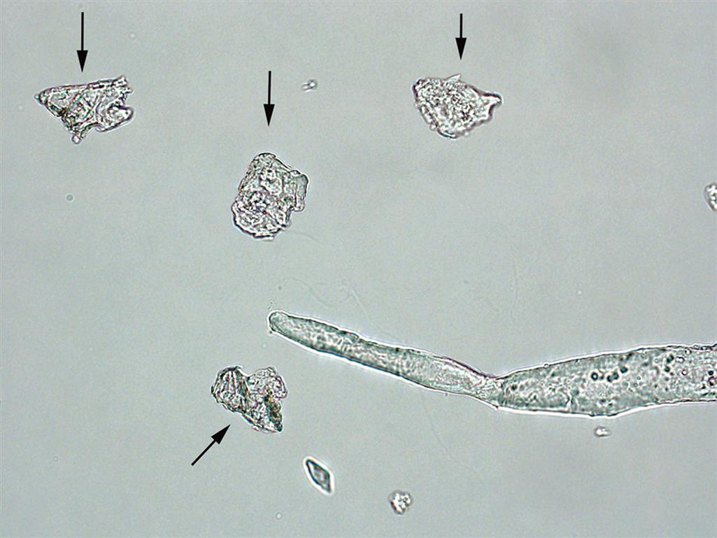
Fig. 4Three starch granules, all highly refractile, with slightly differing appearances, yet each has a centrally located dimple. Fragments of plastic (red arrows) are also present.
Fig. 5A starch granule (black arrow) demonstrating a characteristic dimple. When glass slides and coverslips are used, glass fragments (red arrows) can be present. Numerous white blood cells are also present.
Fig. 6Talc fragments (arrows)—note layering, refractility, and irregular edges. Also a single fiber—its refractility and shape helps distinguish it from a cast. Brightfield.
Fig. 7An absorbent fiber (diaper or hygiene product). Note its flat appearance with perforations and its strong refractility. Brightfield.
Fig. 8A clothing fiber. Its strong refractility, frayed ends, and flatness aid in its proper identification. Brightfield.
Blood Cells
Red Blood Cells
Fig. 9Numerous intact and ghost red blood cells (RBCs) (arrows). Intact RBCs have a characteristic appearance caused by the hemoglobin within them. In contrast, ghost RBCs have lost their hemoglobin but retain an intact cell membrane. This urine was hypotonic (dilute; low specific gravity). Many of the RBCs appear swollen and rounded because of the diffusion of fluid into the cells.
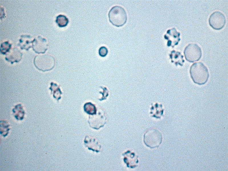
Fig. 10Red blood cells (RBCs) in hypertonic urine (concentrated; high specific gravity). Many of the cells in this field of view have lost their typical biconcave shape and become echinocytes (i.e., crenated). This happens when fluid moves out of the cell in an attempt to achieve balance with the tonicity of the environment. Consequently, the cell membrane shrinks, forming folds or projections, a process that is reversible. Near the center is a single schizocyte form—fragmented RBCs with three pointed extremities. Brightfield.
White Blood Cells
Fig. 11White blood cells and a single squamous epithelial cell. Note the size similarity between the squamous cell nucleus and the diameter of the white blood cells. Brightfield.
Fig. 12Five white blood cells. Note that the lobed nuclei in several of these neutrophils are readily apparent; whereas in degenerating cells, the nucleus has become mononuclear. Brightfield.
Fig. 13(A) Three white blood cells, a single red blood cell, and a squamous epithelial cell. Note the size similarity between the squamous cell nucleus and the diameter of the white blood cells. Brightfield. (B) Numerous white blood cells and two red blood cells (just left of center). Many of the white blood cells show evidence of degeneration. Brightfield.
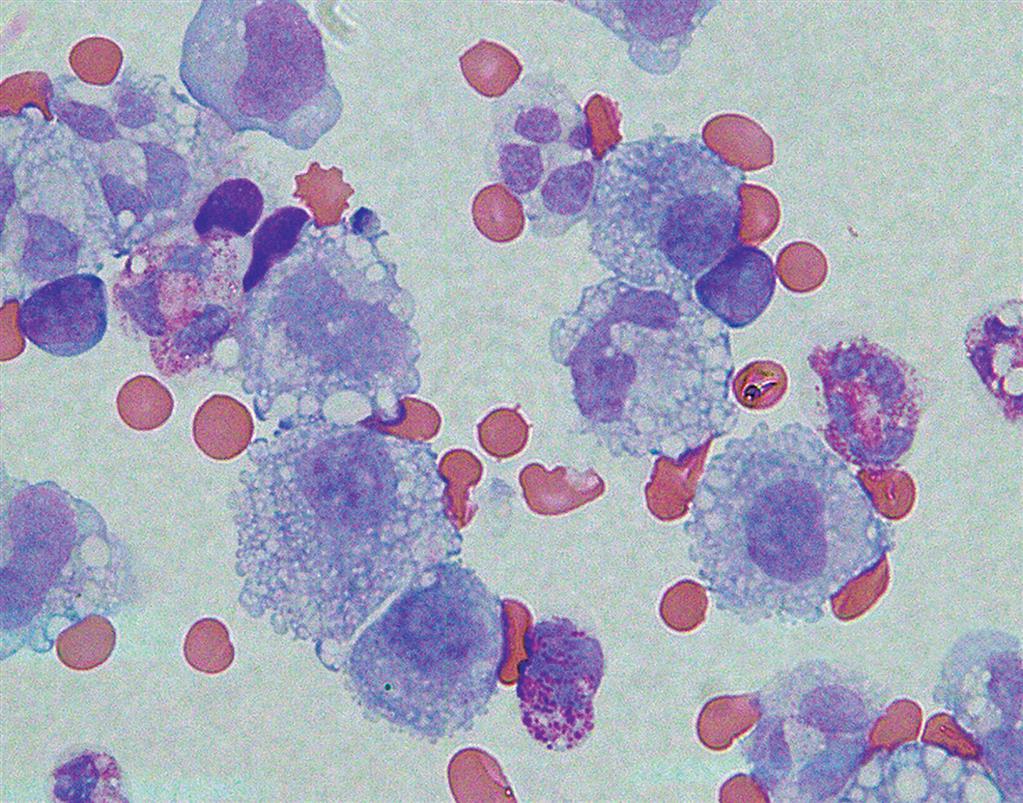
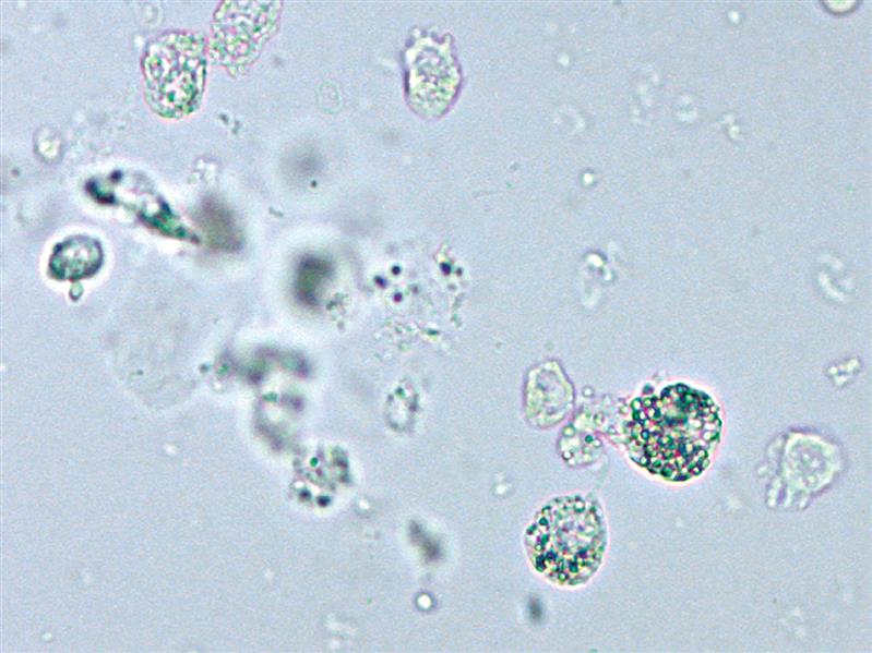
Fig. 14Many macrophages as well as other white blood cells in urine sediment. Cytospin preparation, Wright stained, Brightfield.
Casts
Cellular Casts
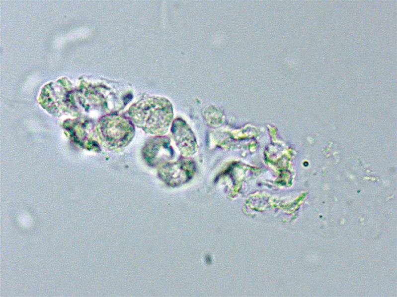
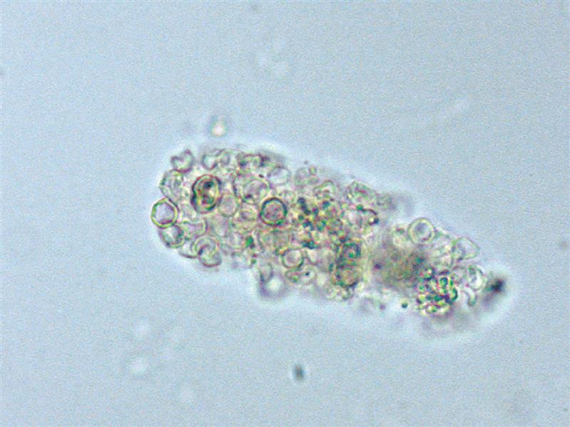
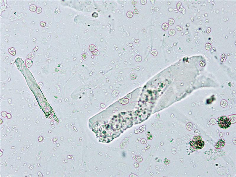
Fig. 15A mixed cellular cast. Brightfield.
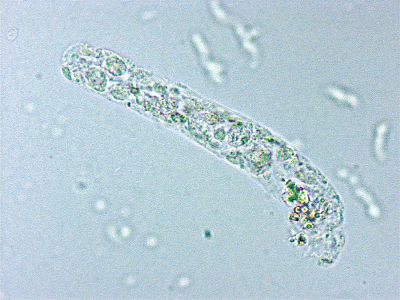
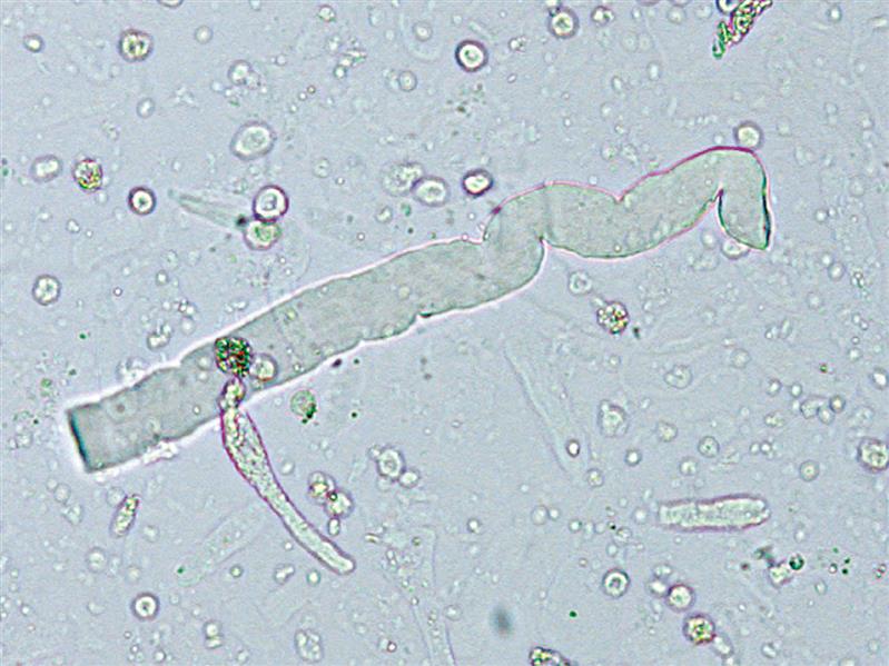
Fig. 16Renal tubular epithelial cell cast with one end broken or incompletely formed.
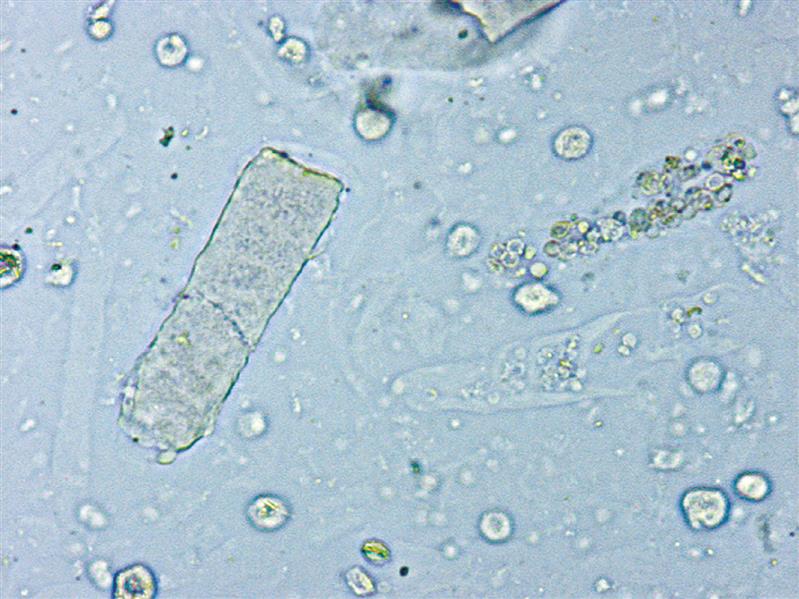
Fig. 17Renal tubular epithelial cell cast. Note the cuboidal shape of the entrapped cells. Also, the nuclei become more apparent when adjusting the fine focus up and down during the microscopic examination.
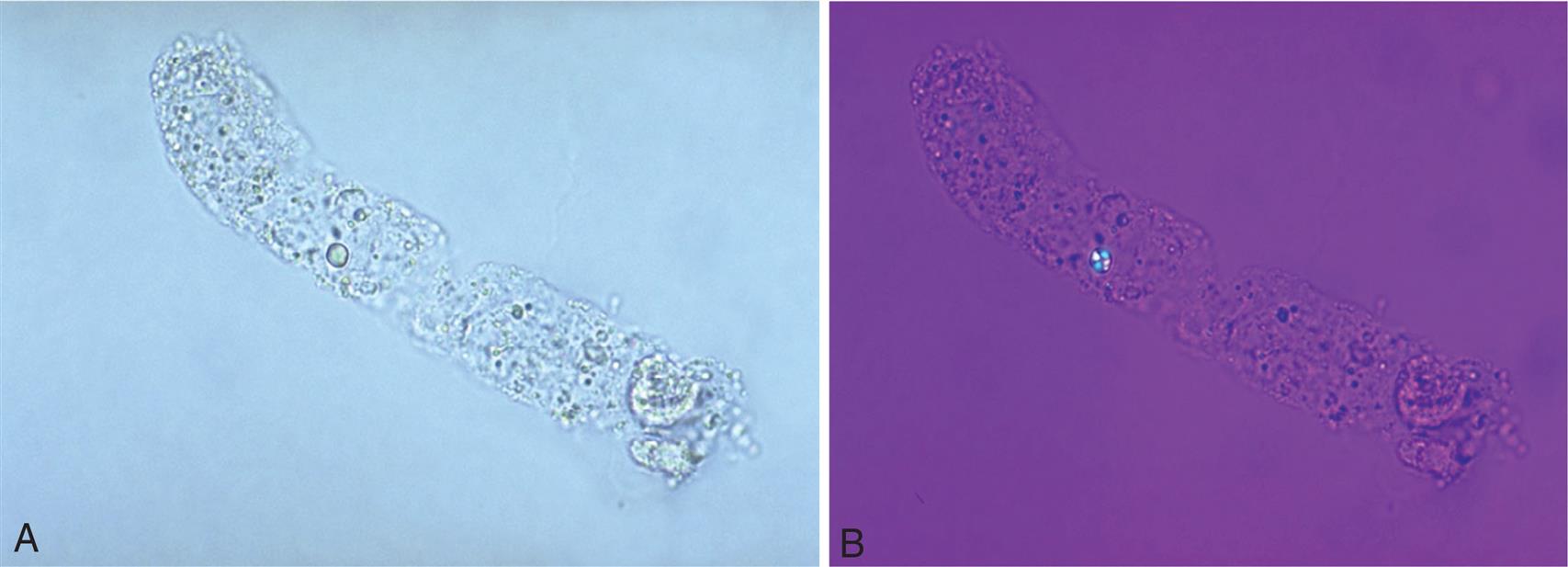
Fig. 18A renal tubular cell cast and several free-floating renal tubular cells in a Sternheimer-Malbin stained sediment. A highly refractile glass fragment is present in the center of this field of view.
Fig. 19A cast with oval fat bodies (i.e., renal tubular cells that contain fat). In this Sternheimer-Malbin stained sediment, the fat droplets take on a yellow or greenish appearance.
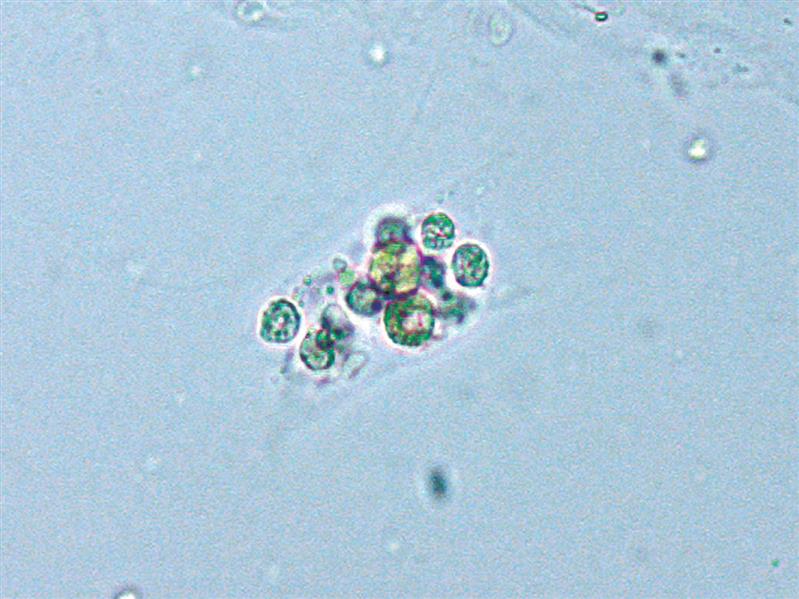
Fig. 20A white blood cell cast. Note the spherical or round shape of entrapped cells.
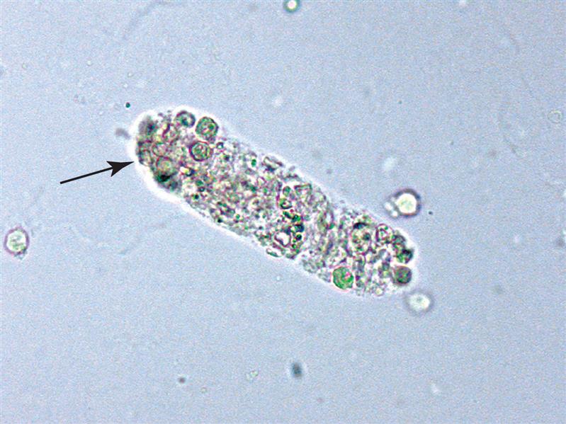
Fig. 21A mixed cell cast. This cast contains both white blood cells and red blood cells (arrow).
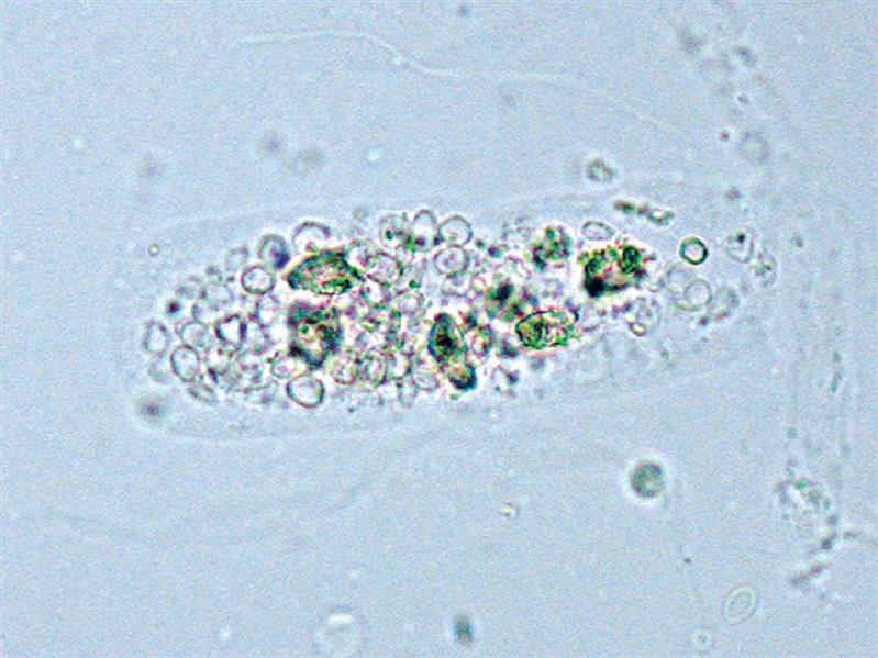
Fig. 22A mixed cell cast, predominantly red blood cells.
Fig. 23A red blood cell cast. Red blood cells are dispersed in the hyaline matrix of this cast.
Fig. 24A red blood cell cast packed with red blood cells.
Fig. 25A fatty cast with fat droplets of varying size within the cast matrix; a fat droplet the size of a red blood cell is most notable mid-cast. Note the deteriorating renal epithelial cell within the cast at its end (lower right). (A) Brightfield. (B) Polarizing microscopy with first-order red compensator. Note that the fat droplet mid-cast demonstrates a Maltese-like cross pattern indicating that it is a cholesterol droplet.
Fig. 26Oval fat bodies in a hyaline matrix (i.e., a fatty cast). (A) Using Sudan III stain, the fat in the oval fat bodies has taken on the characteristic terra-cotta or red-orange color, identifying it as neutral fat (triglycerides). Brightfield. (B) Using phase contrast microscopy, the hyaline matrix of the cast is easy to see.
Granular Casts
Fig. 27Granular cast with several disintegrating renal tubular cells embedded. One cell now appears as a large “coarse” granule that is colored by methemoglobin. Brightfield.
Fig. 28Granular cast. Brightfield.
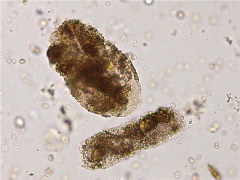
Fig. 29Coarsely granular (and pigmented) cast. The color originates from hemoglobin (now methemoglobin) and the granulation from RBC degeneration. These casts are sometimes called blood casts or “muddy brown” casts. Note the two large coarse granules embedded at the upper end (left) of the cast, which are likely renal tubular cells that have disintegrated and become stained by methemoglobin. Brightfield.
Fig. 30Cast transitioning from cellular to granular to waxy. The intense brown color suggests that pigmentation is derived from hemoglobin (i.e., methemoglobin). This sediment also contains numerous red blood cells and red blood cell casts. Brightfield.
Fig. 31Granular casts. A broad cast indicative of formation in a large collecting duct or in dilated tubules indicates significant renal pathology. The granules most likely originated from red blood cells and coloration from hemoglobin or methemoglobin. Hence, these casts are sometimes referred to as blood casts. Brightfield.
Fig. 32A low-power (100 ×) field of view of urine sediment containing numerous casts: hyaline, granular, red blood cell, and cellular. Brightfield.
Hyaline Casts
Fig. 33A low-power (100 ×) field of view of urine sediment containing numerous hyaline casts. Because their refractive index is similar to that of urine, they can be difficult to observe using brightfield microscopy. Focusing up and down during the microscopic examination aids in the detection of hyaline casts because they are often more apparent when slightly out of focus. Brightfield.
Fig. 34Hyaline cast. Brightfield.
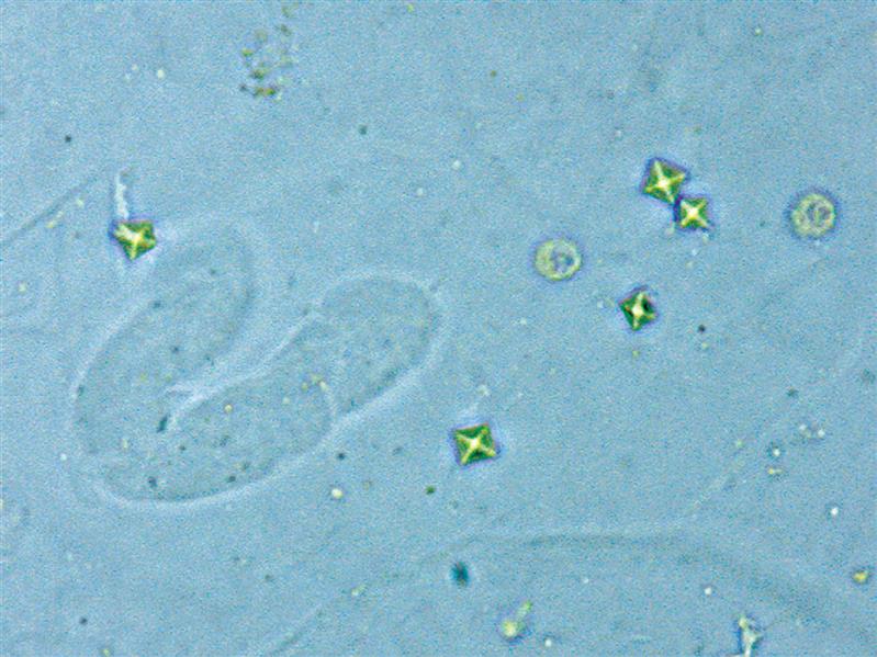
Fig. 35A U-shaped hyaline cast, two white blood cells, and five dihydrate calcium oxalate (weddellite) crystals. Brightfield.
Waxy Casts
Fig. 36A low-power (100 ×) field of view of urine sediment containing numerous casts, particularly hyaline and waxy (three predominate). Brightfield.
Fig. 37A long, broad waxy cast predominates in this field of view. Also present are other waxy and hyaline casts, as well as renal tubular cells and oval fat bodies. Brightfield.
Fig. 38A single “broad” waxy cast and two hyaline casts. Note the difference in refractility between these two types of casts. In this image, the hyaline casts are actually out of focus, which makes them easier to see. Brightfield.
Fig. 39A waxy cast (left) lying almost vertical and two red blood cell casts (right) lying horizontally. Brightfield.
Fig. 40Two waxy casts. One typical in width; one broad and transitioning from granular to waxy. Note the ground-glass appearance and blunt ends, which are characteristic of waxy casts. Brightfield.
Crystals
Ammonium Biurate Crystals
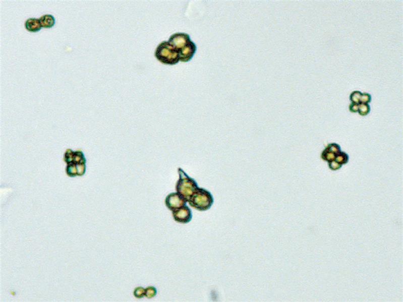
Fig. 41Two ammonium biurate crystals with spicules—“thorny apple” forms. Brightfield.
Fig. 42Ammonium biurate crystals. Note the characteristic yellow to brown color. One crystal (at center) beginning to form a spicule or thorn. Brightfield.
Bilirubin Crystals
Fig. 43Bilirubin crystals—fine needles of characteristic golden yellow color. Bilirubin crystals form in vitro upon refrigeration and storage.
Calcium Carbonate Crystals
Fig. 44Calcium carbonate crystals (arrows) and a single dihydrate calcium oxalate crystal.
Calcium Oxalate Crystals
Fig. 45A single dihydrate calcium oxalate (weddellite) crystal (bi-pyramidal, right of center) and numerous monohydrate calcium oxalate (whewellite) crystals—ovoid and dumbbell (side view of ovoid form) shapes. Brightfield.
Fig. 46(A) A single dihydrate calcium oxalate (weddellite) crystal and numerous monohydrate calcium oxalate (whewellite) crystals that look similar to red blood cells. (B) Same field of view using polarizing microscopy. Rule of thumb: Crystals can polarize light; red blood cells do not.
Fig. 47Calcium oxalate monohydrate (whewellite) crystals, atypical form. (A) Crystals have a yeast-like shape. Brightfield. (B) Polarizing microscopy identifies these structures as crystals, not yeast. Note the stronger birefringence in the “yeast-like” forms compared with the single, ovoid (red blood cell–like) form (at bottom right).
Cholesterol Crystals
Fig. 48Cholesterol crystal (arrow). Brightfield.
Cystine Crystals
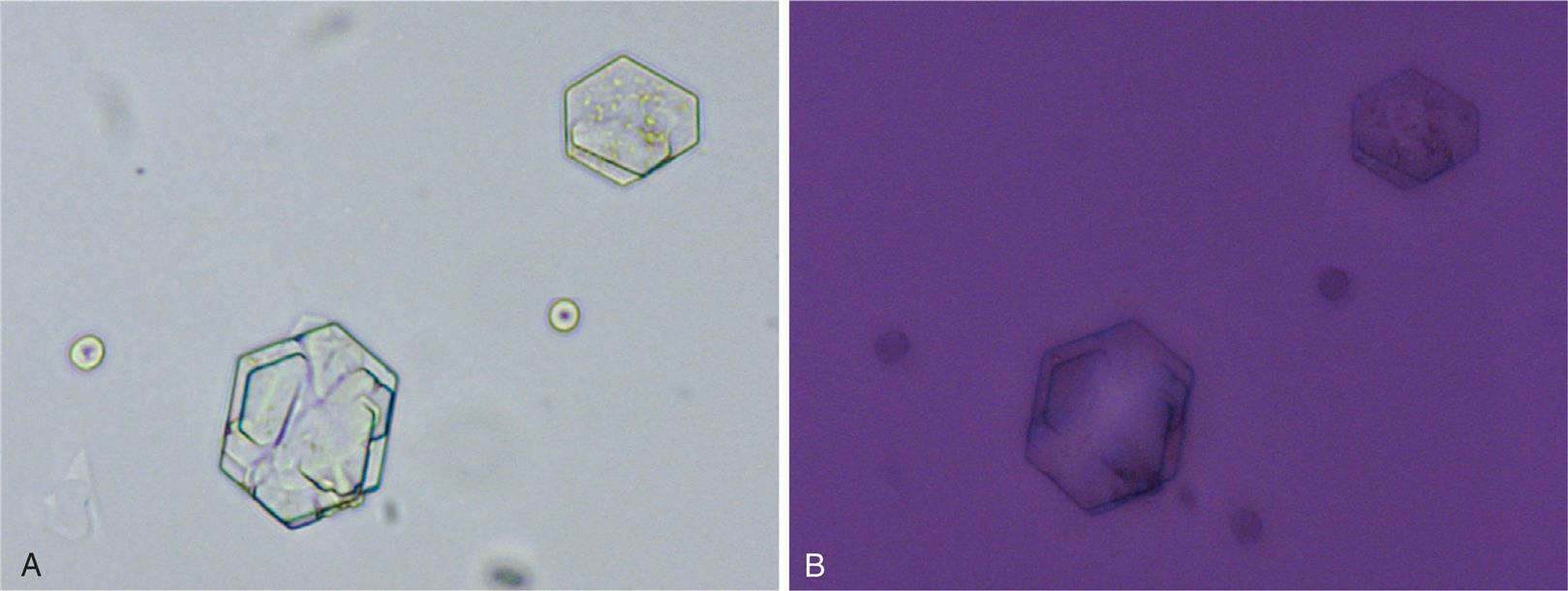
Fig. 49Cystine crystals with layers or lamination. Red blood cells also present. (A) Brightfield. (B) Polarizing microscopy. Note the very weak birefringence of the largest crystal.
Drug Crystals
Fig. 50A “sulfa” crystal, specifically sulfadiazine, surrounded by many yeast cells. Brightfield.
Fig. 51Numerous sulfamethoxazole (Bactrim) crystals surrounding a single barrel-shaped uric acid crystal. Note the yellow-brown color and the similar shape of the many sulfamethoxazole crystals to those of ammonium biurate (Fig. 42). Urine pH aids in differentiating these two crystals. Brightfield.
Fig. 52Indinavir (Crixivan); insoluble at pH >6.0. These crystals can be present as individual needles or prisms, which can aggregate into a variety of bundle forms—wing-like bundles, shocks of wheat, and X-shaped forms. Amorphous phosphates are also present. Brightfield.
Fig. 61Uric acid crystals in the common diamond shape.
Fig. 62Uric acid crystals, barrel or cube forms.
Fig. 63A chunk of a uric acid crystal; note the characteristic color. Also present are a squamous epithelial cell and two dihydrate calcium oxalate crystals.
Fig. 64A single uric acid crystal in an unusual band form and numerous calcium oxalate crystals (mono- and dihydrate forms).
Epithelial Cells
Fig. 65Two squamous epithelial cells covered with bacteria, known as clue cells, and a single typical or “normal” squamous epithelial cell (lower left). In urine that has been contaminated with vaginal secretions, clue cells may be observed. Brightfield.
Fig. 66Three squamous epithelial cells and a single white blood cell. Note the keratohyalin granules evident in the cytoplasm of squamous cells and the similarity in size between the white blood cell and the nuclei of these epithelial cells. Brightfield.
Fig. 67A squamous epithelial cell (lower left cell) and a transitional epithelial cell (upper right cell). Note the similarity in the size and central location of their nuclei. It is the amount of cytoplasm that differs, resulting in different nucleus-to-cytoplasm ratios. Several large rod-shaped bacteria are also present.
Fig. 68A clump of transitional epithelial cells and several individual caudate or club-like transitional epithelial cells. This urine was collected after catheterization and the cells were most likely dislodged during the catheterization process. Note the cytoplasmic blebs forming from some cells of the clump as they degenerate. Phase contrast microscopy.
Fig. 69A fragment of transitional epithelial cells and numerous red blood cells. Note the variation in shape of the transitional epithelial cells—round to caudate.
Fig. 70Transitional epithelial cell or squamous epithelial cell? Reasoning could be used to justify classification into either category. Cells lining the urinary system convert from squamous to transitional (urothelial) epithelium. This cell most likely originated from this area of transition.
Fig. 71A transitional epithelial cell (left) and two typical cuboidal renal tubular (collecting duct) cells (center and right). The center cell is degenerating. Brightfield.
Fig. 72Bilirubin-stained renal tubular epithelial cells. Four cells are free-floating in the urine and two epithelial cell casts—each with three renal tubular cells embedded in their hyaline matrix. Note that renal tubular cells are small; these cells actually measured 11 to 18 μm in diameter. Brightfield.
Fig. 73A single renal tubular cell (arrow) from a large collecting duct. Note the columnar shape and that the size of the nucleus is similar to that of red blood cells, which are also present. Brightfield.
Fat Droplets and Oval Fat Bodies
Fig. 74Several free fat droplets and a fatty cast. Note refractility, variation in size, and greenish hue of the fat droplets. Brightfield.
Fig. 75An oval fat body in the hyaline matrix of a cast. Also present in this field of view are another free-floating oval fat body, a fat droplet, and a hyaline cast. Note the similarity in size and shape of the fat droplet to a red blood cell. Brightfield.
Fig. 76Two oval fat bodies (arrows) loaded with fat, hence their intense refractility. Numerous red blood cells, amorphous material, and debris are also present. Brightfield.
Fig. 77Two oval fat bodies and several renal tubular cells. Brightfield.
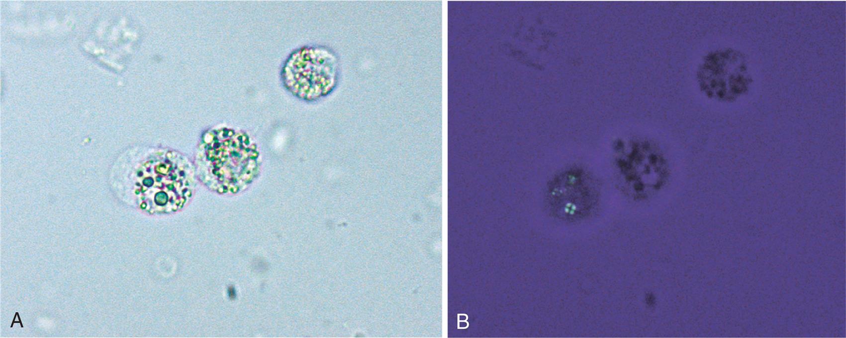
Fig. 78Three oval fat bodies. (A) Note that fat droplets within cells often vary in size, are highly refractile, and may have a greenish sheen (depends on microscope adjustments). Brightfield. (B) Same field as A using polarizing microscopy. Note the characteristic Maltese-style cross of some droplets in the oval fat body on the left, which indicates that these droplets are cholesterol. The non-birefringent fat droplets are neutral fat (triglyceride).
Fig. 79Several oval fat bodies enmeshed within casts and free in the urine sediment. Bacteria and spermatozoa are also present. Brightfield.
Fig. 80An oval fat body engorged with neutral fat (triglycerides) stained using Sudan III. Brightfield.
Microorganisms
Bacteria
Fig. 81Numerous rod-shaped bacteria and a single dihydrate calcium oxalate crystal. Brightfield.
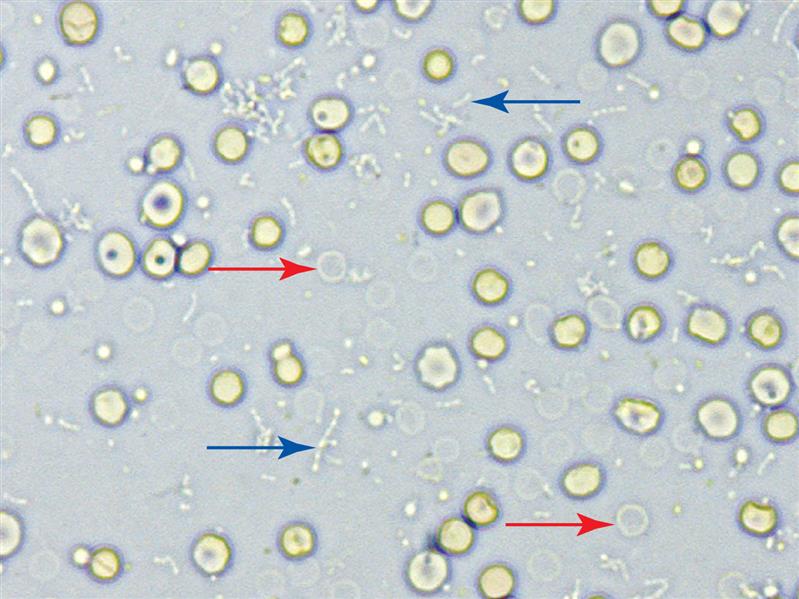
Fig. 82Numerous bacteria, singly and in chains, with several indicated by blue arrows. Many red blood cells, both normal and ghost forms (red arrows), are present. Brightfield.
Trichomonads
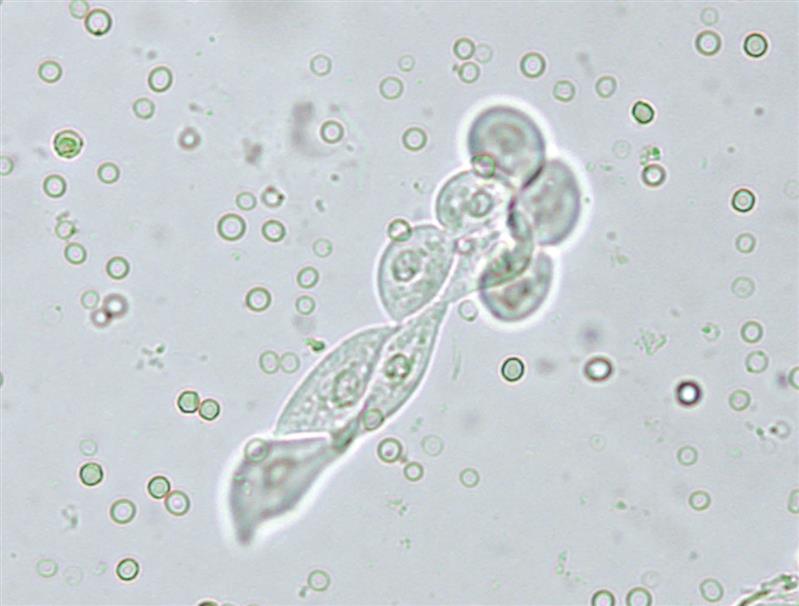
Fig. 83A trichomonad. Their characteristic rapid flitting motion results from their undulating membrane (blue arrow), anterior flagella (two indicated by yellow arrows), and axostyle (red arrow). Because of their size and granular appearance, nonmotile (or dead) trichomonads may be misidentified as white blood cells. Brightfield.
Fig. 84Two trichomonads. Brightfield.
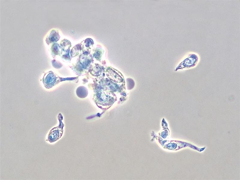
Fig. 85A cluster of four trichomonads. It is common to observe trichomonads clustered together along with white blood cell (WBC) clumps in urine sediment. Brightfield.
Yeast
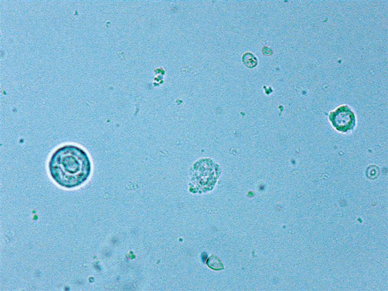
Fig. 86Several budding yeast (blastoconidia), bacteria, and a single ghost red blood cell (arrow). Note the refractility and sheen of the yeast, which is made most evident by focusing up and down during the microscopic examination. Brightfield.
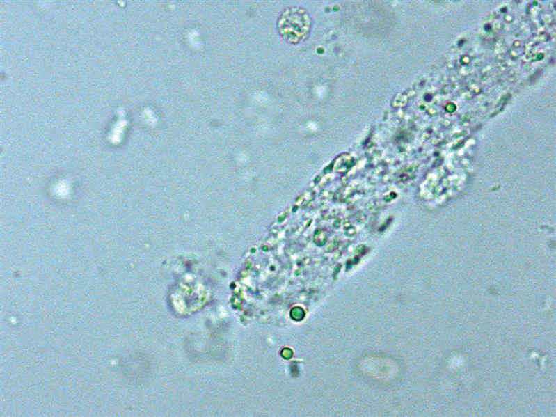
Fig. 87A branch of pseudohyphae (Candida spp.) and a red blood cell demonstrating typical pink-red coloration. Several ovoid yeasts are present in a different focal plane. Brightfield.
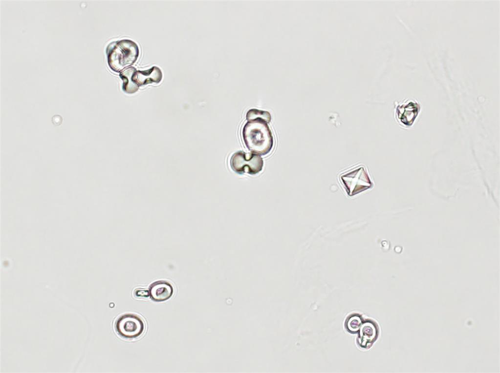
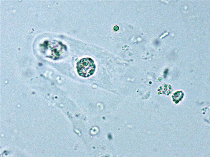
Fig. 88Yeast cells and blastoconidia (budding yeast). These yeast cells appear more round than oval, highlighting the fact that different species of yeast will appear differently. A single dihydrate calcium oxalate crystal is also present. Brightfield.
Fig. 89Early germ tube formation and several yeast cells. A single red blood cell is also present. Brightfield.
Miscellaneous Formed Elements
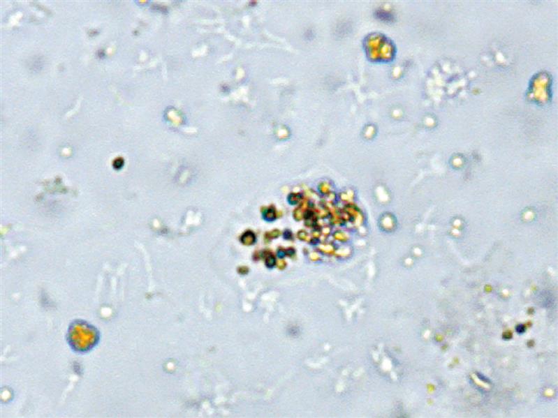
Hemosiderin
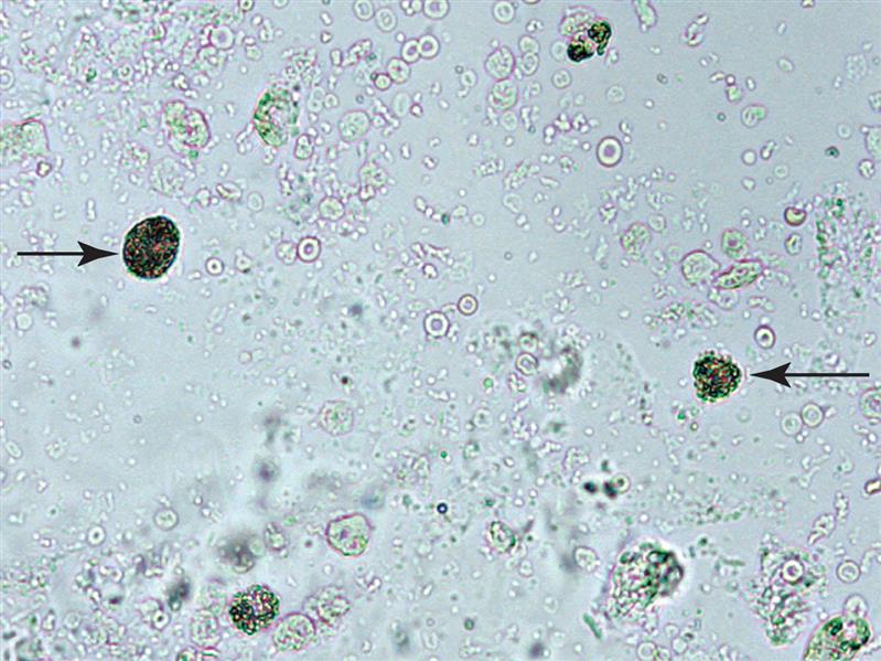
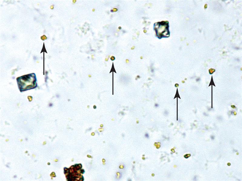
Fig. 90Hemosiderin granules in urine sediment appear yellow-brown. Numerous granules as well as a clump are present in this field of view. Four granules are identified by the arrows. Two dissolving dihydrate calcium oxalate crystals are also present. Brightfield.
Fig. 91Hemosiderin granules in the hyaline matrix of a cast (i.e., a hemosiderin cast). Brightfield.
Mucus
Fig. 92A cluster of mucus threads. Because the refractive index of mucus is similar to that of urine, it can be difficult to observe using brightfield microscopy. Focusing up and down during the microscopic examination aids in the detection of mucus because it is often more apparent when slightly out of focus. A couple of squamous epithelial cells and other elements, on a different focal plane, are also present. Brightfield.
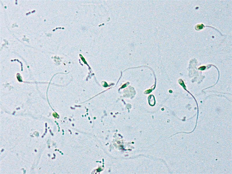
Sperm
Fig. 93A cluster of sperm trapped in mucus. Brightfield.
Fig. 94Sperm and bacteria in urine sediment. Note that several abnormal spermatozoa forms are present. Brightfield.