Seminal Fluid Analysis
After studying this chapter, the student should be able to:
- 1. Discuss the composition of seminal fluid and briefly describe the function of each of the following structures in seminal fluid formation:
- 2. Outline the maturation of sperm (spermatozoa) and identify the morphologic structures in which each maturation phase occurs.
- 3. Summarize the collection of seminal fluid for analysis, including the importance of timing and recovery of the complete specimen.
- 4. Describe the performance of the physical examination (appearance, volume, and viscosity) of seminal fluid and the results expected from a normal specimen.
- 5. Describe the procedures used to evaluate the following characteristics of sperm in seminal fluid, state the normal range for each parameter, and relate each function to male fertility:
- 6. Identify and describe the morphologic appearance of normal and abnormal forms of spermatozoa.
- 7. Discuss the origin and clinical significance of cells other than sperm in the seminal fluid.
- 8. Discuss briefly the role of quantifying the following biochemical substances in seminal fluid and identify the structure evaluated by each substance:
Key Terms1⁎ *
Seminal fluid, or semen, is a complex body fluid used to transport sperm or spermatozoa. It is analyzed routinely to evaluate infertility and to follow up after a vasectomy to ensure its effectiveness. Other reasons for analysis include the evaluation of semen quality for donation purposes and forensic applications (e.g., DNA analysis, detection of semen). Familiarity with the male reproductive tract and its various functions facilitates understanding of the physical, microscopic, and biochemical abnormalities that can occur in semen.
Physiology
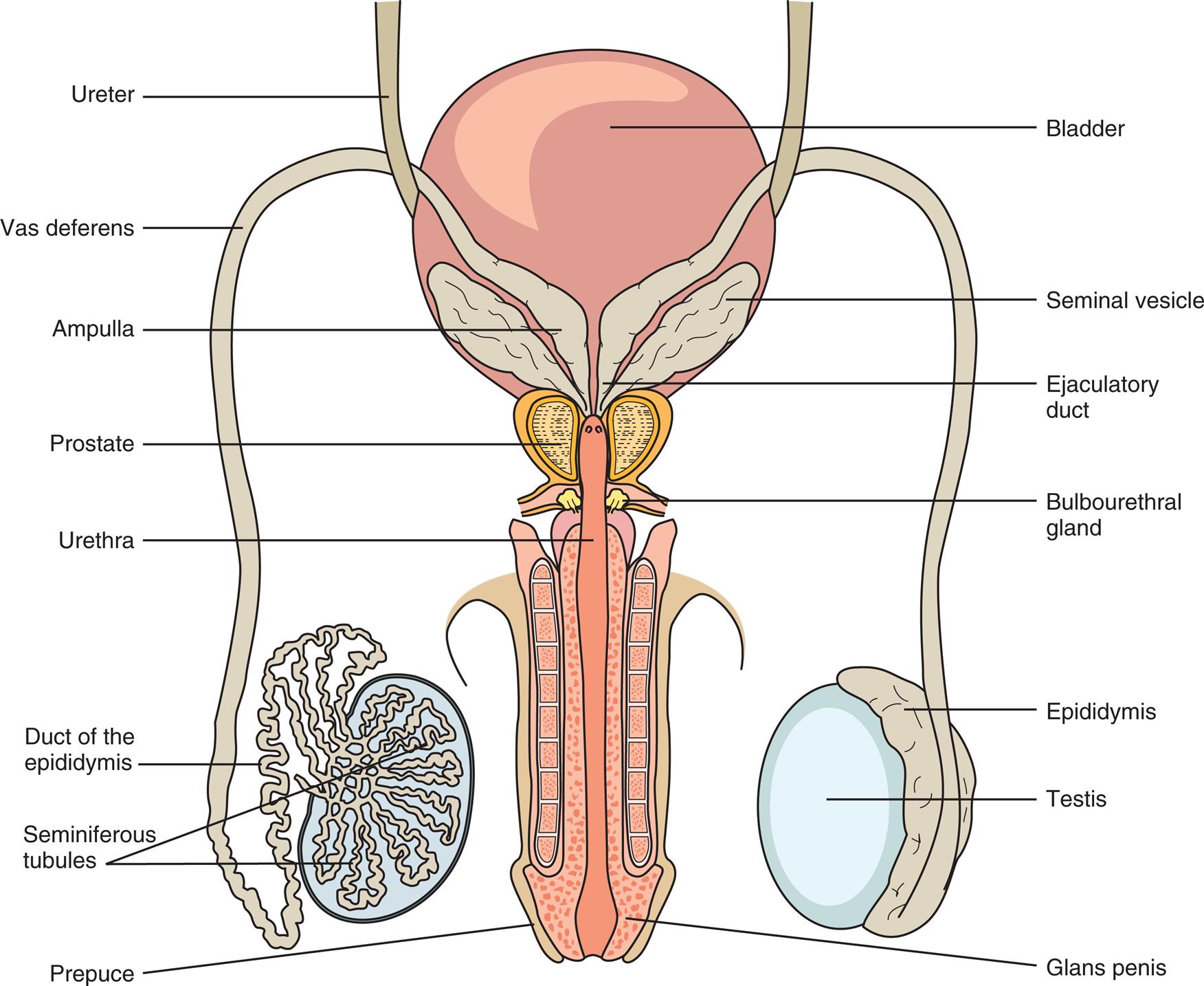
Semen is composed primarily of secretions from the testes, epididymis, seminal vesicles, and prostate gland, with a small amount derived from the bulbourethral glands. The biochemical composition of semen is complex. Although the specific functions of some components (e.g., fructose) are known, the functions of others (e.g., prostaglandins) remain uncertain. The testes are paired glands suspended in the scrotum and located outside the body (Fig. 12.1). Their external location allows for the lower organ temperature necessary for sperm formation.
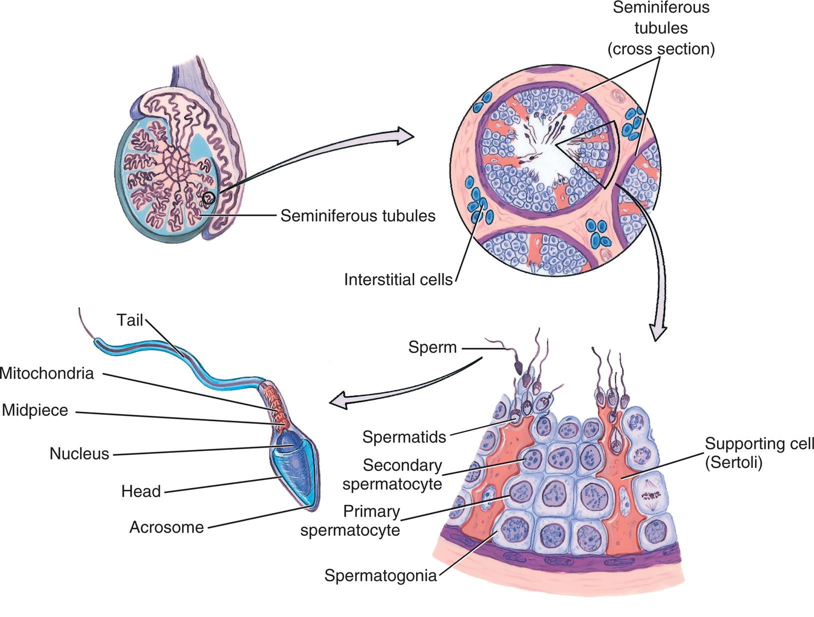
The testes perform both an exocrine function by the secretion of sperm and an endocrine function by the secretion of testosterone. These two functions are interdependent and are regulated by two pituitary hormones: follicle-stimulating hormone and luteinizing hormone. The cells responsible for these two functions are distinctly different. Sperm production is regulated by Sertoli cells in the seminiferous tubules, whereas production and secretion of the male sex hormone, testosterone, is the responsibility of the interstitial cells of Leydig, which are located in the interstitium of the testes, between the seminiferous tubules.
Sertoli cells of the seminiferous tubular epithelium have several functions. Because of their tight interconnections, they essentially form a barrier that separates the epithelium into two distinct compartments: the basal compartment (i.e., germ cell layer) and the adluminal compartment (i.e., epithelium nearest the tubular lumen). As this barrier, or gatekeeper, they limit the movement of chemical substances from the blood into the tubular lumen—playing a role in supplying nutrients, hormones, and other substances necessary for normal spermatogenesis. They also control the movement of spermatocytes from the germ cell layer into the adluminal compartment. Last, they continuously produce a fluid that carries the newly produced immotile sperm into the lumen of the seminiferous tubules and on to the epididymis.
The epithelium of the numerous coiled seminiferous tubules consists of Sertoli and germ cells. The undifferentiated germ cells (spermatogonia) continuously undergo mitotic division to produce more germ cells. At the same time, some of them move slowly toward the tubular lumen, changing in size and undergoing meiotic (reduction) division until they form spermatids. Fig. 12.2 depicts spermatogenesis in the seminiferous tubular epithelium with all stages of spermatogenesis depicted. Spermatogonia (germ cells) evolve into spermatocytes and then spermatids. With nuclear modification and cellular restructuring, spermatids ultimately differentiate into immotile sperm.
When Sertoli cells release sperm into the lumen of the seminiferous tubules, they are nonmotile and still immature. Luminal fluid from Sertoli cells carries the sperm into the tubular network of the epididymis, where they undergo final maturation and become motile. The epididymis also adds carnitine and acetylcarnitine to the lumen fluid. Although the exact function of these chemicals remains to be elucidated, abnormal levels of them have been associated with infertility. Other functions of the epididymis include the concentration of sperm by the absorption of lumen fluid and their storage until ejaculation. After a vasectomy, the epididymis is the site of leukocyte infiltration and phagocytization of accumulated sperm.
The epididymis ultimately forms a single duct that joins the vas deferens. The vas deferens is a thick-walled muscular tube that transports sperm from the epididymis to the ejaculatory duct, and the dilated end of the vas deferens is located inferior to the bladder. Secretions from the seminal vesicles are added at the ejaculatory duct. Both ejaculatory ducts then pass through the prostate gland and empty into the prostatic urethra along with secretions from the prostate. All structures preceding the prostate gland are paired (e.g., two ejaculatory ducts, two seminal vesicles, two testes).
The seminal vesicles and the prostate gland are considered accessory glands of the male reproductive system and are testosterone dependent. They produce and store fluids that provide the principal transport medium for sperm. Seminal vesicle fluid accounts for approximately 70% of the ejaculate and is high in flavin. Flavin imparts the characteristic gray or opalescent appearance to semen and is responsible for its green-white fluorescence under ultraviolet light.1 Another characteristic of seminal vesicle fluid is its high concentration of fructose, believed to serve as a nutrient for spermatozoa. The various proteins secreted by the seminal vesicles play a role in coagulation of the ejaculate, whereas the function of prostaglandins remains under investigation. (Prostaglandins were originally thought to be a prostatic gland secretion, hence their misnaming.)
Prostatic fluid secretions account for approximately 25% of the ejaculate volume. The principal components of this milky, slightly acidic fluid are citric acid; enzymes, particularly acid phosphatase and proteolytic enzymes; proteins; and zinc. Semen is unique in its high concentration of the enzyme acid phosphatase, hence acid phosphatase activity can be used to positively identify the presence of this body fluid. Proteins and some enzymes in prostatic secretions play a role in coagulation of the ejaculate, whereas the proteolytic enzymes are responsible for its liquefaction. Zinc is primarily added to semen by the prostate gland; however, the testes and sperm also contribute zinc. Semen zinc levels can be used to evaluate prostate function; a decreased level is associated with prostate gland disorders.
In summary, semen is a highly complex transport medium for sperm. The paired seminal vesicles and the single prostate gland are the major fluid contributors to semen. Sperm produced by the testes are matured and concentrated in the epididymis and make up only a small percentage of an ejaculate. Dilution of sperm by the relatively large volume of seminal fluid at ejaculation enhances sperm motility. Without adequate dilution, sperm motility is significantly reduced. The entire process of spermatogenesis and maturation (i.e., from primary spermatocyte to mature motile spermatozoon) takes approximately 90 days.
Specimen Collection
Because sperm concentration in normal seminal fluid can vary significantly, two or more samples should be analyzed to evaluate male fertility. Specimen collections should take place within a 3-month period and at least 7 days apart. Sexual abstinence for at least 2 days (48 hours), but not exceeding 7 days, should precede the collection. The patient collects the specimen through masturbation, and the entire ejaculate is collected in a clean, wide-mouth sterile plastic or glass container. Although some plastic containers are toxic to spermatozoa, others are not. Sterile urine specimen or similar containers are often satisfactory, but the laboratory must evaluate them before their use.2 The collection container should be kept at room temperature or warmed (to approximately body temperature) before the collection to avoid the possibility of cold shock to the sperm. The container can be warmed easily by holding it next to the patient’s body or under the arm for several minutes before the collection. This technique can also be used to control the temperature of specimens being transported in cold climates. Specimen containers and request forms must be labeled with the patient’s name, the period of sexual abstinence, and the date and time of specimen collection. The time of actual specimen collection is crucial in evaluating liquefaction and sperm motility.
During specimen collection, lubricants and ordinary condoms should not be used because they have spermicidal properties. For patients unable to collect a specimen through masturbation, special nonspermicidal (e.g., Silastic) condoms can be provided for specimen collection.
The collection of seminal fluid requires sensitivity and professionalism. Written and verbal instructions should be provided to the patient, as well as a comfortable and private room near the laboratory. If the specimen is to be collected elsewhere and delivered to the laboratory, clearly written instructions regarding specimen transport conditions must be provided. Specimens must be received in the laboratory within 1 hour after the collection, and they must be protected from extreme temperatures, that is, maintained at 20°C to 40°C.3 If these criteria are not met, the specimen will not be satisfactory for sperm function tests and an abnormally low sperm motility can result. Because the ejaculate differs in its composition, only complete collections are acceptable for analysis. Patient instructions must state this clearly, and patients should be asked whether any portion of the specimen was not collected. When a portion of the initial ejaculate is not collected, the sperm concentration will be falsely decreased and, owing to a reduction in prostate secretions, the pH is falsely increased and the coagulum will fail to liquefy. Conversely, when the last portion of an ejaculate is missing (primarily seminal vesicle fluid), the semen volume will be decreased, the sperm concentration falsely increased, the pH falsely decreased, and a coagulum will not form.
As with all body fluids, seminal fluid represents a potential biohazard and must be handled accordingly. Because seminal fluid can contain infectious agents such as hepatitis virus, human immunodeficiency virus, herpes virus, and others, all personnel must adhere to standard precautions (see Chapter 1) when handling these specimens.
Physical Examination
Appearance
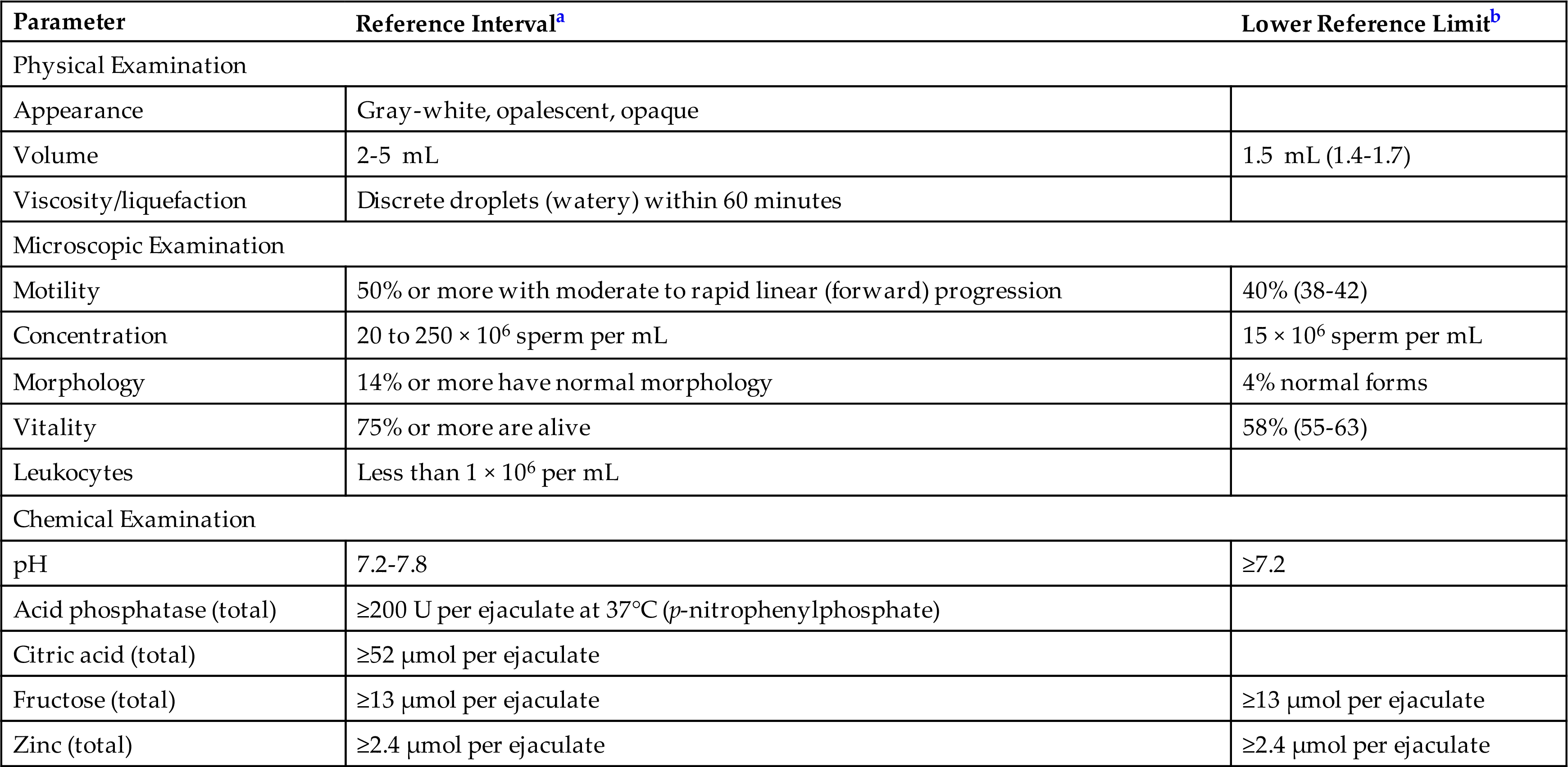
Normal semen is gray-white and opalescent in appearance. A brown or red hue may indicate the presence of blood, whereas yellow coloration has been associated with certain drugs. If large numbers of leukocytes are present, the semen appears more turbid with less translucence. When the specimen appears almost clear, the sperm concentration is usually low. Mucus clumps or strands can be present. Semen has a distinctive odor that is sometimes described as musty. Although infections in the male reproductive tract can modify this odor, a change is rarely noted or reported. Table 12.1 (and Appendix C) summarizes the semen characteristics (physical, microscopic, and chemical parameters) associated with fertility.
Table 12.1
| Parameter | Reference Intervala | Lower Reference Limitb |
|---|---|---|
| Physical Examination | ||
| Appearance | Gray-white, opalescent, opaque | |
| Volume | 2-5 mL | 1.5 mL (1.4-1.7) |
| Viscosity/liquefaction | Discrete droplets (watery) within 60 minutes | |
| Microscopic Examination | ||
| Motility | 50% or more with moderate to rapid linear (forward) progression | 40% (38-42) |
| Concentration | 20 to 250 × 106 sperm per mL | 15 × 106 sperm per mL |
| Morphology | 14% or more have normal morphology | 4% normal forms |
| Vitality | 75% or more are alive | 58% (55-63) |
| Leukocytes | Less than 1 × 10⁶ per mL | |
| Chemical Examination | ||
| pH | 7.2-7.8 | ≥7.2 |
| Acid phosphatase (total) | ≥200 U per ejaculate at 37°C (p-nitrophenylphosphate) | |
| Citric acid (total) | ≥52 μmol per ejaculate | |
| Fructose (total) | ≥13 μmol per ejaculate | ≥13 μmol per ejaculate |
| Zinc (total) | ≥2.4 μmol per ejaculate | ≥2.4 μmol per ejaculate |
aBased on the strict criteria evaluation recommended by the World Health Organization WHO (1999) for assessing sperm morphology for fertility purposes.
bThe one-sided 5th centile lower reference limit recommended by the WHO (2010) for assessing semen characteristics.
Semen is a homogeneous, viscous fluid that immediately coagulates after ejaculation to form a coagulum. Within 30 minutes, the coagulum liquefies (becomes watery). The actual time of specimen collection must be known to evaluate liquefaction. Although liquefaction can take longer, any delay beyond 60 minutes is considered abnormal and must be noted. Because complete liquefaction is necessary to perform analysis, semen specimens that do not liquefy completely can be chemically treated (see Appendix D, “Semen Pretreatment Solution”). After normal liquefaction, undissolved gel-like granules or particles can be present in the specimen, with a small amount considered normal.
Volume
The physical and microscopic analyses of seminal fluid should take place immediately after liquefaction or within 1 hour after collection (for specimens collected away from the laboratory). Specimen volume is measured to one decimal place (0.1 mL) using a sterile serologic pipette (5.0 mL or 10.0 mL). If a semen culture for bacteria is requested, the volume measurement should be performed first using sterile technique. Normally, a complete ejaculate collection recovers 2 to 5 mL of seminal fluid. Volumes less than and greater than this range are considered abnormal and have been associated with infertility.
Viscosity
After complete liquefaction, the viscosity of the semen is evaluated using a Pasteur pipette and observing the droplets that form when the fluid is allowed to fall by gravity. A normal specimen is watery and forms into discrete droplets. Abnormal viscosity or fluid thickness is indicated by the formation of a string or thread greater than 2 cm in length.3 A high content of mucus can increase the viscosity. Other conditions associated with increased viscosity include the production of antisperm antibodies and oligoasthenospermia (i.e., decreased concentration and motility of sperm).4–7
Grading viscosity varies among laboratories. Numeric terms can be used, with 0 indicating a normal, watery (i.e., forms discrete drops) specimen and 4 indicating a specimen with gel-like consistency.8 An alternate reporting format uses descriptive terms, such as normal, slightly viscous (thick), moderately viscous, and extremely viscous (unable to be aspirated into the pipette).
Microscopic Examination
As in other laboratory areas, standardization of procedures and techniques is necessary to enhance the precision and reproducibility of semen analysis. Once achieved, this standardization enables intralaboratory and interlaboratory comparisons of data. Appropriate quality control measures must also be in place whenever applicable. The World Health Organization (WHO) publication WHO Laboratory Manual for the Examination and Processing of Human Semen is an excellent and necessary reference for any laboratory performing semen analysis.3 Microscopic examination includes the determination of sperm motility, concentration, morphology, and viability; the concentration of other cells present; and the presence of sperm agglutination. Some laboratories use a single stain for the evaluation of several parameters, such as eosin-nigrosin stain for sperm vitality, morphology, and the identification of other cells, whereas others use different stains that specifically enhance each parameter to aid in the identification and evaluation of sperm and other cells.
Motility
Motility is one of the most important characteristics of sperm because immotile sperm, even in high concentrations, are unable to reach and fertilize an ovum. Traditionally, the evaluation of sperm motility has been assessed subjectively by experienced technologists. Today, computerized systems that use electro-optical techniques or videography have been developed for semen evaluation. This advanced technology enables objective evaluation of sperm motility and morphology; however, the cost of the equipment precludes many laboratories from acquiring it.
Without an automated system, sperm motility is evaluated subjectively and semiquantitatively using phase-contrast microscopy (brightfield microscopy can also be used with appropriate condenser adjustments). After complete liquefaction, the semen sample is mixed well to ensure homogeneity. A consistent volume of each specimen is evaluated by pipetting a fixed volume (e.g., 10 or 20 μL) of semen onto a microscope slide using a calibrated positive-displacement pipette. The sample is covered with a coverslip of predetermined size (e.g., 18 × 18 mm), and the slide is allowed to settle for about 1 minute before evaluation. To enhance the accuracy and precision of results, wet mounts of each sample should be prepared and evaluated in duplicate.
Because sperm motility is affected adversely by temperature, some laboratories control the temperature of the microscope slide at 37°C using an air curtain incubator.8 Others perform the analysis at room temperature (i.e., 22 ± 2°C).
Initially, each wet mount is screened to ensure uniformity in sperm movement throughout the preparation. Next, sperm motility is graded subjectively from 0 to 4 under ×200 (or ×400) magnification. Table 12.2 shows typical grading criteria used to evaluate sperm motility. Some laboratories use a manual cell counter and evaluate the motility characteristics in 100 sperm, whereas others grade the sperm encountered in 6 to 10 high-power fields (×400).
Table 12.2
The speed and forward progression of each sperm are evaluated. In normal semen evaluated within 60 minutes of collection, 50% or more of the sperm will show moderate to strong linear or forward progression. The practice in some laboratories of reassessing sperm motility at additional time intervals serves no purpose and has no clinical significance. Physiologically or in vivo, sperm leave the seminal fluid within minutes after ejaculation and enter the cervical mucus. Therefore motility on a microscope slide at later time intervals is irrelevant.
Concentration and Sperm Count
For fertility purposes, the actual number of sperm is not as important as other characteristics. This fact is supported by studies of fertile men despite low sperm counts (less than 1 million per mL).9 The concentration of sperm in an ejaculate is considered normal when 20 million to 250 million per mL of sperm are present; values less than or greater than this range are considered abnormal and are associated with infertility. The variation in the sperm concentration within a single individual can be significant and depends partially on the period of sexual abstinence but can also be affected by viral infection and stress. For these reasons, multiple semen specimens should be evaluated to reliably assess the quantity and quality of an individual’s sperm.
Manually, the concentration of sperm is determined by using a hemacytometer after preparing an appropriate dilution of the semen. Frequently, a 1:20 dilution is prepared. If during initial microscopic examination the sperm concentration is noted to be exceptionally high or low, a new dilution can be prepared and mounted. All dilutions should be made using a calibrated positive-displacement pipette to deliver the semen quantitatively to a premeasured amount of diluent (see Appendix D for diluents). Note that a hematology white blood cell (WBC) pipette is not accurate for use with seminal fluid because of its viscosity and should not be used.3 After the hemacytometer is filled with the well-mixed dilution of semen, it is placed in a humidifying chamber and allowed to settle for 3 to 5 minutes before counting. The type of hemacytometer, the specimen dilution used, and the areas counted determine the conversion factor necessary to obtain the concentration of sperm in millions per mL (see Chapter 17 for procedural details).
Several alternative manual counting methods have been developed, such as the Makler chamber (Sefi Medical Instruments, Ltd., Haifa, Israel), Cell VU chambers (Fertility Technology Inc., Murphy, NC), Horwell (Horwell Ltd., London, UK), Microcell slides (Vitrolife Inc., San Diego, CA), and Leja counting chambers (Leja Products B.V., The Netherlands). Studies vary in their outcomes—some supporting the manual hemacytometer method as the method of choice for sperm counting, with other studies finding better accuracy and precision using an alternative counting chamber.4,5,10 Regardless of the method used, the dilution of the semen is always a potential source for error and requires the utmost attention to ensure an accurate and reproducible technique. The counting of motile sperm and high sperm concentrations have also been identified as two sources of error. Therefore the WHO states that the “validity of these alternative counting chambers must be established by checking chamber dimensions, comparing results with the improved Neubauer haemocytometer method, and obtaining satisfactory performance as shown by an external quality control program.”3
In contrast to sperm concentration (sperm per milliliter), the sperm count is the total number of sperm present in the entire ejaculate. This value, often requested by clinicians, is calculated by multiplying the sperm concentration (sperm/mL) by the total volume of the ejaculate.
 Equation 12.1
Postvasectomy Sperm Counts
After a vasectomy, the sperm count in semen ideally should be zero—no sperm present (azoospermia)—within 12 weeks after the procedure. However, studies have shown that nonmotile sperm can be present for as long as 21 months post vasectomy regardless of the number of ejaculations. It is postulated that the persistence or reappearance of nonmotile sperm in semen collections results from the release of nonviable residual sperm in the seminal vesicles and the abdominal portion of the vas deferens. Studies have further demonstrated that despite the presence of low numbers (<1 × 10⁶) of nonmotile sperm post vasectomy, these individuals have a very low risk of causing pregnancy (i.e., comparable with azoospermic men).11
In clinical practice, most men (≈66%) demonstrate azoospermia within 12 weeks, regardless of the number of ejaculations. Note that the most important feature is not the number of sperm present post vasectomy but the status of their motility. The presence of even a single “motile” spermatozoon is evidence of an unsuccessful vasectomy (i.e., recanalization of the vas deferens has occurred), whereas low numbers of “immotile” sperm can persist for months in some men (≈33%).11
Morphology
Sperm morphology, like motility, is routinely assessed subjectively, hence this qualitative determination is subject to intralaboratory and interlaboratory variations. To minimize these variations, standardized procedures and grading criteria must be established by each laboratory and adhered to by all laboratorians. Because the technical ability to identify and classify various morphologic forms requires experience, new staff members must be trained appropriately and their initial work reviewed to ensure accuracy and consistency in reporting. Sperm morphology is complicated by the wide variation in abnormal forms that can be encountered, and an inexperienced observer can easily miss subtle abnormalities in sperm. The computerized systems used to assess sperm motility can also evaluate sperm morphology.
Sperm morphometry—measurement of the sperm head length, width, circumference, and area—enables the generation of objective data. To be considered normal, sperm must meet strict criteria regarding their size and shape, which can be determined by computerized systems or manually using a microscope with a calibrated ocular micrometer.
Human sperm have three distinct areas: head, midpiece, and tail. When viewed from the side, sperm appear to be arrowhead-shaped (Fig. 12.3). When viewed from the top, normal human sperm have oval heads that are 2.5 to 3.5 μm in width and 4.0 to 5.0 μm in length. Only sperm lying flat should be evaluated and their head length-to-width ratio should be 1.5 to 1.75. Spermatozoa with values outside these ranges are considered abnormal, and studies have shown statistically significant differences in the head length-to-width ratios of sperm from ejaculates of fertile and infertile men.6
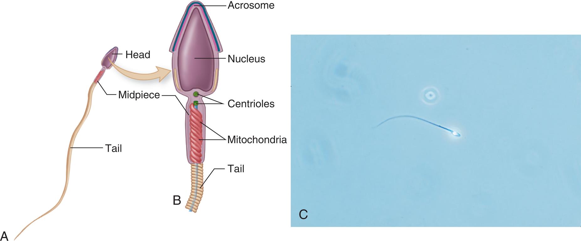
The midpiece, located immediately behind the head, is 6 to 7.5 μm long and is thicker than the tail but not greater than 1 μm in width. The tail should be slender, uncoiled, and at least 45 μm long. When a “basic” morphology evaluation is performed, each spermatozoon (single sperm cell) is identified simply as normal or abnormal with the percent of normal forms reported. If a “complete” morphology evaluation is performed, then each spermatozoon is classified using five categories: normal, head defects, midpiece defects, tail defects, and cytoplasmic droplet present. Cytoplasmic droplets are usually located in the midpiece region and are considered abnormal if this region is greater than one-third the area of a normal sperm head. The head can contain vacuoles, but they are not considered abnormal unless they occupy more than 20% of the head. Note that a single spermatozoon can have multiple defects, and each defect is documented. Figure 12.4 depicts a normal spermatozoon and a variety of abnormal forms.
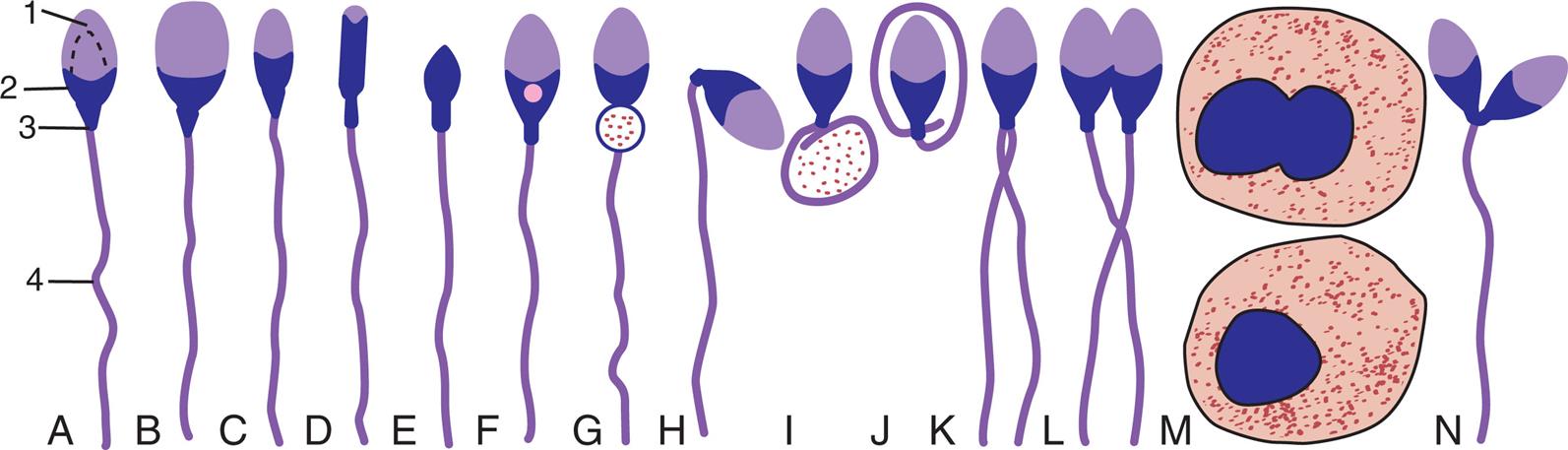
To manually evaluate sperm morphology, smears of fresh semen are made, air dried, and stained. The smears can be made similar to those for traditional blood smears by placing a drop (10–15 μL) of semen near one end of a clean microscope slide. Using the edge of another slide, the drop is allowed to spread along the edge of the second slide, and then the edge of the second slide is moved forward, dragging the semen sample across the surface of the first slide and producing a smear. An alternate technique involves placing the second slide over the first and allowing the semen to spread between them. Once spreading is complete, the slides are pulled apart and allowed to air dry. Staining enhances the visualization of sperm morphology and enables the identification and differentiation of WBCs, epithelial cells of the urethra, and immature spermatogenic cells (i.e., spermatids, spermatocytes, and spermatogonia). Giemsa, Wright’s, and Papanicolaou stains are frequently used. These stains differ with respect to complexity and turnaround time, hence laboratories select the stain that best suits their needs and resources.
Using oil immersion (×1000) and an area of the slide where sperm are evenly distributed, 200 sperm are classified. Note that morphologically abnormal sperm are found in all semen specimens. Abnormalities may involve all or only one region of the spermatozoon and can affect its size or shape, or both. In addition, numerous sperm variations are found within a single ejaculate. Although some morphologic abnormalities have been associated with particular disorders (e.g., tapered heads with varicocele), most abnormalities are nonspecific.
The reference range associated with normalcy varies with the criteria and the rigor used to evaluate sperm morphology. In some laboratories, a normal sperm morphology of 50% or greater is considered “normal.” However, when strict evaluation criteria are used for fertility purposes as in studies of fertile and subfertile individuals, the number of sperm with normal morphology is significantly lower. In these studies, normal sperm morphology of less than 5% is a strong predictor of infertility, whereas fertility is associated with normal sperm morphology values of 12% to 15% or greater.12 Between fertile and subfertile individuals, wide overlap exists in the percentage of sperm with normal morphology. Other variables, particularly sperm concentration and progressive motility, combined with sperm morphology provide the greatest predictive value in assessing male fertility.
Automated Semen Analysis Systems
Several automated systems for the analysis of semen are available, and studies comparing their performance with the conventional manual method have demonstrated acceptable agreement.13,14 Two systems are the SQA-V GOLD analyzer (Medical Electronic Systems Ltd., Caesarea Industrial Park, Israel) and the CEROS computer-aided sperm analysis (CASA) systems (Hamilton Thorne, Beverly, MA). These analyzers measure sperm concentration, total sperm number (sperm count), total motility, progressive motility, nonprogressive motility, normal morphology, motile sperm concentration, and progressively motile sperm concentration. The SQA-V analyzer is a fully automated system that determines semen parameters using electro-optical signals generated by moving spermatozoa and proprietary computer algorithms, as well as spectrophotometry. In contrast, CASA systems require operator interaction to determine a variety of settings, and these systems are image-based—rapid, successive frames of microscopic images are captured and processed by proprietary computer software to detect motile and immotile spermatozoa.
Another difference in automated systems is the amount of semen sample required for analysis. The SQA-V requires 0.5 mL, whereas CASA systems use volumes of 10 to 50 μL. It has been suggested that the larger volume used by the SQA-V is most likely responsible for its improved precision compared with the CASA systems.13
Significant amounts of debris or a high WBC concentration in the semen sample can potentially interfere with accurate analysis by automated analyzers. However, if these features are determined to be present before analysis—by visualizing the sample microscopically—analyzer adjustments can be made to compensate. Despite these issues and the inability to assess abnormal sperm morphology compared with manual methods, automated semen analyzers provide standardization, faster turnaround times, better precision, and reduced potential for human error, as well as automated data recording.
Vitality
Vital staining of a fresh semen smear enables rapid differentiation of live and dead sperm. Because dead sperm have damaged plasma membranes, these cells take up stain; living sperm do not (Fig. 12.5). When a large percentage of immotile sperm are observed, this evaluation determines whether the sperm are immotile because they are dead or because of a structural abnormality (e.g., defective tail).
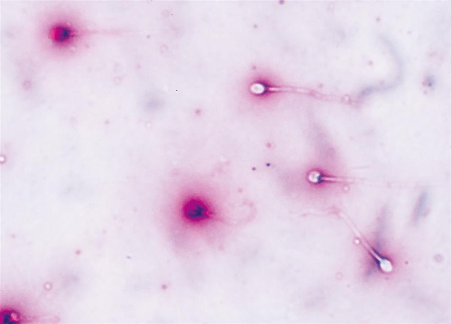
Eosin alone or an eosin-nigrosin (a modification of Blom’s technique) combination is frequently used to determine sperm vitality. Using brightfield or phase-contrast microscopy and ×1000 (or ×400) magnification, 100 sperm on a stained smear are evaluated. The percentage of dead sperm cells should not exceed the percentage of immotile sperm. In other words, if 65% of the sperm in a semen specimen are dead, the motility cannot exceed 35%. Hence the vitality evaluation provides a convenient quality or cross-check of the motility evaluation. In fresh normal semen, 50% or more of the sperm are alive.
Cells Other Than Spermatozoa
An ejaculate is a complex mixture biochemically and cellularly. Ejaculates normally contain cells other than sperm, such as urethral epithelial cells, WBCs, and immature spermatogenic cells (i.e., spermatids, spermatocytes, and spermatogonia), as well as particulate matter and cellular debris. The spermatogenic cells can be difficult to differentiate from WBCs because of size and nuclear pattern similarities. A peroxidase stain can aid in this evaluation because neutrophils are peroxidase-positive cells, whereas lymphocytes and spermatogenic cells are peroxidase-negative cells. However, owing to the carcinogenicity of the chemicals used in many peroxidase stains and the special handling required, Wright’s stain may be preferred.
The presence of greater than 1 million WBCs per milliliter of ejaculate indicates an inflammatory process, most often involving the male accessory glands (e.g., seminal vesicle, prostate). However, a normal WBC count does not rule out infection. Note that the concentrations of WBC and spermatogenic cells can be determined after the sperm count using the same hemacytometer preparation (see Chapter 17). When the concentrations of these cells exceed 1 million per milliliter, a stained smear (e.g., Wright’s stain, peroxidase stain) of the fresh ejaculate is evaluated. Using this smear, the numbers of WBCs and immature spermatogenic cells are counted in the same fields used to count 100 mature sperm. With the sperm count (S) and by using the following equation, the concentration (C) of these cell types (N) is determined (Eq. 12.2)3:
 Equation 12.2
Immature spermatogenic cells are present in the semen when they are exfoliated prematurely from the germinal epithelium of the seminiferous tubules. Distinguishing between an increase in WBCs and an increase in immature spermatogenic cells is necessary to evaluate infection and infertility.
Red blood cells normally are not present in seminal fluid. If their presence is apparent during various aspects of the microscopic evaluation, it should be reported. Similarly, the finding of bacteria in semen should be reported. Bacteria do not normally reside in the male reproductive tract. However, collection of semen by masturbation makes bacterial contamination difficult to avoid.
Agglutination
Agglutination, the sticking together of motile sperm, is evident by microscopic examination of a wet preparation. Although some clumping of immotile sperm may occur in normal semen specimens, the observation of a distinct head-to-head, head-to-tail, or tail-to-tail orientation of sperm is associated with the presence of sperm-agglutinating antibodies. Clumping of sperm with other entities, such as mucus and other cell types, is not identified as agglutination. The extent of true agglutination is often graded as “few,” “moderate,” or “many.” Even a small amount of true agglutination is significant and indicates the need for further evaluation.
Immunoglobulin G and immunoglobulin A antibodies bound to sperm have been identified and correlated with reduced fertility. This is known as immunologic infertility where the man or the woman produce antisperm antibodies and the individual producing them can be identified. When the man is the source, the antibodies are present on the surface of the sperm before intercourse; when the woman is producing them, the sperm are coated with antibodies after they enter the cervical mucus.
Macroscopic and microscopic tests are available to detect and determine the immunoglobulin class of sperm antibodies ([Ig]G, IgA).3 Both tests produce comparable results, but the mixed agglutination reaction (MAR) test is rapid (≈3 minutes) and easy to perform, whereas the immunobead test is time-consuming (≈45 minutes), technically more complicated, and more expensive. The cutoff values for these tests vary among laboratories. The WHO defines agglutination as clinically significant (abnormal) when antisperm antibodies coat 50% or more of the spermatozoa, whereas other institutions use lower cutoffs (e.g., 20%, 10%).15
Chemical Examination
pH
The pH of fresh normal semen is alkaline and ranges from 7.2 to 7.8. Fresh specimens with a pH less than 7.2 can be obtained from individuals with abnormalities of the epididymis, the vas deferens, or the seminal vesicles. In contrast, fresh specimens exceeding pH 7.8 suggest an infection in the male reproductive tract. Specimens not tested within 1 hour of collection can show changes in the pH for several reasons. An increase in pH can occur because of loss of carbon dioxide; conversely, a decrease in pH can occur because of the accumulation of lactic acid, particularly in specimens with a high sperm count.2
Despite the limited usefulness of a seminal fluid pH, the measurement is easy to determine and is usually included in a seminal fluid analysis. Commercial pH paper strips with a range from 4.0 to 10.0 should be used and results recorded to the nearest 0.1 pH unit. Appropriate quality control solutions should be used to ensure the accuracy of the pH strips.
Fructose
The determination of fructose in semen is a commonly performed chemical test. Because fructose is produced and secreted by the seminal vesicles, its presence in semen reflects the secretory function of this gland and the functional integrity of the ejaculatory ducts and vas deferens. The fructose level is most often determined when the sperm count reveals azoospermia (i.e., no sperm). Obstruction of the ejaculatory ducts or abnormalities of the seminal vesicles or vas deferens can cause low fructose levels and azoospermia.
Normally, semen fructose levels are equal to or greater than 13 μmol per ejaculate. Several quantitative, spectrophotometric procedures are available for fructose determinations. A rapid and easy qualitative tube test based on the development of an orange-red color in the presence of fructose can also be performed.2 With this test, failure of the specimen to develop an orange-red color indicates the absence of fructose. Although this technique is qualitative, relies on the visual assessment of color, and lacks sensitivity to decreased fructose levels, its ease of performance and rapid turnaround time make it a useful tool.
Other Biochemical Markers
Quantitative determinations of zinc and citric acid levels in semen can be used to evaluate the secretory function of the prostate gland. The usefulness of zinc and citric acid measurements as markers of biochemical function is ongoing; clinicians are attempting to establish correlations with disease processes (e.g., low zinc levels with prostatitis). Quantitation of zinc can be performed by spectrophotometric or atomic absorption spectroscopy techniques. In normal semen, the total zinc concentration is equal to or greater than 2.4 mmol per ejaculate.
Citric acid, the major anion in semen, can be quantitated using spectrophotometric methods.1 Decreased levels indicate dysfunction of the prostate gland. The total citric acid concentration in normal semen is equal to or greater than 52 mmol per ejaculate.
Acid phosphatase activity is a useful marker to assess the secretory function of the prostate gland. Normally, seminal fluid contains 200 units of enzyme activity or more per ejaculate, whereas other body fluids contain insignificant amounts. Because of this uniquely high concentration, prostatic acid phosphatase measurements are often used to determine whether semen is present in vaginal fluid specimens obtained from women after an alleged rape or sexual assault. Even washings of the skin or stained clothing can reveal significant levels of prostatic acid phosphatase, which positively identifies the presence of semen.
Other biochemical substances are being investigated in an attempt to identify and establish specific markers for male reproductive tract abnormalities. For example, L-carnitine and α-glucosidase are being evaluated as indicators of epididymal function, whereas specific lactate dehydrogenase isoenzymes of sperm are being examined for their clinical use in the evaluation of male fertility.
Study Questions
- 3. Which of the following structures performs an endocrine and an exocrine function?
- 4. The primary function of semen is to
- 5. Match the number of the structure to the feature that best describes it. Only one structure is correct for each feature.
- 6. Which of the following is a requirement when collecting semen specimens?
- A. The patient should abstain from sexual intercourse for at least 2 days after the collection.
- B. Only complete collections of the entire ejaculate are acceptable for analysis.
- C. A single semen specimen is sufficient for the evaluation of male fertility.
- D. Semen specimens must be evaluated within 3 hours after collection.
- 7. Which of the following conditions adversely affects the quality of a semen specimen?
- 8. Which of the following statements regarding semen is true?
- A. Semen usually coagulates within 30 minutes after ejaculation.
- B. For semen to liquefy before 60 minutes is abnormal.
- C. After liquefaction, the viscosity of normal semen is similar to that of water.
- D. After liquefaction, the presence of particulate matter is highly indicative of a bacterial infection.
- 9. Which of the following statements regarding the manual evaluation of sperm motility is not
true? - 10. Which of the following statements regarding sperm concentration is true?
- A. Sperm concentration within a single individual is usually constant.
- B. Sperm concentration depends solely on the period of abstinence.
- C. In a normal ejaculate, sperm concentration ranges from 20 million to 250 million per mL.
- D. For fertility purposes, sperm concentration is more important than sperm motility.
- 11. Which of the following statements regarding sperm morphology is true?
- 12. Which of the following parameters directly relates to and provides a check of the sperm motility evaluation?
- 13. Microscopically, immature spermatogenic cells are often difficult to distinguish from
- 14. A semen pH greater than 7.8 is associated with
