Chapter 1:
 Parts of the Human Body

Section 1.1. Major Divisions of the Human Body
- • The human body can be divided into two major sections (Figure 1-1):
-
- • The axial body
- • The appendicular body 1
- • When we learn how to name the location of a structure of the body or a point on the body (see Chapter 2), it will be crucial that we understand the difference between the axial body and the appendicular body.
Axial Body
Appendicular Body
- • The appendicular body is made up of appendages that are “added onto” the axial body.
- • The appendicular body can be divided into the right and left upper extremities and the right and left lower extremities.
- • An upper extremity contains the following body parts:
-
- • Shoulder girdle (scapula and clavicle)
- • Arm
- • Forearm
- • Hand 2
- • A lower extremity contains the following body parts:
-
- • Pelvis (pelvic girdle)
- • Thigh
- • Leg
- • Foot 2
- • The pelvis is often considered to be part of the axial body. However, it is a transitional body part of both the axial body and the appendicular body 3 ; the sacrum and coccyx are axial body bones and the pelvic bones are appendicular body bones. For symmetry, we will consider the pelvis to be part of the lower extremity (i.e., the appendicular body), because the shoulder girdle is part of the upper extremity. Note: The word girdle is used because the pelvic and shoulder girdles resemble a girdle in that they encircle the body as a girdle does (actually, the shoulder girdle does not completely encircle the body because the two scapulae do not meet in back).

(A) shows the axial body and the appendicular body of the human body in anterior view. The axial body is represented in the image by head, neck, and trunk (all shaded in green). (B) shows the axial body and the appendicular body of the human body in posterior view. The axial body is represented in the image by head, neck, and trunk (all shaded in green). (C) shows the axial body and the appendicular body of the human body in lateral view. The axial body is represented in the image by head, neck, and trunk (all shaded in green).
Section 1.2. Major Body Parts
- • A body part is a part of the body that can move independently of another body part that is next to it.
- • Generally, it is the presence of a bone (sometimes more than one bone) within a body part that defines the body part.
- • For example, the humerus defines the arm; the radius and ulna define the forearm.
- • The human body has 11 major body parts (Figure 1-2):
-
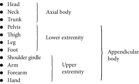
- • It is important to distinguish the thigh from the leg. The thigh is between the hip joint and the knee joint, whereas the leg is between the knee joint and the ankle joint. 4 In our terminology, the thigh is not part of the leg.
- • It is important to distinguish the arm from the forearm. The arm is between the shoulder joint and the elbow joint, whereas the forearm is between the elbow joint and the wrist joint. In our terminology, the forearm is not part of the arm.
- • The shoulder girdle contains the scapulae and the clavicles. 4
- • The pelvis as a body part includes the pelvic girdle of bones.
-
- • The pelvic girdle contains the two pelvic bones, the sacrum, and the coccyx. 4


(A) shows the axial, upper extremity, and lower extremity parts of the human body in anterior view. The axial body parts are head (violet shade), neck (pink shade), and trunk (light green shade). The upper extremity body parts are shoulder girdle (pale blue shade), arm (pale orange shade), forearm (pale pink shade), and hand (yellow shade). The lower extremity body parts are pelvis (blue shade), thigh (light orange shade), leg (pink shade), and foot (yellow shade). (B) shows the axial, upper extremity, and lower extremity parts of the human body in posterior view. The axial body parts are head (violet shade), neck (pink shade), and trunk (light green shade). The upper extremity body parts are shoulder girdle (pale blue shade), arm (pale orange shade), forearm (pale pink shade), and hand (yellow shade). The lower extremity body parts are pelvis (blue shade), thigh (light orange shade), leg (pink shade), and foot (yellow shade). (C) shows the axial, upper extremity, and lower extremity parts of the human body in lateral view. The axial body parts are head (violet shade), neck (pink shade), and trunk (light green shade). The upper extremity body parts are shoulder girdle (pale blue shade), arm (pale orange shade), forearm (pale pink shade), and hand (yellow shade). The lower extremity body parts are pelvis (blue shade), thigh (light orange shade), leg (pink shade), and foot (yellow shade).
Section 1.3. Joints Between Body Parts
- • What separates one body part from the body part next to it is the presence of a joint between the bones of the body parts. A joint is located between two adjacent body parts (Figure 1-3). 3
- • When we say that a body part moves, our general rule will be that the body part moves relative to an adjacent body part.
- • This movement occurs at the joint that is located between these two body parts (Figure 1-4).

(A) shows the various types of joints in a human body in anterior view. The labels of the illustration from top to bottom are shoulder joint, elbow joint, wrist joint, hip joint, knee joint, and ankle joint. (B) shows the various types of joints in a human body in posterior view. The labels of the illustration from top to bottom are shoulder joint, spinal joint, elbow joint, lumbosacral spinal joint, wrist joint, hip joint, knee joint, and ankle joint. (C) shows the various types of joints in a human body in lateral view . The labels of the illustration from top to bottom are shoulder joint, elbow joint, hip joint, wrist joint, knee joint, and ankle joint.

(A) shows lifting of the right leg (indicated with an arrow) with respect to the position of the pelvis; the labels included in the image from top to bottom are pelvis, hip joint, and thigh. (B) shows flexion of the right leg (indicated with an arrow) in relation to the ground position. The labels included in the image from top to bottom are thigh, knee joint, and leg.
Section 1.4. Movement of a Body Part Relative to an Adjacent Body Part
- • When movement of our body occurs, we see the following:
-
- • It is a body part that is moving.
- • That movement is occurring at a joint that is located between that body part and an adjacent body part. 5
- • To name this movement properly and fully, two things must be stated:
-
- 1. The name of the body part that is moving
- 2. The joint where the movement is occurring 5
- • Most texts describe a movement of the body by stating only the body part that is moving or by stating only the joint where the motion is occurring. However, to be complete and to fully describe and understand what is happening, both aspects should be stated. By doing this every time you describe a movement of the body, you will gain a better visual picture and understanding of the movement that is occurring.
- • Figures 1-5, 1-6, and 1-7 show examples of movements of body parts relative to adjacent body parts.

An illustration indicates stretching of the right arm (direction of the stretching indicated with an arrow). The labels included in the illustration0 from top to bottom are shoulder joint, scapula, and the arm.

An illustration shows stretching of the right arm parallel to the trunk region in a lateral view (an arrow marks the direction of stretching). The labels included in the illustration from top to bottom are forearm, arm and elbow joint.

An illustration shows lifting of the right foot in relation to the left foot, which is in ground state. An arrow indicates the direction of the movement of the right foot. The labels included in the illustration from top to bottom are leg, foot, and ankle joint.
Section 1.5. Movement Within a Body Part
- • We have seen that when a major body part moves, the movement occurs at the joint that is located between that body part and an adjacent body part.
- • Because that joint is located between two different major body parts, when one body part moves relative to another body part, it can be said that the movement occurs between body parts.
- • However, sometimes movement can occur within a major body part.
- • This can occur whenever the major body part has two or more smaller body parts (i.e., bones) located within it. When this situation exists, movement can occur at the joint that is located between these smaller body parts (i.e., bones) within the major body part. 5
-
- • The simplest example of this is the hand. The hand is considered to be a major body part, and motion of the hand is described as occurring between it and the forearm at the wrist joint (Figure 1-8A). However, the hand has other body parts, the fingers, within it. Each finger is a body part in its own right, because a finger can move relative to the palm of the hand (Figure 1-8B). Furthermore, each finger has three separate parts (i.e., bones), and each part can move independently (Figure 1-8C).
-

FIGURE 1-8 A, Lateral view showing the hand moving relative to the forearm at the wrist joint. B, Depiction of motion within the hand. This is a lateral view showing a finger moving relative to the palm of the hand at the joint that is located between them. C, Illustration of movement of one part of a finger relative to another part of the finger at the joint that is located between them. Note: B and C both illustrate the concept of movement occurring within a major body part because smaller body parts are within it. (A) in lateral view indicates the downward and upward movement of the right hand; a bidirectional arrow shows the direction of the movement on both ways. The labels included in the illustration from left to right are forearm, wrist joint, and hand. (B) shows flexion of the forefinger and the middle finger; a bidirectional arrow indicates the direction of the movement of both the fingers. (C) shows flexion of the forefinger in relation to the middle finger; a bidirectional arrow indicates the direction of flexion.
-

FIGURE 1-9 A, Lateral view showing the forearm moving (flexing) relative to the arm at the elbow joint. B, Movement of one of the bones (i.e., the radius) within the forearm relative to the other bone (i.e., the ulna) of the forearm; this motion occurs at the radioulnar joints located between the two bones. (A) shows flexion of the right forearm in a direction that is parallel to the trunk in a lateral view; an arrow indicates the direction of flexion. The labels included in the illustration from top to bottom are forearm, arm, and elbow joint. (B) shows location of various bones and joints in anterior in ventral and dorsal views of the forearm. The labels included in the illustration in both views from top to bottom are proximal radioulnar joint, ulna, radius, and distal radioulnar joint.
- • A second example is the forearm. The forearm is usually described as moving relative to the arm at the elbow joint (Figure 1-9A). However, the forearm has two bones within it, and joints are located between these two bones. Motion of one of these bones can occur relative to the other (Figure 1-9B). In this case each of the two bones would be considered to be a separate, smaller body part.
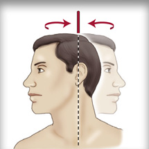
- • A third, more complicated example is the cervical spine. The cervical spine has seven vertebrae within it. The neck may be described as moving relative to the trunk that is beneath it (Figure 1-10A). However, each of the seven vertebrae can move independently. Therefore, motion can occur between vertebrae within the neck at the joints located between the vertebrae (Figure 1-10B).
Section 1.6. True Movement of a Body Part Versus “Going Along For The Ride”
- • In lay terms, when we say that a body part has moved, it does not always mean that true movement of that body part has occurred (according to the terminology that is used in the musculoskeletal field for describing joint movements).
- • A distinction must be made between true movement of a body part and what we will call “going along for the ride.”
- • For true movement of a body part to occur, the body part must move relative to an adjacent body part (or the body part must have movement within it).
-

FIGURE 1-10 A, Lateral view of the neck showing the neck moving relative to the trunk at the spinal joint between them (C7–T1). B, Motion within the neck that is occurring between several individual vertebrae of the neck. This motion occurs at the spinal joints located between these bones. (A) shows lateral view of skeletal regions of head, neck, and trunk. The illustration shows bending of the neck region with respect to the trunk region. The labels included in the illustration from top to bottom are neck (with vertebrae marked in blue shade), C7-T1 joint (immediately below the neck region), and trunk (below the C7-T1 joint). (B) shows bending of the neck region in lateral view. The cervical spinal joints associated with the neck region are indicated in blue shade.
- • For example, in Figure 1-11 we see that a person is moving the right upper extremity.
- • In lay terms, we might say that the person’s right hand is moving because it is changing its position in space.
- • However, in our terminology the right hand is not moving because the position of the hand relative to the forearm is not changing (i.e., the right hand is not moving relative to the forearm [and motion is not occurring within the hand]).
- • The movement shown in Figure 1-11 is flexion of the forearm at the elbow joint. It is the forearm that is moving relative to the arm at the elbow joint.
-
• The hand is not moving in this scenario. We could say that the hand is merely “going along for the ride.”

FIGURE 1-11 A and B, Illustration of the concept that the forearm is moving (because its position relative to the arm is changing). The motion that is occurring here is flexion of the forearm at the elbow joint. The hand is not moving, because its position relative to the forearm is not changing; the hand is merely “going along for the ride.” (A) shows flexion of the right forearm in a lateral view in the direction that is parallel to the thigh region. (B) shows flexion of the right forearm in a lateral view in a direction that is parallel to the trunk region.
- • Figure 1-12 depicts true movement of the hand relative to the forearm.

(A) shows flexion of the right hand in a lateral view in a direction that is parallel to the thigh region. (B) shows flexion of the right hand in lateral view in a direction that is in midline with the thigh region.
Section 1.7. Regions of the Body
- • Within the human body, areas or regions are given names. Sometimes these regions are located within a body part; sometimes they are located across two or more body parts. The various regions of the body are shown in Figure 1-13. 6

(A) shows major regions of the human body in an anterior view. The labels included in the illustration from left to right are axillary, brachial, antecubital, antebrachial, carpal, palmar, femoral, crural, plantar, facial, mandibular, supraclavicular, pectoral, abdominal, pubic, inguinal, and patellar. (B) shows in posterior view the major regions of the human body with labels marked from left to right as interscapular (spine), scapular (upper back), cubital (elbow), pelvic (lower hip), gluteal (buttocks), sural (calf), cranial (back of head), cervical (neck), thoracic (back), lumbar (lower back), sacral (between hips), and popliteal (back of knee).