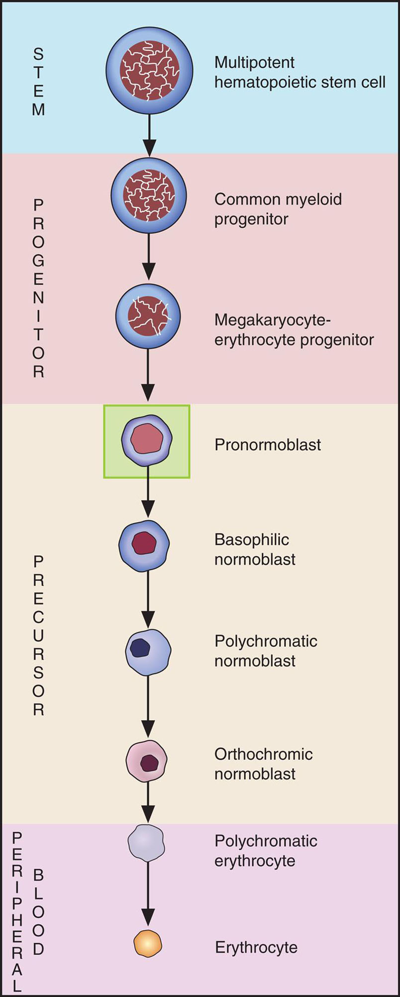
Erythrocyte Maturation
All photomicrographs are ×1000 original magnification with Wright-Giemsa staining unless stated otherwise.
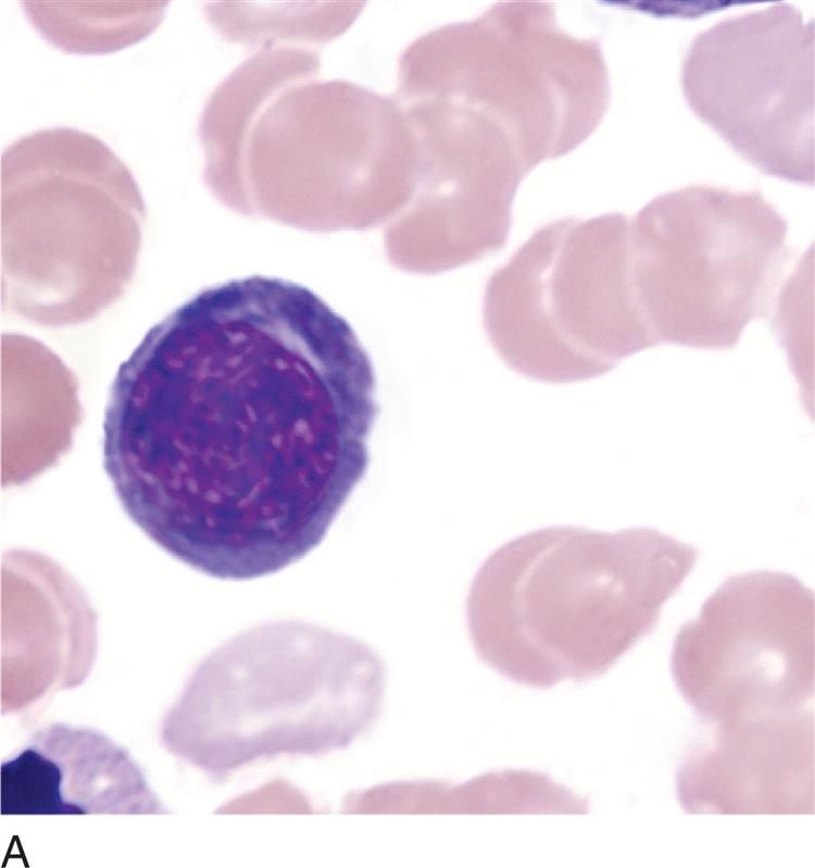
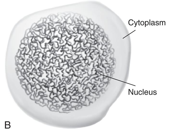
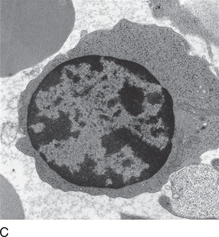
Pronormoblast
| Proerythroblast |
| Rubriblast |

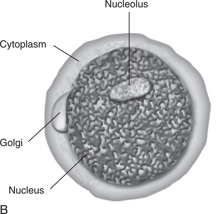
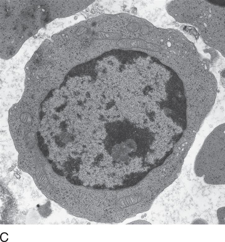
SIZE: 12 to 20 μm
NUCLEUS: Round to slightly oval
Nucleoli: 1 to 2
Chromatin: Fine
CYTOPLASM: Dark blue; may have prominent Golgi
N:C RATIO: 8:1
REFERENCE INTERVAL:
Bone Marrow: 1%
Peripheral Blood: 0%

Basophilic normoblast
| Basophilic Erythroblast |
| Prorubricyte |
SIZE: 10 to 15 μm
NUCLEUS: Round to slightly oval
Nucleoli: 0 to 1
Chromatin: Slightly condensed
CYTOPLASM: Dark blue
N:C RATIO: 6:1
REFERENCE INTERVAL:
Bone Marrow: 1% to 4%
Peripheral Blood: 0%
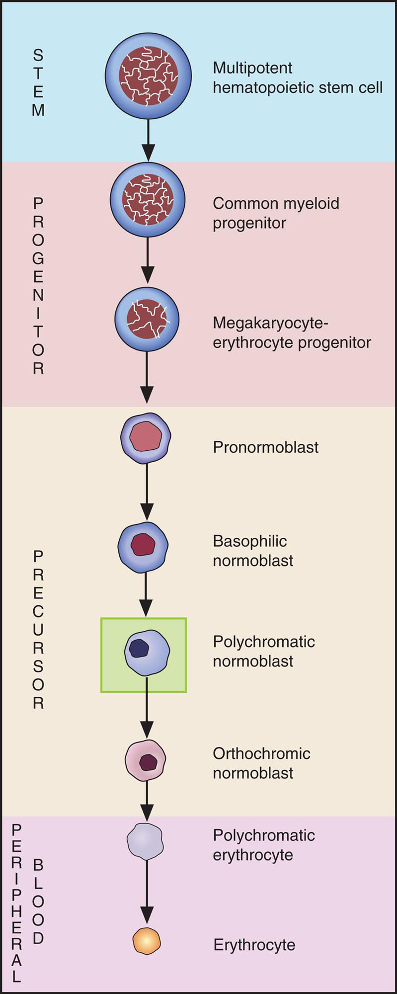
Polychromatic normoblast
| Polychromatic Erythroblast |
| Rubricyte |


SIZE: 10 to 12 μm
NUCLEUS: Round
Nucleoli: None
Chromatin: Quite condensed
CYTOPLASM: Gray-blue as a result of hemoglobinization
N:C RATIO: 4:1
REFERENCE INTERVAL:
Bone Marrow: 10% to 20%
Peripheral Blood: 0%

Orthochromic normoblast
| Orthochromic Erythroblast |
| Metarubricyte |
SIZE: 8 to 10 μm
NUCLEUS: Round
Nucleoli: 0
Chromatin: Fully condensed
CYTOPLASM: More pink or salmon than blue
N:C RATIO: 0.5:1
REFERENCE INTERVAL:
Bone Marrow: 5% to 10%
Peripheral Blood: 0%
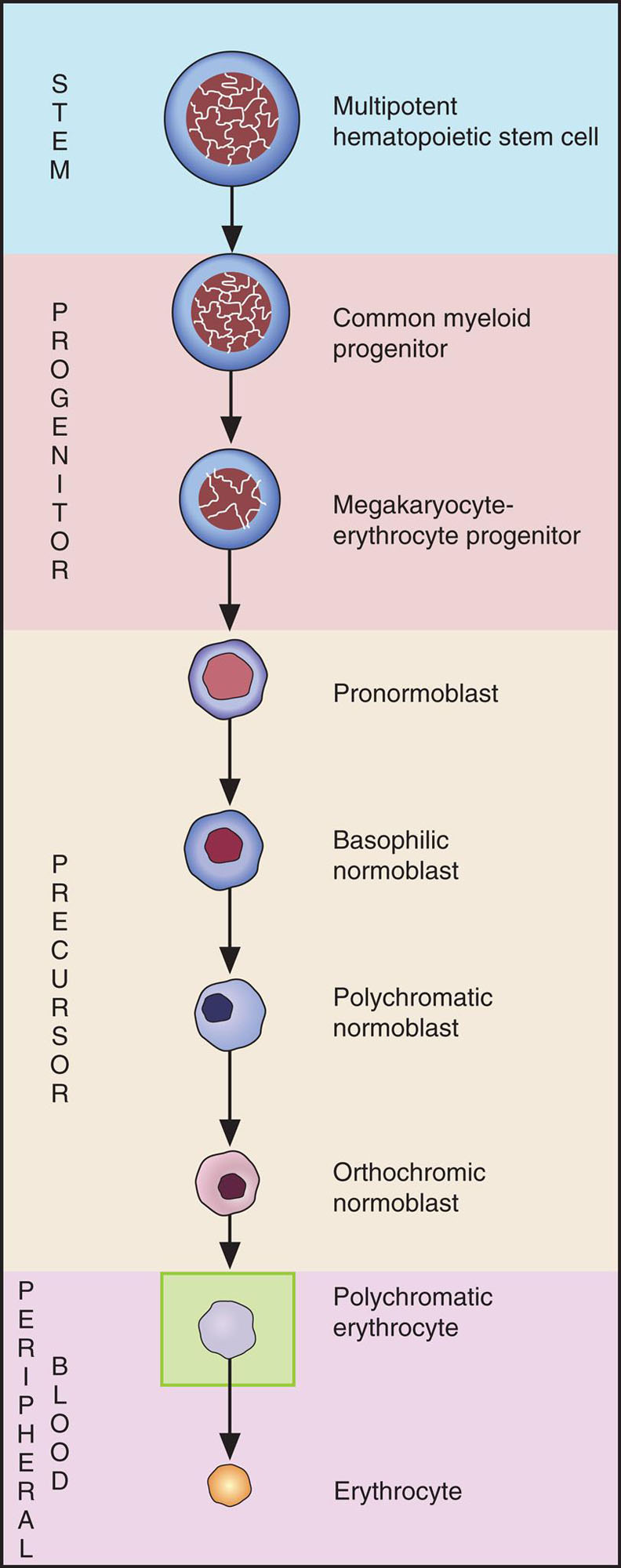
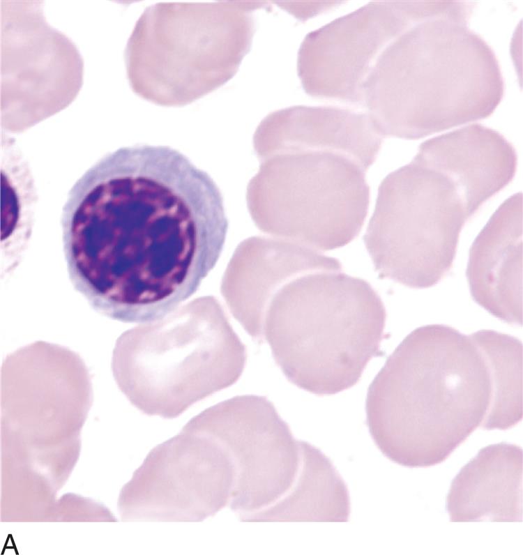
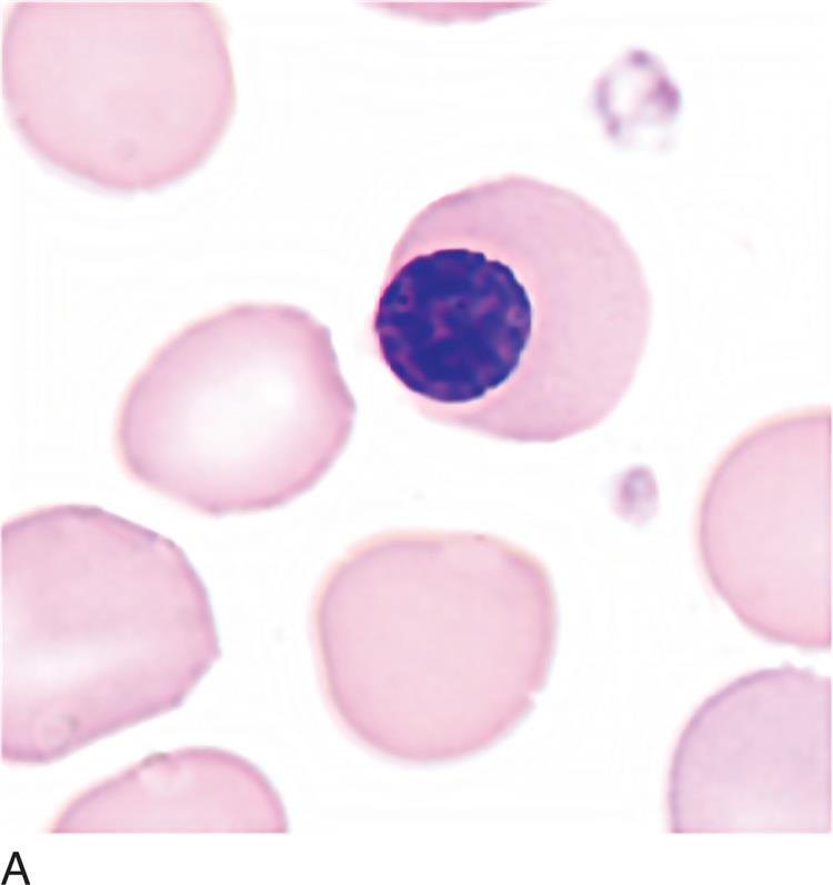
Polychromatic erythrocyte
| Diffusely Basophilic Erythrocyte |
| Reticulocyte |
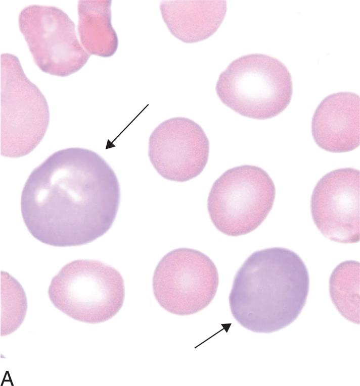

SIZE: 8 to 8.5 μm
NUCLEUS: Absent
Nucleoli: NA
Chromatin NA
CYTOPLASM: Color is slightly more blue/purple than the mature erythrocyte
N:C RATIO: NA
REFERENCE INTERVAL:
Bone Marrow: 1%
Peripheral Blood: 0.5% to 2.0%
NOTE: When stained with supravital stain (e.g., new methylene blue), polychromatic erythrocytes appear as reticulocytes (contain precipitated ribosomal material; see Figure 12.5A).
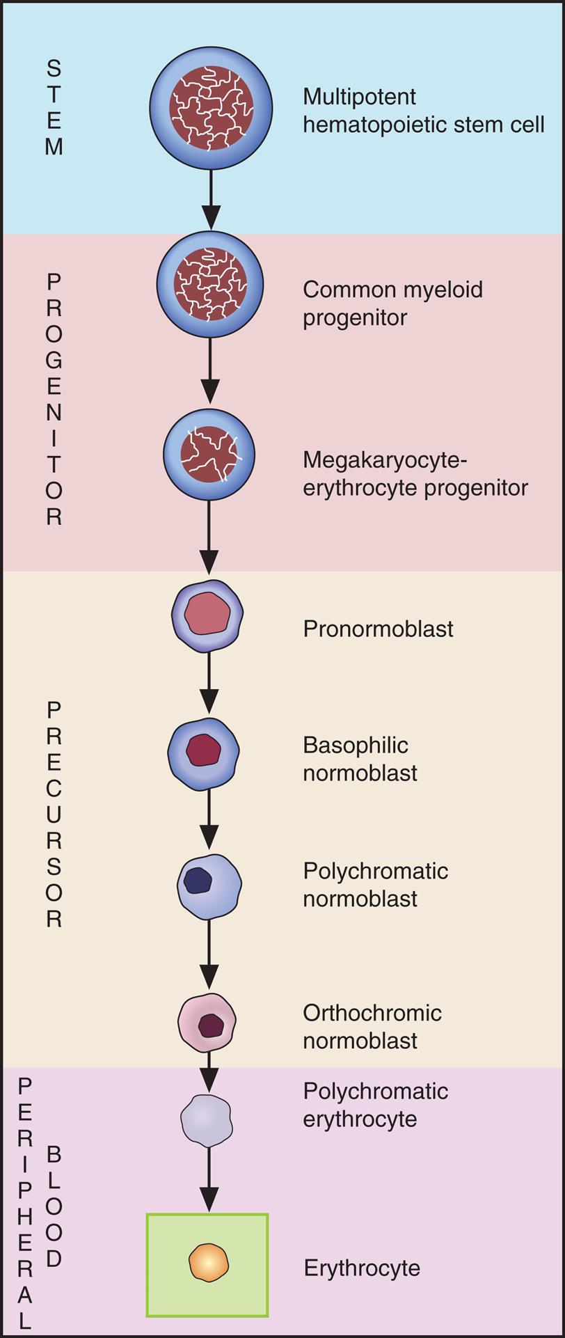
Erythrocyte
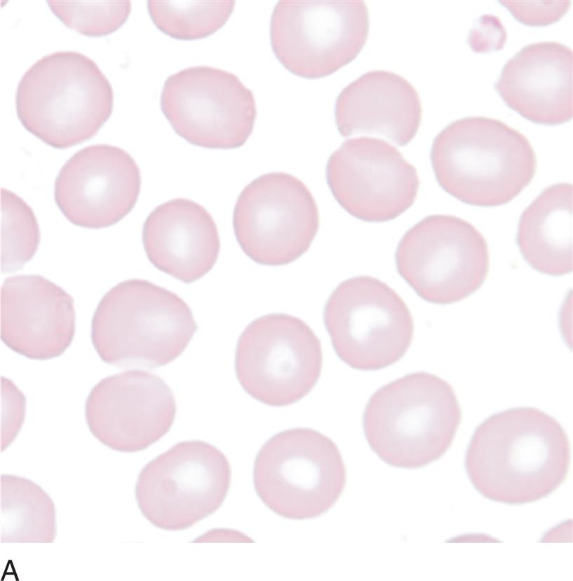

SIZE: 7 to 8 μm
NUCLEUS: Absent
Nucleoli: NA
Chromatin: NA
CYTOPLASM: Salmon with central pallor of about one-third of the diameter of the cell
N:C RATIO: NA
REFERENCE INTERVAL
Bone Marrow: NA
Peripheral Blood: Predominant cell type
