Nuclear and Cytoplasmic Changes in Leukocytes
Hyposegmentation of neutrophils
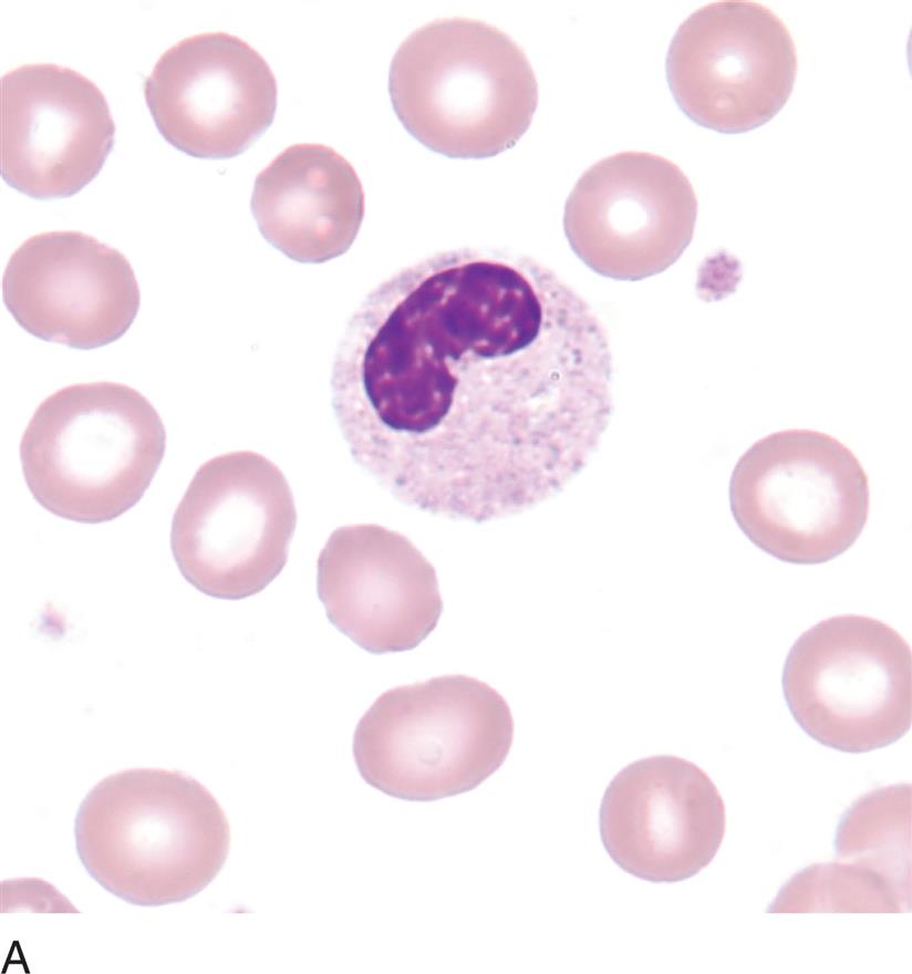
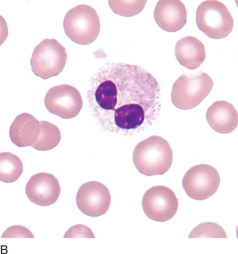

DESCRIPTION: Peanut-shaped, bilobed or non-segmented, granulocyte nucleus with the coarse chromatin of a mature cell
ASSOCIATED WITH: Pelger-Hüet anomaly, pseudo-Pelger-Hüet anomaly
NOTE: Pelger-Hüet anomaly is inherited and affects the majority of granulocytes. Pseudo-Pelger-Hüet is acquired, affects less than 50% of granulocytes, and is usually accompanied by other morphologic indications of malignancy, such as those seen in myeloproliferative or myelodysplastic disorders (see Chapter 17, Myeloproliferative Neoplasms and Chapter 18 Myelodyspastic Syndromes.).
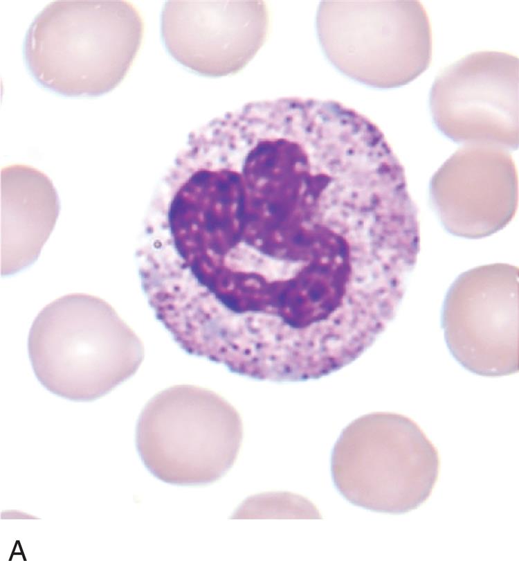
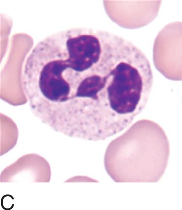
Hypersegmentation of neutrophils
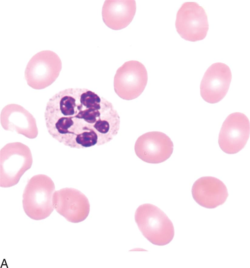
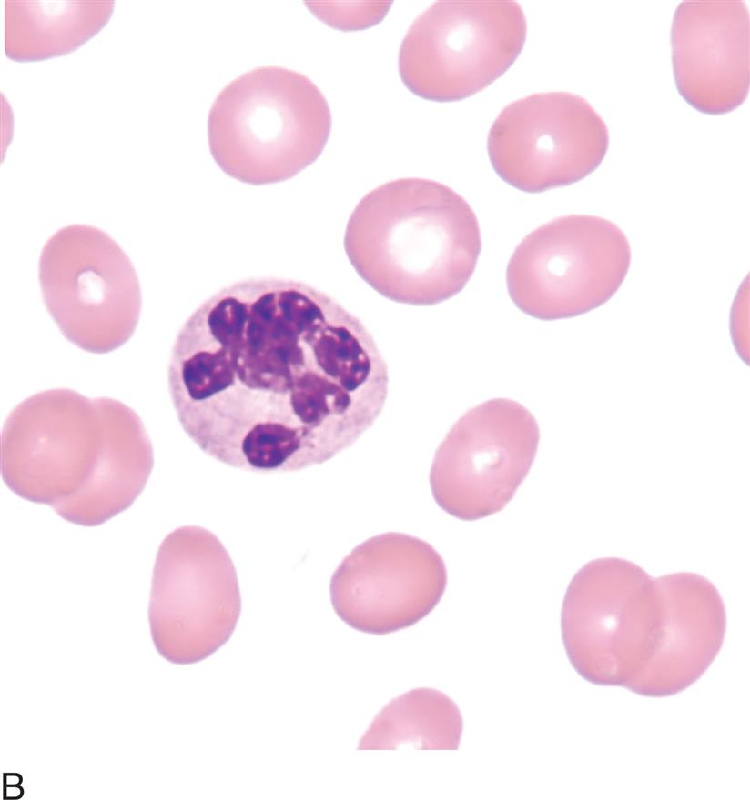
DESCRIPTION: Six or more lobes in granulocyte nucleus
ASSOCIATED WITH: Megaloblastic anemias, chronic infections, myelodysplastic syndrome; rarely inherited
Vacuolation in Neutrophils


DESCRIPTION: Unstained circular area within the cytoplasm
NUMBER: Few to many
ASSOCIATED WITH: Bacterial or fungal infection, poisoning, burns, chemotherapy, artifact
NOTE: Vacuoles rarely may contain microorganisms or pigment. Vacuoles are seen in normal monocytes and do not suggest infection.
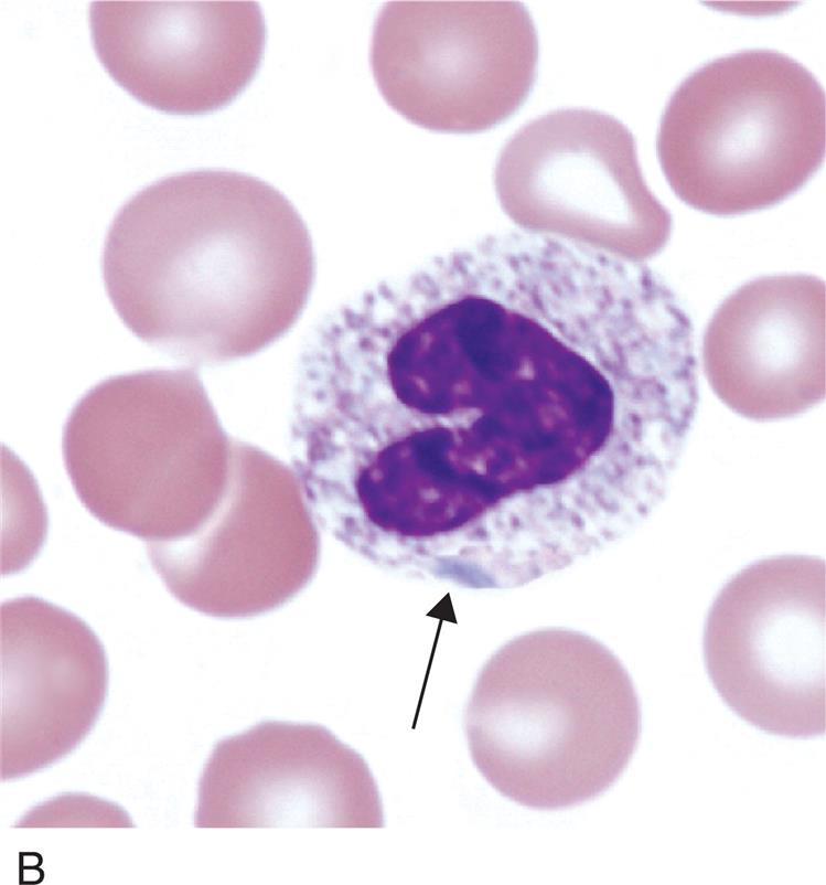
Döhle body


DESCRIPTION: Gray-blue, variably shaped inclusion in cytoplasm
COMPOSITION: Ribosomal RNA
NUMBER: Single or multiple
ASSOCIATED WITH: Wide range of conditions, including bacterial infection, sepsis, and normal pregnancy
NOTE: May be seen in cells with toxic granulation or on same slide with toxic granulation. (See Figure 14.5B.)
Blue-green inclusion
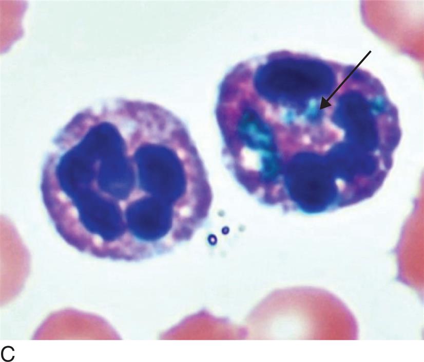
DESCRIPTION: Amorphous blue-green granules in cytoplasm of neutrophils
COMPOSITION: Lipid-rich, possibly lipofusion-like material
NUMBER: Single or multiple
ASSOCIATED WITH: Severe liver disease, lactic acidosis, sepsis
NOTE: Also known as the blue-green crystals of death.
Toxic granulation

DESCRIPTION: Prominent dark purple-black granules in the cytoplasm of neutrophils, unevenly distributed
COMPOSITION: Primary granules
NUMBER: Few to many
ASSOCIATED WITH: Wide range of conditions including bacterial infection, sepsis and following administration of granulocyte colony-stimulating factor
Hypogranulation/agranulation in Neutrophils


DESCRIPTION: Decreased number or absence of specific granules giving the cytoplasm a colorless appearance
ASSOCIATED WITH: Myelodysplastic syndrome, myeloproliferative neoplasms, infection
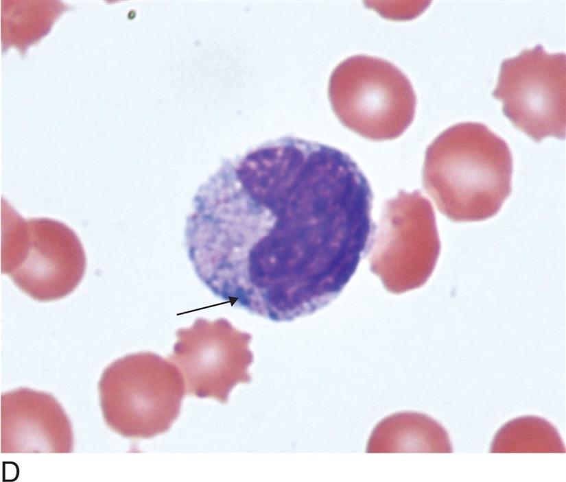
Reactive lymphocytes
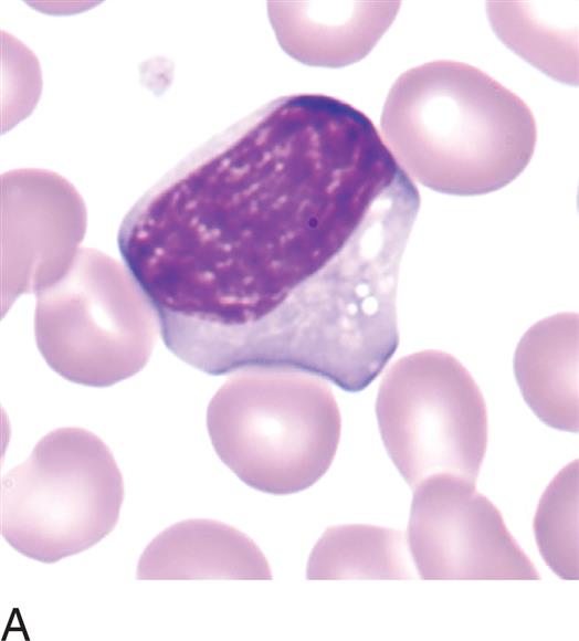
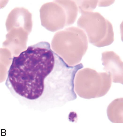
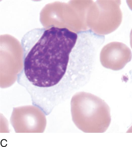
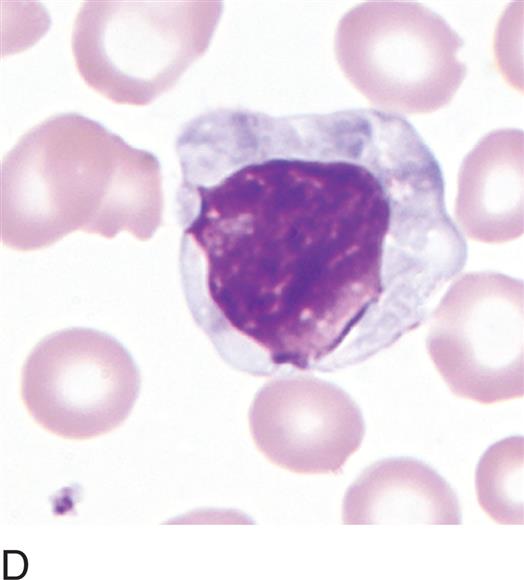
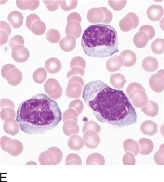
DESCRIPTION: 10 to 30 μm; pleomorphic; easily indented by surrounding cells
NUCLEUS: Irregular
Nucleoli: Occasionally present
Chromatin: When compared with that of a resting lymphocyte, chromatin is coarse to fine and dispersed
CYTOPLASM: Pale blue to deeply basophilic, may stain unevenly with peripheral or radial basophilia
Granules: May have increased numbers of azurophilic granules
Vacuoles: Occasional
ASSOCIATED WITH: Viral infections and other antigenic stimulation, including organ transplantation