Precursor Lymphoid Neoplasms
The World Health Organization classifies precursor lymphoid neoplasms into two major groups: B lymphoblastic leukemia/lymphoma and T lymphoblastic leukemia/lymphoma. Leukemia is primarily a disease of peripheral blood and bone marrow, whereas the primary site of involvement for lymphoma is the lymph system. Because this is an atlas of blood cells, only the leukemia morphology will be presented. Acute lymphoblastic leukemia (ALL) is not classified morphologically or by cytochemistry but by a combination of cytogenetic profiles, genotype, and immunophenotype. B lymphoblastic leukemia is subdivided into nine subtypes that are associated with recurrent genetic abnormalities (Box 16.1). Those cases of B-ALL that do not fall within one of these groups are classified as B lymphoblastic leukemia, not otherwise specified. Although 50% to 70% of patients with T-ALL do have abnormal karyotypes, none of the abnormalities is clearly associated with distinctive biologic features, and thus T-ALL is not further subdivided.
Lymphoblasts may be either small and homogeneous or large and heterogeneous. Further testing is needed to determine the phenotype and genotype.
Acute lymphoblastic leukemia, small blasts

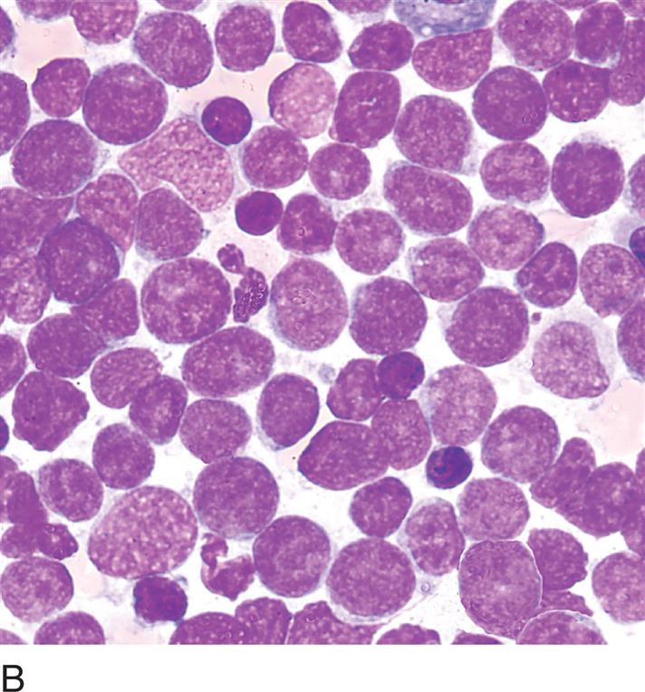

MORPHOLOGY:
Peripheral Blood: ± Blasts, small blasts (about one to two-and-a-half times the size of a resting lymphocyte) with scant blue cytoplasm, condensed chromatin and indistinct nucleoli, thrombocytopenia
Bone Marrow: 20% or more of all nucleated cells make up a homogeneous population of blasts
NOTE: Hematogones (immature B cells) may be seen in bone marrow and peripheral blood of newborns or in patients during bone marrow recovery. Care must be taken not to confuse hematogones with small lymphoblasts (see Figs. 16.1D and 23.4).
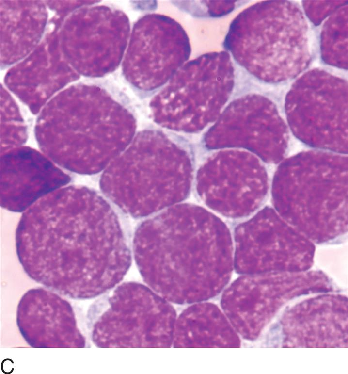
Acute lymphoblastic leukemia, large blasts


MORPHOLOGY:
Peripheral Blood: Blasts two to three times the size of a resting lymphocyte, moderate cytoplasm, irregular nuclear membrane, prominent nucleoli, thrombocytopenia, morphologically difficult to distinguish from acute myeloid leukemia
Bone Marrow: 20% or more of all nucleated cells comprise a heterogeneous population of blasts