Anomalies of the Collecting System
Calyceal Diverticulum
A calyceal diverticulum is a cystic cavity within the kidney that is lined by transitional epithelium and communicates with a calyx or less commonly with the renal pelvis through a narrow isthmus (Estrada et al, 2009). This abnormality, first described by Rayer in 1841, may be multiple, with the upper calyx most frequently affected.
In the past, an incidence of 4.5 per 1000 excretory urograms had been reported (Timmons et al, 1975). A similar incidence was noted in both children and adults, with no predilection for either side or sex. Most diverticula, labeled type I, occur adjacent to an upper or, occasionally, a lower pole calyx. Type II diverticula are larger, communicate with the renal pelvis, and tend to be symptomatic (Wulfsohn, 1980).
Congenital and acquired factors have been suggested to explain the formation of calyceal diverticula. The similar incidence in children and adults is consistent with an embryologic etiology (Abeshouse, 1950; Mathieson, 1953; Devine et al, 1969; Middleton and Pfister, 1974). At the 5-mm stage of the embryo, ureteral branches of the third and fourth generation, which ordinarily degenerate, may persist as isolated branches, resulting in the formation of a calyceal diverticulum (Lister and Singh, 1973).
A localized cortical abscess draining into a calyx has also been postulated as an etiologic factor. Other proposed causes include obstruction from stones or infection within a calyx, progressive fibrosis of an infundibular stenosis, renal injury, achalasia, and spasm or dysfunction of one of the supposed sphincters surrounding a minor calyx (Amar, 1975; Patriquin et al, 1985; Siegel and McAlister, 1979). Vesicoureteral reflux has also been thought be an etiologic factor. Amar (1975) postulated that calyceal tubular backflow of infected urine could result in abscess formation and parenchymal injury leading to diverticular formation. Small diverticula are usually asymptomatic and are found incidentally by ultrasonography, CT, or MRI. These diverticula tend to progressively distend with trapped urine (Amar, 1975; Patriquin et al, 1985; Siegel and McAlister, 1979). Infection, milk of calcium (i.e., crystallization of calcium salts without actual stone formation) (Patriquin et al, 1985), or true stone formation are complications of stasis or obstruction that can produce symptoms (Lister and Singh, 1973; Siegel and McAlister, 1979) (Fig. 117–19). Hematuria, pain, and UTI may be seen in the presence of stones, which may be present in almost 40% of patients. In the Mayo Clinic series (Timmons et al, 1975), 39% of patients with calyceal diverticula had calculi.
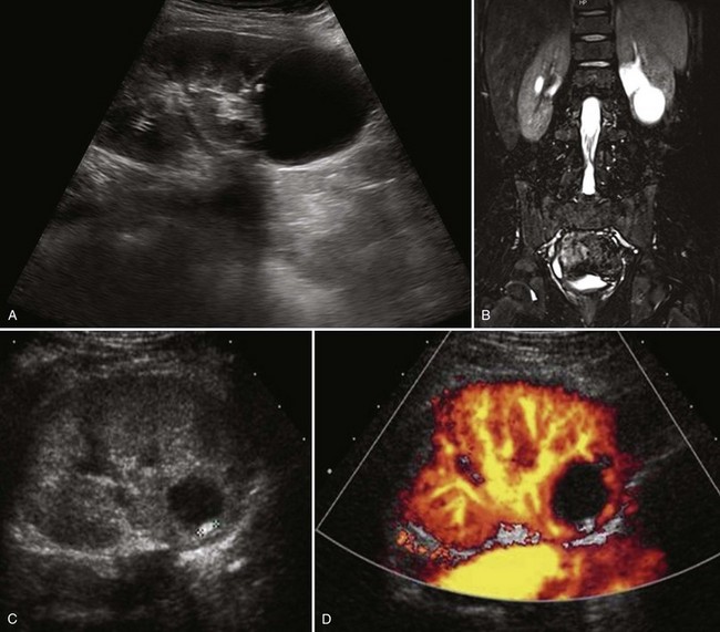
Figure 117–19 Lower pole calyceal diverticulum. A, Sagittal ultrasonogram. B, Sequence of coronal T2 images. C, Sagittal ultrasonogram shows small stone within a diverticulm. D, Color Doppler ultrasonogram demonstrates absence of flow.
(B, Courtesy of Dr. Sara Milla; C and D, courtesy of Dr. Shpetim Telegrafi.)
Key Points: Calyceal Diverticulum
The diagnosis is suggested on ultrasonography but confirmed on CT scan and MR urography (Ellis et al, 1990; Estrada et al, 2009) (see Fig. 117–19). Delayed films demonstrate pooling of contrast material in the diverticulum. Retrograde pyelography and delayed imaging CT with contrast medium enhancement and MR urography are used to diagnose and define the precise anatomy. Ultrasonography delineates a fluid-filled area more centrally located near the collecting system than a simple renal cyst (see Fig. 117–19). When it is filled with microcalculi, ultrasonography characteristically demonstrates a layering effect, within the diverticulum, between clear fluid above and echo-dense debris without shadowing below (Patriquin et al, 1985). Ultrasonography will image the milk of calcium within the diverticulum as the patient changes position.
Patients who are asymptomatic do not require treatment but should be followed periodically with ultrasonography. Estrada and colleagues (2009) reported 10 of 23 calyceal diverticula (43%) that required treatment at a mean of 27 ± 25 months after initial treatment. Most commonly, these children presented with febrile UTI. The indications for surgery included enlargement of the diverticulum associated with pain or infection, abscess formation, urosepsis, and symptomatic calculus formation. Percutaneous ablation of the communication and fulguration of the diverticular lining was used until 1995. Although percutaneous ablation remains a viable treatment alternative, the availability of pediatric laparoscopic equipment has led to the retroperitoneal laparoscopic approach for marsupialization of the diverticulum and fulguration of the epithelial lining (Estrada et al, 2009). Ureteroscopy with enlargement of the diverticular communication and removal of the stones has also been reported (Baldwin et al, 1998).
Hydrocalycosis
Hydrocalycosis is a very rare cystic dilation of a major calyx with a demonstrable connection to the renal pelvis (Fig. 117–20). It may be caused by a congenital or acquired intrinsic obstruction, such as a parapelvic cyst.
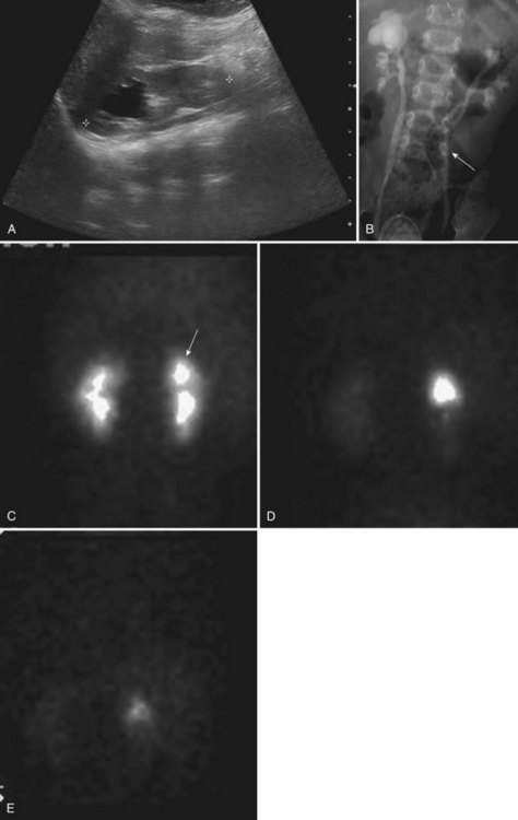
Figure 117–20 Two-month-old girl with prenatal history of right upper pole renal cyst. A, Sagittal ultrasonogram shows right kidney and a 2.5-cm cystic structure in the right upper pole (space between calipers denotes renal length). B, Voiding cystourethrogram demonstrates bilateral vesicoureteral reflux with marked dilation of the right upper pole compound calyx suggestive of hydrocalycosis. Note on the right, triplication of the pelvis, and, on the left, early division of the ureter (arrow) with triplication of the pelvis associated with the uppermost ureter. C, MAG3 scan in prone view shows prompt symmetrical uptake of the isotope. D, Prefurosemide injection with retained isotope in right upper pole compound calyx. E, Prompt drainage of the calyx following furosemide injection.
Dilation of the upper calyx due to obstruction of the upper infundibulum by vessels or stenosis has been described (Fraley, 1966; Johnston and Sandomirsky, 1972). Cicatrization of an infundibulum may result from infection or trauma. Conversely, hydrocalycosis has been reported to occur without an obvious cause (Williams and Mininberg, 1968). It has been postulated that achalasia of a ring of muscle at the entrance of the infundibulum into the renal pelvis causes a functional obstruction (Moore, 1950; Williams and Mininberg, 1968).
Mild upper calyceal dilation caused by partial infundibular obstruction is relatively common but usually asymptomatic. Although the most frequent presenting symptom is upper abdominal or flank pain, hydrocalycosis may be detected on prenatal ultrasonography (see Fig. 117–20). On occasion, a mass may be palpated. Stasis can lead to hematuria or urinary infection, or both.
Hydrocalycosis must be differentiated from multiple dilated calyces secondary to ureteral obstruction, calyceal clubbing as a result of recurrent pyelonephritis or medullary necrosis, renal tuberculosis, a large calyceal diverticulum, and megacalycosis. These entities can be differentiated by a combination of radiographic studies, surgical findings, histology, and cultures of the tissue.
Hydrocalycosis due to vascular obstruction is usually treated by dismembered infundibulopyelostomy. If the cystic dilation is caused by an intrinsic stenosis of the infundibulum, an intubated infundibulotomy or partial nephrectomy has been performed (Lang, 1991). Although clinical improvement is apparent in most instances, the radiologic appearance often is not altered significantly.
Megacalycosis
Megacalycosis is defined as nonobstructive enlargement of calyces resulting from malformation of the renal papillae (Fig. 117–21). It was first described by Puigvert in 1963. The calyces are generally dilated and malformed and may be increased in number (12 to 20) (Gittes, 1984; Pieretti-Vanmarcke et al, 2009). The renal pelvis is not dilated, nor is its wall thickened, and the UPJ is normally funneled without evidence of obstruction. The cortical tissue around the abnormal calyx is normal in thickness. The medulla, however, is underdeveloped and assumes a falciform (crescent) or semilunar appearance instead of its normal pyramidal shape. The collecting tubules are not dilated but are definitely shorter than normal, and they are oriented transversely rather than vertically from the corticomedullary junction (Puigvert, 1963). A mild disorder of maximal concentrating ability has been reported (Gittes and Talner, 1972), but acid excretion is normal after an acid load (Vela-Navarrete and Garcia Robledo, 1983). Other functions of the kidney-glomerular filtration, renal plasma flow, and isotope uptake also are not altered (Gittes, 1984).
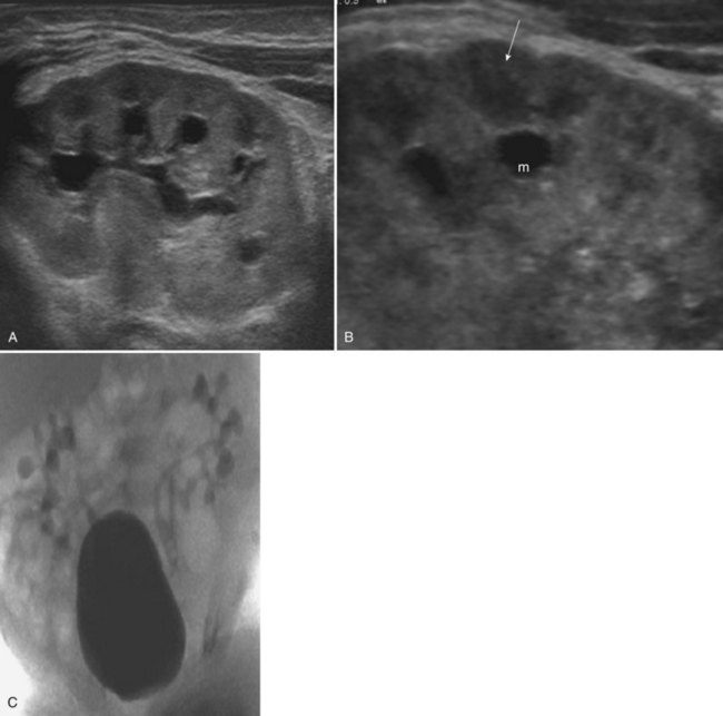
Figure 117–21 Megacalycosis. A, Sagittal ultrasonogram shows dilated calyces and normal-appearing pyramids. B, Enlarged view of A demonstrates distinct megacalyx (m) and normal pyramid (arrow). C, Voiding cystourethrogram shows bilateral vesicoureteral reflux into megacalyces.
Megacalycosis is congenital and has been diagnosed prenatally (Vidal Company et al, 2001). It occurs predominantly in males with a ratio of 6 : 1 and has been found only in whites. Bilateral disease has been seen almost exclusively in males, whereas segmental unilateral involvement occurs only in females (Cacciaguerra et al, 1996), suggesting an X-linked partially recessive gene with reduced penetrance in females (Gittes, 1984). Except for one report of two affected brothers, the entity has not been thought to be familial (Briner and Thiel, 1988).
It was theorized by Puigvert (1964) and endorsed by Johnston and Sandomirsky (1972) that there is transient delay in the recanalization of the upper ureter after the branches of the UB hook up with the metanephric blastema. This produces transient obstruction when the glomeruli start producing urine. The fetal calyces may dilate and retain their obstructed appearance, despite the lack of evidence of obstruction in postnatal life (Gittes and Talner, 1972). The increased number of calyces may be an aborted response by the branching UB to the obstruction.
The abnormality is usually diagnosed during radiographic evaluation of a UTI or other congenital anomalies (Arambasic et al, 2003). Adults frequently present with hematuria secondary to renal calculi.
The calyces are dilated and usually increased in number, but the infundibuli and pelvis may not be enlarged. Although the UPJ does not appear obstructed, there may be segmental dilation of the distal third of the ureter (Kozakewich and Lebowitz, 1974). Megacalycosis associated with an ipsilateral segmental megaureter was described in 12 children (Mandell et al, 1987), mostly boys, and predominantly left-sided. A normal-caliber ureter was interposed between the two entities. This anatomic picture may be mistaken for congenital ureteropelvic, ureterovesical junction obstruction, or infundibular stenosis (Redman and Neeb, 2005; Pieretti-Vanmanrcke et al, 2009). Diuretic renography reveals a normal pattern for uptake and washout of the isotope (Gomez Tellado et al, 1997). Long-term follow-up of these patients does not reveal progression of the anatomic derangement or functional impairment of the kidney (Gittes, 1984).
Key Points: Megacalycosis
Unipapillary Kidney
The unipapillary kidney is a rare anomaly with only 18 cases reported (Neal and Murphy, 1960; Sakatoku and Kitayama, 1964; Harrison et al, 1976; Morimoto et al, 1979; Toppercer, 1980; Kaneto et al, 1997). This anomaly is present not uncommonly in monkeys, rabbits, dogs, marsupials, insectivores, and monotremes. The cause is thought to be a failure of progressive branching after the first three to five generations (which create the pelvis) of the UB (Potter, cited by Harrison et al, 1976). The solitary calyx drains a ridgelike papilla. Nephrons attach to fewer collecting tubules, which then drain directly into the pelvis. Biopsies of these kidneys reveal glomerulosclerosis, tubular atrophy, and increased fibrosis (Bischel et al, 1978).
The kidney is smaller than normal, often with reduced function, but usually is in its correct location (Smith et al, 1984; Kaneto et al, 1997). The sparse arterial tree has a normal configuration. The contralateral kidney is frequently absent. Genital anomalies are often present. The condition is frequently asymptomatic. Often there are abnormalities of the proximal ureter (i.e., megaureter, reflux, or ectopic insertion), suggesting an underlying UB defect as the cause (Smith et al, 1984; Kaneto et al, 1997).
Extrarenal Calyces
Extrarenal calyces are an uncommon congenital anomaly in which the major calyces and the renal pelvis are outside the parenchyma of the kidney, with only about 20 cases reported in the literature (Nataraju et al, 2009). This entity was originally reviewed by Eisendrath in 1925 and then more extensively by Malament and colleagues in 1961. The kidney is usually discoid, with the pelvis and the major and minor calyces located outside the renal parenchyma. The calyces may be blunted. The renal vessels have an anomalous distribution into the kidney, usually at the circumferential edge of the flat, widened hilus. Malament considered this condition to be the result of abnormally slow development of the nephrogenic anlage or a too early and rapidly developing UB, both resulting in abnormal coalescence of the collecting system with the nephrogenic mass. Extrarenal calyces have been associated with ectopic and horseshoe kidneys and renal dysplasia. They usually do not produce symptoms, although hydronephrosis with impairment of normal drainage may lead to stasis, infection, and calculi, requiring surgical intervention (Nataraju et al, 2009).
Anomalous Calyx (Pseudotumor of the Kidney)
A number of normal variants of the pyelocalyceal system in the kidney have been described. One such entity manifests as a localized mass, usually situated between the infundibula of the upper and middle calyces, and is called a hypertrophied column of Bertin (Fig. 117–22). The column may be sufficiently large to compress and deform the adjacent pelvis and calyces, suggesting a mass or “pseudotumor.” The individual calyces, however, are normally shaped and developed.
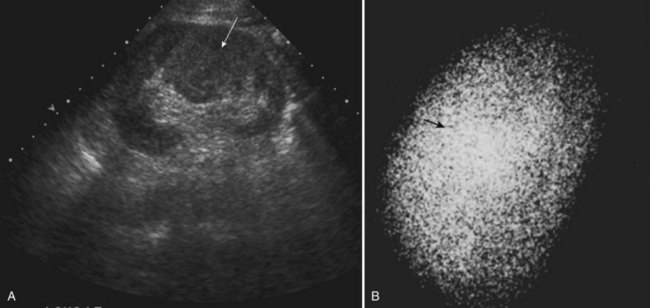
Figure 117–22 Column of Bertin. A, Ultrasonogram shows sagittal view of left kidney and possible midpolar mass. B, DMSA scan demonstrates normal uptake in the area of the mass (arrow), suggesting the pseudotumor, but it is actually a hypertrophied column of Bertin.
(A, Courtesy of Dr. Shpetim Telegrafi.)
It is important to differentiate this calyceal anomaly from true disease of the calyx and from a parenchymal tumor. A renal ultrasound study shows a normal echogenic pattern of parenchyma in the area in question, and a renal scan shows normal uptake of the radioisotope in this area (Parker et al, 1976).
Infundibulopelvic Stenosis
Infundibulopelvic stenosis most likely forms a link between cystic dysplasia of the kidney and the grossly hydronephrotic organ (Kelalis and Malek, 1981; Uhlenhuth et al, 1990). This condition includes a variety of radiographically dysmorphic kidneys with varying degrees of infundibular or infundibulopelvic stenosis that may be associated with renal dysplasia (Fig. 117–23). The first case involving the entire pelvis and all the infundibuli of both kidneys was reported by Boyce and Whitehurst in 1976. Rayer had described a focal form of the disease in 1841, and several reports noting narrowing of one or two infundibuli appeared between 1949 and 1976 (Uhlenhuth et al, 1990). These authors tried to link the focal form to cystic dysplasia secondary to obstruction, in which multicystic kidney disease is the severest form in the spectrum. Uhlenhuth and colleagues (1990) believed that this phenomenon is the result of extensive dysgenesis of the pyelocalyceal system but with preservation of renal function. Reflux is not commonly observed in these patients.
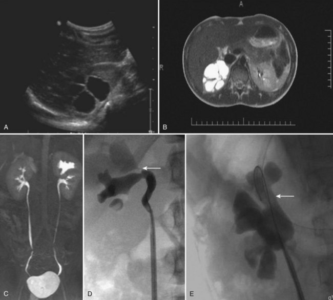
Figure 117–23 Infundibular stenosis of right upper pole calyx. A, Sagittal ultrasonogram. B, Axial T2 images. C, Coronal MR urogram. D, Right retrograde pyelogram shows no filling of the stenotic infundibulum (arrow). E, Right retrograde pyelogram following laser incision and balloon dilation of the infundibulum (arrow).
(Courtesy of Dr. Pasquale Casale.)
Key Points: Infundibulopelvic Stenosis
Infundibulopelvic stenosis is usually bilateral and is commonly associated with vesicoureteral reflux, suggesting an abnormality of the entire UB (Kelalis and Malek, 1981). Patients usually present with urinary infection, hypertension, or flank pain. Sometimes, an asymptomatic child with multiple anomalies is found to have this condition. Despite extensively dysmorphic kidney features, the function is either normal or only slightly affected (Kelalis and Malek, 1981). In Husmann’s study of 21 patients with the longest follow-up to date (median of 11 years), 90% had bilateral renal disease (Husmann et al, 1994). Ten patients had bilateral infundibulopelvic stenosis, 6 had a contralateral dysplastic renal unit, and 3 had contralateral URA. Renal insufficiency or end-stage renal disease developed in 8 (37%), all of whom had bilateral kidney anomalies. Renal biopsies in those with end-stage renal disease demonstrated renal dysplasia proximal to the stenotic infundibuli and varying degrees of glomerulosclerosis of the glomeruli that were not involved in the regions with dysplasia. They proposed that the decreased total functional renal tissue leads to hyperfiltration injury. They suggested that routine infundibulotomy should not be performed, but endoscopic or percutaneous surgery should be considered for increasing hydronephrosis (see Fig. 117–23). More recently, Nurzia and colleagues (2002) recommend monitoring of renal function to include a baseline and yearly serum creatinine level, estimation of glomerular filtration rate, and urinalysis. Prevention of hyperfiltration injury with calcium channel blocking agents, angiotensin-converting enzyme inhibitors, and dietary restriction of protein should be considered when a decline in renal function with azotemia, proteinuria, or hypertension occurs (Paddu et al, 2009).
Pelvis
Extrarenal Pelvis
An extrarenal pelvis is of clinical importance only when drainage is impaired and may be associated with malposition and malrotation that predispose to urinary stasis, infection, and calculous disease. UPJ obstruction causing dilation of only the renal pelvis and not the calyces has been reported (Johnson, 1999).
Bifid Pelvis
Approximately 10% of normal renal pelves are bifid, the pelvis dividing first at or just within its entrance to the kidney to form two major calyces. A bifid pelvis should be considered a normal variant. When there is an incomplete duplication and a bifid pelvis, a 99mTc-mercaptoacetyltriglycine or MAG3 scan may show the “yo-yo” effect of urine ascending one limb after descending from another, which is thought, but not proven, to infrequently cause flank pain and UTI (Chu et al, 2003). If further division of the renal pelvis occurs, triplication of the pelvis may result, but this is extremely rare (see Fig. 117–20).
Ashley DJB, Mostofi FK. Renal agenesis and dysgenesis. J Urol. 1960;83:211.
Chevalier RL. When is one kidney not enough? Kidney Int. 2009;76:475-477.
Costantini F, Shakya R. GDNF/Ret signaling and the development of the kidney. BioEssays. 2006;28:117-127.
Glodny B, Petersen J, Hofmann KJ, et al. Kidney fusion anomalies revisited: clinical and radiological analysis of 209 cases of crossed fused ectopia and horseshoe kidney. BJU Int. 2008;103:224-238.
Michos O. Kidney development: from ureteric bud formation to branching morphogenesis. Curr Opin in Genet Dev. 2009;19(5):484-490.
Sanna-Cherchi S, Ravani P, Corbani V, et al. Renal outcome in patients with congenital anomalies of the kidney and urinary tract. Kidney Int. 2009;76:528-533.
Uetani N, Bouchard M. Plumbing in the embryo: developmental defects of the urinary tracts. Clin Genet. 2009;75:307-317.
Woolf AS, Hillman KA. Unilateral renal agenesis and the congenital solitary functioning kidney: developmental, genetic and clinical perspectives. BJI Int. 2006;99:17-21.
Abeshouse BS. Serous cysts of the kidney and their differentiation from other cystic diseases of the kidney. Urol Cutan Rev. 1950;54:582.
Abeshouse BS. Renal aneurysm: report of two cases and review of the literature. Urol Cutan Rev. 1951;55:451.
Abeshouse BS, Bhisitkul I. Crossed renal ectopia with and without fusion. Urol Int. 1959;9:63.
Adeniran AJ, Stanek J. Amnion nodosum revisited: clinicopathologic and placental correlations. Arch Pathol Lab Med. 2007;131:1829-1833.
Airik R, Kispert A. Down the tube of obstructive nephropathies: the importance of tissue interactions during ureter development. Kidney Int. 2007;72:1459-1467.
Alexander JC, King KB, Fromm CS. Congenital solitary kidney with crossed ureter. J Urol. 1950;64:230.
Amar A. The clinical significance of renal caliceal diverticulum in children: relation to vesicoureteral reflux. J Urol. 1975;113:255.
Anderson EE, Harrison JH. Surgical importance of the solitary kidney. N Engl J Med. 1965;273:683.
Angulo JC, Lopez JI, Vilanova JR, Flores N. Intrathoracic kidney and vertebral fusion: a model of combined misdevelopment. J Urol. 1992;147:1351.
1998 . [Reflux from a solitary kidney. Reflux and multicystic renal dysplasia or unilateral renal agenesis.], Anonymous. Prog Urol, 1998;8, 883-887.
Anson BJ, Daseler EH. Common variations in renal anatomy, affecting the blood supply, form and topography. Surg Gynecol Obstet. 1961;112:439.
Anson BJ, Kurth LE. Common variations in the renal blood supply. Surg Gynecol Obstet. 1955;100:157.
Anson BJ, Riba LW. The anatomical and surgical features of ectopic kidney. Surg Gynecol Obstet. 1939;68:37.
Arambasic J, Puseljic S, Angebrandt S, Puseljic I. Rare renal anomalies in childhood. Acta Med Croat. 2003;57:83-85.
Arfeen S, Rosborough D, Lugar M, Nolph KD. Familial renal agenesis and focal and segmental glomerulosclerosis. Am J Kidney Dis. 1993;21:663.
Argueso LR, Ritchey ML, Boyle ETJr, et al. Prognosis of patients with unilateral renal agenesis. Pediatr Nephrol. 1992;6(5):412-416.
Arora RS, Pryce R. Is ultrasonography required to rule out renal malformations in babies with isolated preauricular tags? Arch Dis Child. 2004;89(5):492-493.
Ashley DJB, Mostofi FK. Renal agenesis and dysgenesis. J Urol. 1960;83:211.
Atiyeh B, Husmann D, Baum M. Contralateral renal anomalies in patients with renal agenesis and noncystic renal dysplasia. Pediatrics. 1993;91:812.
Baggenstoss AH. Congenital anomalies of the kidney. Med Clin North Am. 1951;35:987.
Bain AD, Scott JS. Renal agenesis and severe urinary tract dysplasia: a review of 50 cases, with particular reference to associated anomalies. BMJ. 1960;1:841.
Baldwin DD, Beaghler MA, Ruckle HC, et al. Ureteroscopic treatment of symptomatic caliceal diverticular calculi. Tech Urol. 1998;4:92.
Barnewolt CE, Lebowitz RL. Absence of a renal sinus echo complex in the ectopic kidney of a child: a normal finding. Pediatr Radiol. 1996;26:18-23.
Barroeta JE, Stopyra GA. 47,XXY with associated bilateral renal agenesis. Arch Pathol Lab Med. 2004;128:e44-e45.
Barry JE, Auldist AW. The VATER association. Am J Dis Child. 1974;128:769.
Basar H, Basar R, Basar MM, Erbil M. The comparison of the incidence of horseshoe kidney in autopsy cases versus urologic patient population. Okajimas Folia Anat Jpn. 1999;76:137-139.
Battin J, Lacombe D, Leng JJ. Familial occurrence of hereditary renal adysplasia with müllerian anomalies. Clin Genet. 1993;43:23.
Beck WC, Hlivko AE. Wilms’ tumor in the isthmus of a horseshoe kidney. Arch Surg. 1960;81:803.
Bell ET. Renal diseases. Philadelphia: Lea & Febiger; 1946.
Bell R. Horseshoe kidney in pregnancy. J Urol. 1946;56:159.
Benchekroun A, Kasmaoui EH, Jira H, et al. Pathological pelvic kidney. Ann Urol. 2002;36:231-235.
Benchekroun A, Lachkar A, Soumana A, et al. Pathological horseshoe kidney. Ann Urol. 1998;32:279-282.
Benjamin JA, Schullian DM. Observation on fused kidneys with horseshoe configuration: the contribution of Leonardo Botallo (1564). J Hist Med Allied Sci. 1950;5:315.
Bentson JR, Crandall PH. Use of the Fogarty catheter in arteriovenous malformations of the spinal cord. Radiology. 1972;105:65.
Berlin HS, Stein J, Poppel MH. Congenital superior ectopia of the kidney. AJR Am J Roentgenol. 1957;78:508.
Bernik TR, Ravnic DJ, Bernik S, Wallack MK. Ectopic supernumerary kidney, a cause of para-aortic mass: case report and review. Am Surg. 2001;67(7):657-659.
Bianca S, Ingegnosi C, Ettore G. Reproductive risk factors in unilateral and bilateral renal agenesis. Congen Anom. 2003;43:79-80.
Biedel CW, Pagon RA, Zapata JO. Müllerian anomalies and renal agenesis: autosomal dominant urogenital adysplasia. J Pediatr. 1984;104:861.
Bischel MD, Blustein WC, Kinnas NC, et al. Solitary renal calix. JAMA. 1978;240:2467-2468.
Blackard CE, Mellinger GT. Cancer in a horseshoe kidney. Arch Surg. 1968;97:616.
Boatman DL, Cornell SH, Kolln CP. The arterial supply of horseshoe kidney. AJR Am J Roentgenol. 1971;113:447.
Boatman DL, Culp DAJr, Culp DA, Flocks RH. Crossed renal ectopia. J Urol. 1972;108:30.
Boatman DL, Kolln CP, Flocks RH. Congenital anomalies associated with horseshoe kidney. J Urol. 1972;107:205.
Boijsen E, Kohler R. Renal arteriovenous fistulae. Acta Radiol. 1962;57:433.
Bookstein JJ, Goldstein HM. Successful management of post biopsy arteriovenous fistula with selective arterial embolization. Radiology. 1973;109:535.
Borer JG, Bauer SB, Peters CA, et al. Single system ectopic ureter draining an ectopic dysplastic kidney: delayed diagnosis in the young female with continuous urinary incontinence. Br J Urol. 1998;81:474-478.
Borer JG, Corgan FJ, Krantz R, et al. Unilateral single vaginal ectopic ureter with ipsilateral hypoplastic pelvic kidney and bicornuate uterus. J Urol. 1993;149:1124.
Boullier J, Chehval MJ, Purcell MH. Removal of a multicystic half of a horseshoe kidney: significance of pre-operative evaluation in identifying abnormal surgical anatomy. J Pediatr Surg. 1992;27:1244.
Boyce WH, Whitehurst AW. Hypoplasia of the major renal conduits. J Urol. 1976;116:352.
Boyden EA. Description of a horseshoe kidney associated with left inferior vena cava and disc-shaped suprarenal glands, together with a note on the occurrence of horseshoe kidneys in human embryos. Anat Rec. 1931;51:187.
Brenner BM, Lawler EV, Mackenzie HS. The hyperfiltration theory: a paradigm shift in nephrology. Kidney Int. 1996;49(6):1774-1777.
Brenner BM, Mackenzie HS. Nephron mass as a risk factor for progression of renal disease. Kidney Int Suppl. 1997;63:S1240-S1247.
Bridge RAC. Horseshoe kidneys in identical twins. Br J Urol. 1960;32:32.
Briner V, Thiel G. Hereditares Poland Syndrom mit Megacalicose der Rechten niere. Schweitz Med Wochenschr. 1988;118:898.
Bryan AL, Nigro JA, Counseller VS. One hundred cases of congenital absence of the vagina. Surg Gynecol Obstet. 1949;88:79.
Bulbul MA, Farrow GA. Renal artery aneurysms. Urology. 1992;40:124.
Bunchman TE, Walker HS, Joyce PF, et al. Sonographic evaluation of renal artery aneurysm in childhood. Pediatr Radiol. 1991;21:312.
Buntley D. Malignancy associated with horseshoe kidney. Urology. 1976;8:146.
Burke EC, Wenzl JE, Utz DC. The intrathoracic kidney: report of a case. Am J Dis Child. 1967;113:487.
Cacciaguerra S, Bagnara V, Arena C, et al. Megacalycosis on duplex system upper moiety. Eur J Pediatr Surg. 1996;6:42.
Caine M. Crossed renal ectopia without fusion. Br J Urol. 1956;28:257.
Caldamone AA, Rabinowitz R. Crossed fused renal ectopia, orthotopic multicystic dysplasia and vaginal agenesis. J Urol. 1981;126:105.
Camassei FD, Francalanci P, Ferro F, et al. Cystic dysplasia of the rete testis: report of two cases and review of the literature. Pediatr Dev Pathol. 2002;5:206-210.
Campbell MF. Renal ectopy. J Urol. 1930;24:187.
Campbell MF. Anomalies of the kidney, 2nd ed. Campbell MF, editor. Urology, vol 2. WB Saunders, Philadelphia, 1963, 1589.
Campbell MF. Anomalies of the kidney, 3rd ed. Campbell MF, Harrison JH, editors. Urology, vol 2. WB Saunders, Philadelphia, 1970, 1447-1452.
Candiani GB, Fedele L, Candiani M. Double uterus, blind hemivagina, and ipsilateral renal agenesis: 36 cases and long-term followup. Obstet Gynecol. 1997;90:26.
Carbillon L, Seince N, Largilliere C, et al. First-trimester diagnosis of sirenomelia: a case report. Fetal Diagn Ther. 2001;16:284-288.
Carlson HE. Supernumerary kidney: a summary of fifty-one reported cases. J Urol. 1950;64:221.
Carpentier PJ, Potter EL. Nuclear sex and genital malformation in 48 cases of renal agenesis with special reference to nonspecific female pseudohermaphroditism. Am J Obstet Gynecol. 1959;78:235.
Carroll TJ, Park JS, Hayashi S, et al. Wnt9b plays a central role in the regulation of mesenchymal to epithelial transitions underlying organogenesis of the mammalian urogenital system. Dev Cell. 2005;9:283-292.
Cascio S, Paran S, Puri P. Associated urological anomalies in children with unilateral renal agenesis. J Urol. 1999;162:1081-1083.
Cascio S, Sweeney B, Granata C, et al. Vesicoureteral reflux and ureteropelvic junction obstruction in children with horseshoe kidney: treatment and outcome. J Urol. 2002;167:2566-2568.
Castor JE, Green NA. Complications of horseshoe kidney. Urology. 1975;6:344.
Cerny JC, Chang CY, Fry WJ. Renal artery aneurysms. Arch Surg. 1968;96:653.
Charny CW, Gillenwater JY. Congenital absence of the vas deferens. J Urol. 1965;93:399.
Chevalier RL. When is one kidney not enough? Kidney Intl. 2009;76:475-477.
Chevalier RL, Roth JA. Renal function in the fetus, neonate, and child. In: Wein AJ, Kavoussi LR, Novick AC, editors. Campbell-Walsh urology. 9th ed. Philadelphia: Saunders Elsevier; 2007:3149-3162.
Chi X, Michos O, Shakya R, Riccio P, et al. Ret-dependent cell rearrangements in the wolffian duct epithelium initiate ureteric bud morphogenesis. Dev Cell. 2009;17:199-209.
Cho JY, Moon MH, Lee YH, et al. Measurement of compensatory hyperplasia of the contralateral kidney: usefulness for differential diagnosis of fetal unilateral empty renal fossa. Ultrasound Obstet Gynecol. 2009;34:515-520.
Cho KJ, Stanley JC. Non-neoplastic congenital and acquired renal arteriovenous malformations and fistula. Radiology. 1978;129:333.
Chow JS, Benson CB, Lebowitz RL. The clinical significance of an empty renal fossa on prenatal sonography. J Ultrasound Med. 2005;24(8):1049-1054.
Chu WC, Chan KW, Metreweli C. Scintigraphic detection of “yo-yo” phenomenon in incomplete ureteric duplication. Pediatr Radiol. 2003;33:59-61.
Cilento BGJr, Benacerraf BR, Mandell J. Prenatal and postnatal findings in monochorionic, monoamniotic twins discordant for bilateral renal agenesis-dysgenesis (perinatal lethal renal disease). J Urol. 1994;151:1034-1035.
Clarke DA. Times of first void and first stool in 500 newborns. Pediatrics. 1977;60:457.
Clemmons JJW. Embryonic renal injury: a possible factor in fetal malnutrition. Pediatr Res. 1977;11:404.
Cohen JR, Shamash FS. Ruptured renal artery aneurysms during pregnancy. J Vasc Surg. 1987;6:51.
Collins DC. Congenital unilateral renal agenesis. Ann Surg. 1932;95:715.
Collura G, De Dominicis M, Patricolo M, Caione P. Hydronephrosis due to malrotation in a pelvic ectopic kidney with vascular anomalies. Urol Int. 2004;72:349-351.
Colquhoun-Kerr JS, Gu W-X, Jameson JL, et al. X-Linked Kallmann syndrome and renal agenesis occurring together and independently in a large Australian family. Am J Med Genet. 1999;83:22-27.
Conrad GR, Loes DJ. Ectopic supernumerary kidney: functional assessment using radionuclide imaging. Clin Nucl Med. 1987;12:253.
Cook WA, Stephens FD. Fused kidneys: morphologic study and theory of embryogenesis. In: Bergsma D, Duckett JW, editors. Urinary system malformations in children. New York: Allen R Liss, 1977.
Cope JR, Trickey SE. Congenital absence of the kidney: problems in diagnosis and management. J Urol. 1982;127:10.
Correa RJJr, Paton RR. Polycystic horseshoe kidney. J Urol. 1976;116:802.
Costantini F, Shakya R. GDNF/Ret signaling and the development of the kidney. BioEssays. 2006;28:117-127.
Craver RD, Ortenberg J, Baliga R. Glomerulocystic disease: unilateral involvement of a horseshoe kidney in trisomy 18. Pediatr Nephrol. 1993;7:375.
Crummy ABJr, Atkinson RJ, Caruthers SB. Congenital renal arteriovenous fistulas. J Urol. 1965;93:24.
Currarino G. Single vaginal ectopic ureter and Gartner’s duct cyst with ipsilateral renal hypoplasia and dysplasia (or agenesis). J Urol. 1982;128:988.
Currarino G, Weisbruch GJ. Transverse fusion of the renal pelves and single ureter. Urol Radiol. 1989;11:88.
Curtis JA, Sadhu V, Steiner RM. Malposition of the colon in right renal agenesis, ectopia and anterior nephrectomy. AJR Am J Roentgenol. 1977;129:845.
D’Alberton A, Reschini E, Ferrari N, Candiani P. Prevalence of urinary tract abnormalities in a large series of patients with uterovaginal atresia. J Urol. 1981;126:623.
Dajani AM. Horseshoe kidney: a review of twenty-nine cases. Br J Urol. 1966;38:388.
Das BB, Rajegowda BK, Bainbridge R, Giampietro PF. Caudal regression syndrome versus sirenomelia: a case report. J Perinatol. 2002;22:168-170.
Das S, Amar AD. Ureteropelvic junction obstruction with associated renal anomalies. J Urol. 1984;131:872.
David RS. Horseshoe kidney: a report of one family. BMJ. 1974;4:571.
Davidson WM, Ross GIM. Bilateral absence of the kidneys and related congenital anomalies. J Pathol Bacteriol. 1954;68:459.
deBrito CJ, Silva LA, Fonseca Filho VL, Fernandes DC. Abdominal aortic aneurysm in association with horseshoe kidney. Int Angiol. 1991;10:122.
DeCastro FJ, Shumacher H. Asymptomatic thoracic kidney. Clin Pediatr. 1969;8:279.
Dees J. Clinical importance of congenital anomalies of upper urinary tract. J Urol. 1941;46:659.
Dees JE. Prognosis of the solitary kidney. J Urol. 1960;83:550.
Degani S, Leibovitz Z, Shapiro I, Ohel G. Variations of the origin of renal arteries in the fetus identified on power Doppler and 3D sonography. J Clin Ultrasound. 2010;38(2):59-65.
Dell’Acqua A, Mengozzi E, Rizzo F, et al. Ultrafast MR imaging of the foetus: a study of 25 non-central nervous system anomalies. Radiol Med. 2002;104:75-86.
Delson B. Ectopic kidney in obstetrics and gynecology. N Y State J Med. 1975;75:2522.
DeNoronha LL, Costa MFE, Godinho MTM. Ectopic thoracic kidney. Am Rev Respir Dis. 1974;109:678.
Desai MR, Jasani A. Percutaneous nephrolithotripsy in ectopic kidneys. J Endourol. 2000;14:289-292.
DeSai SG, DeSautels RE. Congenital arteriovenous malformation of the kidney. J Urol. 1973;110:17.
DeSanto NG, Anastasio P, Spitali L, et al. Renal pressure is normal in adults born with unilateral renal agenesis and is not related to hyperfiltration or renal failure. Miner Electrolyte Metab. 1997;23:283-286.
Deshpande SA, Jog S, Watson H, Gornall A. Do babies with isolated single umbilical artery need routine postnatal renal ultrasonography? Dis Child Fetal Neonatal Ed. 2009;94(4):F265-F267. [Epub Jan 8, 2009.]
Deshpande SA, Watson H. Renal ultrasonography not required in babies with isolated minor ear anomalies. Arch Dis Child Fetal Neonatal Ed. 2006;91(1):F29-F30.
Devine CJJr, Guzman JA, Devine PC, Poutasse EF. Calyceal diverticulum. J Urol. 1969;101:8.
Diaz G. Renal ectopy: report of a case with crossed ectopy without fusion, with fixation of kidney in normal position by the extraperitoneal route. J Int Coll Surg. 1953;19:158.
Dicker D, Samuel N, Feldberg D, Goldman JA. The antenatal diagnosis of Potter syndrome: a lethal and not so rare malformation. Eur J Obstet Gynecol Reprod Biol. 1984;18:17.
Dische MR, Johnston R. Teratoma in horseshoe kidneys. Urology. 1979;13:435.
Domenech-Mateu JM, Gonzales-Compta X. Horseshoe kidney: a new theory on its embryogenesis based on the study of a 16-mm human embryo. Anat Rec. 1988;222:408.
Donat SM, Donat PE. Intrathoracic kidney: a case report with a review of the world’s literature. J Urol. 1988;140:131.
Donohue RE, Fauver HE. Unilateral absence of the vas deferens. JAMA. 1989;261:1180.
Doroshow LW, Abeshouse BS. Congenital unilateral solitary kidney: report of 37 cases and a review of the literature. Urol Surv. 1961;11:219.
Downs RA, Lane LW, Burns E. Solitary pelvic kidney: its clinical implications. Urology. 1973;1:51.
Dowton SB, Hing AV, Sheen-Kaniecki V, Watson MS. Chromosome 18q22.2→qter deletion and a congenital anomaly syndrome with multiple vertebral segmentation defects. J Med Genet. 1997;34:414-417.
Drop A, Czekajska-Chehab E, Maciejewski R. Thoracic ectopic kidney in adults. Folia Morphol (Warszawa). 2003;62:313-316.
Duncan PA, Shapiro LR, Stangel JJ, et al. The MURCS association: müllerian duct aplasia, renal aplasia, and cervicothoracic somite dysplasia. J Pediatr. 1979;95(3):399-402.
Dursun H, Bayazit AK, Büyükçelik M, et al. Associated anomalies in children with congenital solitary functioning kidney. Pediatr Surg Int. 2005;21(6):456-459.
Eberle J, Schwarz E, Abbrederis K. Kidney calculus episode in a supernumerary. Urologe. 2002;41:362-363.
Eidelman A, Yuval E, Simon D, Sibi Y. Retrocaval ureter. Eur Urol. 1978;4:279.
Eisendrath DN. Report of case of hydronephrosis in kidney with extrarenal calyces. J Urol. 1925;13:51.
Ellis JH, Patterson SK, Sonda LP, et al. Stones and infection in renal caliceal diverticula: treatment with percutaneous procedures. AJR Am J Roentgenol. 1990;156:995.
Emanuel B, Nachman RP, Aronson N, Weiss H. Congenital solitary kidney: a review of 74 cases. Am J Dis Child. 1974;127:17.
Estrada CR, Datta S, Schneck FX, et al. Caliceal diverticula in children: natural history and management. J Urol. 2009;181(3):1306-1311.
Evans WP, Resnick MI. Horseshoe kidney and urolithiasis. J Urol. 1981;125:620.
Exley M, Hotchkiss WS. Supernumerary kidney with clear cell carcinoma. J Urol. 1944;51:569.
Felix W. The development of the urogenital organs. In: Keibel F, Mall FP, editors. Manual of human embryology, vol. 2. Philadelphia: JB Lippincott; 1912:752.
Fitch N, Lachance RC. The pathogenesis of Potter’s syndrome of renal agenesis. Can Med Assoc J. 1972;107:653.
Fortune CH. The pathological and clinical significance of congenital one-sided kidney defect with the presentation of three new cases of agenesis and one of aplasia. Ann Intern Med. 1927;1:377.
Forys S, Wilczynski J, Oszukowski P, Respondek-Liberska M. Bilateral renal agenesis at the Department for Diagnosis and Prophylaxis of Fetal Malformation at the Institute Polish Mother’s Memorial Hospital with fetal echocardiography in years 1994-2002. Ginekologia Polska. 2003;74:1083-1087.
Fraley EE. Vascular obstruction of superior infundibulum causing nephralgia: a new syndrome. N Engl J Med. 1966;275:1403.
Fraley EE. Dismembered infundibulopyelostomy: improved technique for correcting vascular obstruction of the superior infundibulum. J Urol. 1969;101:144.
Franciskovic V, Martincic N. Intrathoracic kidney. Br J Urol. 1959;31:156.
Fryns JP, van Schoubroeck D, Vandenberghe K, et al. Diagnostic echographic findings in cryptophthalmos syndrome (Fraser syndrome). Prenat Diagn. 1997;17:582-584.
Fusonie D, Molnar W. Anomalous pulmonary venous return, pulmonary sequestration, bronchial atresia, aplastic right upper lobe, pericardial defect and intrathoracic kidney: an unusual complex of congenital anomalies in one patient. AJR Am J Roentgenol. 1966;97:350.
Garritano AP. Aneurysm of the renal artery. Am J Surg. 1957;94:638.
Gearhart JP, Matthews R. Exstrophy-epispadias complex. In: Wein AJ, Kavoussi LR, Novick AC, et al, editors. Campbell-Walsh urology. 9th ed. Philadelphia: Saunders Elsevier; 2007:3497-3553.
Geisinger JF. Supernumerary kidney. J Urol. 1937;38:331.
Georgieff MK, Petry CD, Wobken JD, Oyer CE. Liver and brain iron deficiency in newborn infants with bilateral renal agenesis. Pediatr Pathol Lab Med. 1996;16:509-519.
Gerber WL, Culp DA, Brown RC, et al. Renal mass in crossed-fused ectopia. J Urol. 1980;123:239.
Geyer JR, Poutasse EF. Incidence of multiple renal arteries on aortography. JAMA. 1962;183:118.
Gilliland B, Dick F. Uterus didelphys associated with unilateral imperforate vagina. Obstet Gynecol. 1976;48(Suppl. 1):5S.
Gittes RF. Congenital megacalices. Monogr Urol. 1984;5:1.
Gittes RF, Talner LB. Congenital megacalyces vs. obstructive hydronephrosis. J Urol. 1972;108:833.
Glass PM, Uson AC. Aneurysms of the renal artery: a study of 20 cases. J Urol. 1967;98:285.
Glassberg KI. Normal and abnormal development of the kidney: a clinician’s interpretation of current knowledge. J Urol. 2002;167:2339-2351.
Gleason PE, Kelalis PP, Husmann DA, Kramer SA. Hydronephrosis in renal ectopia: incidence, etiology and significance. J Urol. 1994;151:1660.
Glenn JF. Analysis of 51 patients with horseshoe kidney. N Engl J Med. 1959;261:684.
Glodny B, Petersen J, Hofmann KJ, et al. Kidney fusion anomalies revisited: clinical and radiological analysis of 209 cases of crossed fused ectopia and horseshoe kidney. BJU Int. 2008;103:224-238.
Gomez Tellado M, Pais E, et al. [Use of Tc 99m DTPA in the follow-up of 2 pediatric patients diagnosed with megacalycosis or Puigvert’s disease.]. Arch Esp Urol. 1997;50:762-766.
Govindarajan M, Rajan RS, Kalyanpur A, Ravikumar. Magnetic resonance imaging diagnosis of Mayer-Rokitansky-Kuster-Hauser syndrome. J Hum Reprod Sci. 2008;1:83-85.
Grandone CH, Haller JO, Berdon WE, Friedman AP. Asymmetric horseshoe kidney in the infant: value of renal nuclear scanning. Radiology. 1985;154:366.
Graves FT. The anatomy of the intrarenal arteries and its application to segmental resection of the kidney. Br J Surg. 1954;42:132.
Graves FT. The aberrant renal artery. J Anat. 1956;90:553.
Gray SW, Skandalakis JE. Anomalies of the kidney and ureter. In: Gray SW, Skandalakis JE, editors. Embryology for surgeons. Philadelphia: WB Saunders; 1972:485.
Gray SW, Skandalakis JE. The kidney and ureter. In: Gray SW, Skandalakis JE, editors. Embryology for surgeons. Philadelphia: WB Saunders; 1972:480.
Greenberg LW, Nelsen CE. Crossed fused ectopia of the kidneys in twins. Am J Dis Child. 1971;122:175.
Grieshammer U, Ma Le, Plump AS, et al. SLIT2 mediated ROBO2 signaling restricts kidney induction to a single site. Dev Cell. 2004;6:709-717.
Grotas AB, Phillips JL. Renal mass in solitary, crossed, ectopic pelvic kidney. Urology. 2009;73(6):1223-1224.
Gruenwald P. The relation of the growing mullerian duct to the wolffian duct and its importance for the genesis of malformations. Anat Rec. 1941;81:1-19.
Gu L, Alton DJ. Crossed solitary renal ectopia. Urology. 1991;38:556.
Guarino N, Tadini B, Camardi P, et al. The incidence of associated urological abnormalities in children with renal ectopic. J Urol. 2004;172(4 Pt. 2):1757-1759.
Guerrier D, Mouchel T, Pasquier L, Pellerin I. The Mayer-Rokitansky-Küster-Hauser syndrome (congenital absence of uterus and vagina)—phenotypic manifestations and genetic approaches. J Negat Results BioMed. 2006;5:1.
Guggemos E. A rare case of an arterial connection between the left and right kidneys. Ann Surg. 1962;156:940.
Guioli S, Ryohei S, Lovell-Badge R. The origin of the mullerian duct in chick and mouse. Devel Bio. 2007;302:389-398.
Gunterberg B. Renal arteriovenous malformation. Acta Radiol. 1968;7:425.
Gutierrez R. Surgical aspects of renal agenesis: with special reference to hypoplastic kidney, renal aplasia and congenital absence of one kidney. Arch Surg. 1933;27:686.
Gutierrez R. The clinical management of horseshoe kidney: a study of horseshoe kidney disease, its etiology, pathology, symptomatology, diagnosis and treatment. New York: Paul B Hoeber; 1934.
Hageman JH, Smith RF, Szilagyi DE, Elliot JP. Aneurysms of the renal artery: problems of prognosis and surgical management. Surgery. 1978;84:563.
Hampton LJ, Borden TA. Ureteropelvic junction obstruction in a thoracic kidney treated by dismembered pyeloplasty. Urology. 2002;60:164.
Harrison RB, Wood JL, Gillenwater JY. A solitary calyx in a human kidney. Radiology. 1976;121:310.
Hawes CJ. Congenital unilateral ectopic kidney: a report of two cases. J Urol. 1950;64:453.
Hefferman JC, Lightwood RG, Snell ME. Horseshoe kidney with retrocaval ureter: second reported case. J Urol. 1978;120:358.
Hegde S, Coulthard MG. Renal agenesis and unilateral nephrectomy: what are the risks of living with a single kidney? Pediatr Nephrol. 2009;24:439-446.
Heinonen PK. Unicornuate uterus and rudimentary horn. Fertil Steril. 1997;68:224-230.
Heinonen PK. Gestational hypertension and preeclampsia associated with unilateral renal agenesis in women with uterine malformations. Eur J Obstet Gynecol Reprod Biol. 2004;114:39-43.
Heling KS, Tennstedt C, Chaoui R, et al. Reliability of prenatal sonographic lung biometry in the diagnosis of pulmonary hypoplasia. Prenat Diagn. 2001;21:649-657.
Hendren WH, Donahoe PK, Pfister RC. Crossed renal ectopia in children. Urology. 1976;7:135.
Henke PK, Stanley JC. Renal artery aneurysms: diagnosis, management and outcomes. Min Chir. 2003;58:305-311.
Hertz M, Rabenstein ZJ, Shairin N, Melzer M. Crossed renal ectopia: clinical and radiologic findings in 22 cases. Clin Radiol. 1977;28:339.
Hertz M, Shahin N. Ectopic thoracic kidney. Isr J Med Sci. 1969;5:98.
Hildreth TA, Cass AS. Cross renal ectopia with familial occurrence. Urology. 1978;12:59.
Hill LM, Nowak A, Hartle R, Tush B. Fetal compensatory renal hypertrophy with a unilateral functioning kidney. Ultrasound Obstet Gynecol. 2000;15:191-193.
Hiraoka M, Tsukahara H, Ohshima Y, et al. Renal aplasia is the predominant cause of congenital solitary kidneys. Kidney Int. 2002;61:1840-1844.
Hislop A, Hey EJ, Reid L. The lungs in congenital bilateral renal agenesis and dysplasia. Arch Dis Child. 1979;54:32.
Hitchcock R, Burge DM. Renal agenesis: an acquired condition. J Pediatr Surg. 1994;29:454.
Hoffman CK, Filly RA, Allen PW. The “lying down” adrenal sign: a sonographic indicator of renal agenesis or ectopia in fetuses and neonates. J Ultrasound Med. 1992;11:533.
Hoffman RI, McMillan TE. Discussion. Trans South Central Sec, American Urology Assoc. 1948:82.
Hohenfellner M, Schultz-Lampel D, Lampel A, et al. Tumor in the horseshoe kidney: clinical implications and review of embryogenesis. J Urol. 1992;147:1098.
Holt SA, Peterson NE. Ectopia of seminal vesicle: associated with agenesis of ipsilateral kidney. Urology. 1974;4:322.
Huang HM, Yeh RM, Tan CT, et al. Auditory abnormalities associated with unilateral renal agenesis. Int J Pediatr Otorhinolaryngol. 2001;60:113-118.
Huber D, Griffin A, Niesche J, et al. Aortic aneurysm in the presence of a horseshoe kidney. Aust N Z J Surg. 1990;60:963.
Hughson MD, Farris AB, Douglas-Denton R, et al. Glomerular number and size in autopsy kidneys: the relationship to birth weight. Kidney Int. 2003;63:2113-2122.
Husmann DA, Kramer SA, Malek RS, Allen TD. Infundibulopelvic stenosis: a long-term follow-up. J Urol. 1994;152:837.
Hynes DM, Watkin EM. Renal agenesis: roentgenologic problem. AJR Am J Roentgenol. 1970;110:772.
Ishikawa T, Fujisawa M, Kawabata G, Kamidono S. Assessment of availability of magnetic resonance angiography (MRA) in renal arteriovenous fistula. Urol Res. 2004;32:104-106.
Jancu J, Zuckerman H, Sudarsky M. Unilateral renal agenesis associated with multiple abnormalities. South Med J. 1976;69:94.
Janda GM, Nepple KG, Cooper CS, Austin JC. Supernumerary kidney in a child with OEIS complex. Urolology. 2009;74(2):305-307.
Jarmin WD. Surgery of the horseshoe kidney with a postaortic isthmus: report of two cases of horseshoe kidney. J Urol. 1938;40:1.
Jayanthi VR, Churchill BM, Khoury AE, McLorie GA. Bilateral single ureteral ectopia: difficulty attaining continence using standard bladder neck repair. J Urol. 1997;158(2):1933-1936.
Johnson JF. Ureteropelvic junction obstruction associated with extrarenal pelvis: a potential cause of cystic abdominal mass anterior to a normal appearing kidney in the newborn. J Clin Ultrasound. 1999;27:474.
Johnston JH, Sandomirsky SK. Intrarenal vascular obstruction of the superior infundibulum in children. J Pediatr Surg. 1972;7:318.
Joly JS. Fusion of the kidneys. Proc R Soc Med. 1940;33:697.
Kakei H, Kondo A, Ogisu BI, Mitsuya H. Crossed ectopia of solitary kidney: a report of two cases and a review of the literature. Urol Int. 1976;31:40.
Kaneoya F, Gotoh S, Yokokawa M. Unusual duplication of renal collecting system mimicking supernumerary kidney—a case report. Nippon Hinyokika Gakkai Zasshi. 1989;80(2):270-273.
Kaneto H, Metoki R, Fukuzaki A, et al. Unicalyceal kidney associated with ureteral anomalies. Eur Urol. 1997;32:328.
Kaneyama K, Yamataka A, Satake S, et al. Associated urologic anomalies in children with solitary kidney. J Pediatr Surg. 2004;39:85-87.
Kao PF, Sheih CP, Tsui KH, et al. The 99mTc-DMSA renal scan and 99mTc-DTPA diuretic renogram in children and adolescents with incidental diagnosis of horseshoe kidney. Nucl Med Commun. 2003;24:525-530.
Katz SH, Chatten J. The urethra in bilateral renal agenesis. Arch Pathol. 1974;97:269.
Kelalis PP, Malek RS. Infundibulopelvic stenosis. J Urol. 1981;125:568.
Kelalis PP, Malek RS, Segura JW. Observations on renal ectopia and fusion in children. J Urol. 1973;110:588.
Kenney PJ, Robbins GL, Ellis DA, Spert BA. Adrenal glands in patients with congenital renal anomalies: computed tomography appearance. Radiology. 1985;155:181.
Kerecuk L, Schreuder MF, Woolf S. Renal tract malformations: perspectives for nephrologists. Nat Clin Pract Nephrol. 2008;4:312-325.
Kiprov DD, Colvin RB, McCluskey RT. Focal and segmental glomerulosclerosis and porteinuria associated with unilateral renal agenesis. Lab Invest. 1982;46:275-281.
Klinger G, Merlob P, Aloni D, et al. Normal pulmonary function in a monoamniotic twin discordant for bilateral renal agenesis. Am J Med Genet. 1997;73:76-79.
Knutson T, Hawas B. Horseshoe kidney with a circumcaval ureter. Scand J Urol Nephrol. 2004;38:348-350.
Kobayashi A, Behringer RR. Developmental genetics of the female reproductive tract in mammals. Nat Rev Genet. 2003;4(12):969-980.
Kobayashi A, Kwan K-M, Carroll TJ, et al. Distinct and sequential tissue-specific activities of the LIM-class homeobox gene Lim1 for tubular morphogenesis during kidney development. Develelopment. 2005;132:2809-2823.
Kobayashi A, Shawlot W, Kania A, Behringer RR. Requirement of Lim1 for female reproductive tract development. Development. 2004;131:539-549.
Kohler HG. An unusual case of sirenomelia. Teratology. 1972;6:659.
Kohn G, Borns PF. The association of bilateral and unilateral renal aplasia in the same family. J Pediatr. 1973;83:95.
Kolln CP, Boatman DL, Schmidt JD, Flocks RH. Horseshoe kidney: a review of 105 patients. J Urol. 1972;107:203.
Kolon TF, Gray CL, Sutherland RW, et al. Upper urinary tract manifestations of the VACTERL association. J Urol. 2000;163:1949-1951.
Koureas AP, Panourgias EC, Gouliamos AD, et al. Imaging of a supernumerary kidney. Eur Radiol. 2000;10:1722-1723.
Kovacs T, Csecsei K, Toth L, Papp Z. Familial occurrence of bilateral renal agenesis. Acta Paediatr Hung. 1991;31:13.
Kozakewich HPW, Lebowitz FL. Congenital megacalices. Pediatr Radiol. 1974;2:251.
Kretschmer HL. Supernumerary kidney: report of a case with review of the literature. Surg Gynecol Obstet. 1929;49:818.
Kron SD, Meranze DR. Completely fused pelvic kidney. J Urol. 1949;62:278.
Kupeli B, Isen K, Biri H, et al. Extracorporeal shockwave lithotripsy in anomalous kidneys. J Endourol. 1999;13:349-352.
Lacasta Garcia JD, Sanz Velez JI, Abad Roger J. Ectopic thoracic kidney. Acta Urol Esp. 1999;23:536-538.
Lacroix H, Bernaerts P, Nevelsteen A, Hanssens M. Ruptured renal artery aneurysm during pregnancy. J Vasc Surg. 2001;33:188-190.
Lang EK. Percutaneous infundibuloplasty: management of calyceal diverticula and infundibular stenosis. Radiology. 1991;181:871.
Latini JM, Curtis MR, Cendron M, et al. Prenatal failure to visualize kidneys: a spectrum of disease. Urology. 1998;52:306-311.
Lee HP. Crossed unfused renal ectopia with tumor. J Urol. 1949;61:333.
Leiter E. Horseshoe kidney: discordance in monozygotic twins. J Urol. 1972;108:683.
Leitha T. The usefulness of Tc99m-DMSA SPECT and three-dimensional surface rendering in an asymptomatic patient with a single kidney in the pelvis. Clin Nucl Med. 1998;23:414-416.
Levin H. Bilateral renal agenesis. J Urol. 1952;67:86.
Levinson RS, Batourina E, Choi C, et al. Foxd1-dependent signals control cellularity in the renal capsule, a structure required for normal renal development. Development. 2005;132:529-539.
Liatsikos EN, Perimenis P, Dandinis K, et al. Mermaid and Potter’s syndrome occurring simultaneously. Int Urol Nephrol. 1999;31(3):277-281.
Liddell RM, Rosenbaum DM, Blumhaen JD. Delayed radiologic appearance of bilateral thoracic ectopic kidneys. AJR Am J Roentgenol. 1989;152:120.
Limkakeng AD, Retik AB. Unilateral renal agenesis with hypoplastic ureter: observations on the contralateral urinary tract and report of four cases. J Urol. 1972;108:149.
Lippe B, Geffner ME, Dietrich RB, et al. Renal malformations in patients with Turner syndrome: imaging in 141 patients. Pediatrics. 1988;82:852.
Lister J, Singh H. Pelvicalyceal cysts in children. J Pediatr Surg. 1973;8:901.
Longo VJ, Thompson GJ. Congenital solitary kidney. J Urol. 1952;68:63.
Lopez-Garcia JA, Azparren Echevarria J, Garmendia G, et al. Seminal vesicle cyst with renal agenesis. Arch Esp Urol. 1998;51:419-426.
Lorentz WBJr, Browning MC, D’Souza VJ, et al. Intrarenal aneurysm of the renal artery in children. Am J Dis Child. 1984;138:751.
Love L, Wasserman D. Massive unilateral nonfunctioning hydronephrosis in horseshoe kidney. Clin Radiol. 1975;26:409.
Lozano RH, Rodriguez C. Intrathoracic ectopic kidney: report of a case. J Urol. 1975;114:601.
Lundius B. Intrathoracic kidney. AJR Am J Roentgenol. 1975;125:678.
Mace JW, Kaplan JM, Schanberger JE, Gotlin RW. Poland’s syndrome: report of seven cases and review of the literature. Clin Pediatr. 1972;11:98.
Mackie GG, Awang H, Stephens FD. The ureteric orifice: the embryologic key to radiologic status of duplex kidneys. J Pediatr Surg. 1975;10:473.
Macksood MJ, James REJr. Giant hydronephrosis in ectopic kidney in a child. Urology. 1983;22:532.
Macpherson RI. Supernumerary kidney: typical and atypical features. Can Assoc Radiol J. 1987;38(2):116-119.
Magee MC, Lucey DT, Fried FA. A new embryologic classification for uro-gynecologic malformations: the syndromes of mesonephric duct induced müllerian deformities. J Urol. 1979;121:265.
Magri J. Solitary crossed ectopic kidney. Br J Urol. 1961;33:152.
Maizels M, Stephens FD. Renal ectopia and congenital scoliosis. Invest Urol. 1979;17:209.
Majumdar A, Vainio S, Kispert A, et al. Wnt11 and REt/Gdnf pathways cooperate in regulating ureteric branching during metanephric kidney development. Development. 2003;130:3175-3185.
Malament M, Schwartz B, Nagamatsu GR. Extrarenal calyces: their relationship to renal disease. AJR Am J Roentgenol. 1961;86:823.
Maldonado JE, Sheps SG, Bernatz PE, et al. Renal arteriovenous fistula. Am J Med. 1964;37:499.
Malek RS, Kelalis PP, Burke EC. Ectopic kidney in children and frequency of association of other malformations. Mayo Clin Proc. 1971;46:461.
Malek RS, Utz DC. Crossed, fused, renal ectopia with an ectopic ureterocele. J Urol. 1970;104:665.
Mandell GA, Snyder HM3rd, Haymen SK, et al. Association of congenital megacalycosis and ipsilateral segmental megaureter. Pediatr Radiol. 1987;17:28.
Mandell J, Peters CA, Estroff JA, et al. Human fetal compensatory renal growth. J Urol. 1993;150:790-792.
Mascatello V, Lebowitz RL. Malposition of the colon in left renal agenesis and ectopia. Radiology. 1976;120:371.
Masse J, Watrin T, Laurent A, et al. The developing female genital tract: from genetics to epigenetics. Int J Dev Biol. 2009;53:411-424.
Masturzo B, Kalache KD, Cockell A, et al. Prenatal diagnosis of an ectopic intrathoracic kidney in right-sided congenital diaphragmatic hernia using color Doppler ultrasonography. Ultrasound Obstet Gynecol. 2001;18:173-174.
Mathieson AJM. Calyceal diverticulum: a case with a discussion and a review of the condition. Br J Urol. 1953;25:147.
Mauer SM, Dobrin RS, Vernier RL. Unilateral and bilateral renal agenesis in monoamniotic twins. J Pediatr. 1974;84:236.
McAlhany JCJr, Black HC, Hanback LDJr, Yarbrough DR3rd. Renal arteriovenous fistulas as a cause of hypertension. Am J Surg. 1971;122:117.
McCallum T, Milunsky J, Munarriz R, et al. Unilateral renal agenesis associated with congenital bilateral absence of the vas deferens: phenotypic findings and genetic considerations. Hum Reprod. 2001;16:282-288.
McCrea LE. Congenital solitary pelvic kidney. J Urol. 1942;48:58.
McDonald JH, McClellan DS. Crossed renal ectopia. Am J Surg. 1957;93:995.
McDonald R, Donaldson L, Emmett L, Tejani A. A decade of living donor transplantation in North American children: the 1998 Annual Report of the North American Pediatric Renal Transplant Cooperative Study (NAPRTCS). Pediatr Transplant. 2000;4:221-234.
McKeil CFJr, Graf EC, Callahan DH. Renal artery aneurysms: a report of 16 cases. J Urol. 1966;96:593.
McPherson E. Renal anomalies in families of individuals with congenital solitary kidney. Genet Med. 2007;9(5):298-302.
McPherson E, Carey J, Kramer A, et al. Dominantly inherited renal adysplasia. Am J Med Genet. 1987;26:863.
Meek JR, Wadsworth GH. A case of horseshoe kidney lying between the great vessels. J Urol. 1940;43:448.
Mendelsohn C. Using mouse models to understand normal and abnormal urogenital tract development. Organogenesis. 2009;5(1):306-314.
Merklin RJ, Michele NA. The variant renal and suprarenal blood supply with data on the inferior phrenic, ureteral and gonadal arteries: a statistical analysis based on 185 dissections and review of the literature. J Int Coll Surg. 1958;29:41.
Mesrobian HG, Rushton HG, Bulas D. Unilateral renal agenesis may result from in utero regression of multicystic renal dysplasia. J Urol. 1993;150:793.
Mesrobian H-GJ, Kelalis PP, Hrabovsky E, et al. Wilms’ tumor in horseshoe kidneys: a report from the National Wilms’ Tumor Study. J Urol. 1985;133:1002.
Messing E, Kessler R, Kavaney RB. Renal arteriovenous fistula. Urology. 1976;8:101.
Michos O. Kidney development: from ureteric bud formation to branching morphogenesis. Curr Opin in Genet Dev. 2009;19(5):484-490.
Michos O, Goncalves A, Lopez-Rios J, et al. Reduction of BMP4 activity by gremlin 1 enables ureteric bud outgrowth and GDNF/WNT11 feedback signaling during kidney branching morphogenesis. Development. 2007;134:2397-2405.
Michos O, Panman L, Vintersten K, et al. Gremlin-mediated BMP antagonism induces the epithelial-mesenchymal feedback signaling controlling metanephric kidney and limb organogenesis. Development. 2004;131:3401-3410.
Middleton AWJr, Pfister RC. Stone-containing pyelocaliceal diverticulum: embryogenic, anatomic, radiologic and clinical characteristics. J Urol. 1974;111:1.
Miles BJ, Moon MR, Bellville WD, Keesling VJ. Solitary crossed renal ectopia. J Urol. 1985;133:1022.
Mininberg DT, Roze S, Yoon HJ, Parl M. Hypertension associated with crossed renal ectopia in an infant. Pediatrics. 1971;48:454.
Miyagawa Y, Oka T, Takano Y, et al. Renal artery aneurysm causing hydronephrosis. Int J Urol. 2001;8:463-466.
Miyamoto N, Yoshida M, Kuratani S, et al. Defects of urogenital development in mice lacking Emx2. Development. 1997;124(9):1653-1664.
Miyazaki Y, Oshima K, Fogo A, et al. Bone morphogenetic protein 4 regulates the budding site and elongation of the mouse ureter. J Clin Invest. 2000;105:863-873.
Miyazaki Y, Oshima K, Fogo A, et al. Evidence that bone morphogenetic protein 4 has multiple biological functions during kidney and urinary tract development. Kidney Int. 2003;63:835-844.
Moerman P, Fryns JP, Sastrowijoto SH, et al. Hereditary renal adysplasia: new observations and hypotheses. Pediatr Pathol. 1994;14:405.
Mohaupt MG, Perrig M, Vogt B. 3D ultrasound imaging: a useful non-invasive tool to detect A-V fistulas in transplanted kidneys. Nephrol Dial Transplant. 1999;14:943.
Montoya G, Vega J, Moreno O, Huerta JC. Spontaneous renal arteriovenous fistula-caused hematuria. Gaceta Med Mex. 2004;140:85-87.
Moore T. Hydrocalycosis. Br J Urol. 1950;22:304.
Morimoto S, Sangen H, Takamatsu M, et al. Solitary calix in siblings. J Urol. 1979;122:690.
Murphy DM, Zincke H. Transitional cell carcinoma in the horseshoe kidney: report of 3 cases and review of the literature. Br J Urol. 1982;54:484.
Murugasu B, Cole BR, Hawkins EP, et al. Familial renal dysplasia. Am J Kidney Dis. 1991;18:490.
N’Guessan G, Stephens FD. Supernumerary kidney. J Urol. 1983;130:649.
N’Guessan G, Stephens FD. Congenital superior ectopic (thoracic) kidney. Urology. 1984;24:219.
Nakano M, Tada K, Takahashi Y, et al. Unilateral multicystic dysplastic kidney in an adult: report of a case. Hinyokika Kiyo. 1996;42(5):373-376.
Nataraju G, Nandeesh BN, Gayathri MN. Extrarenal calyces: a rare anomaly of the renal collecting system. Indian J Pathol Microbiol. 2009;52(3):368-369.
Nation EF. Horseshoe kidney: a study of thirty-two autopsy and nine surgical cases. J Urol. 1945;53:762.
Neal A, Murphy L. Unipapillary kidney: an unusual developmental abnormality of the kidney. J Coll Radiol Aust. 1960;4:81.
Neville H, Ritchey ML, Shamberger RC, et al. The occurrence of Wilms tumor in horseshoe kidneys. J Pediatr Surg. 2002;37:1134-1137.
Nishimura K, Maeda T, Hiroe T, et al. Left renal artery aneurysm with left accessory renal arteries. Int Angiol. 2002;21:103-106.
Nishinakamura R, Matsumoto Y, Nakao K, et al. Murine homolog of SALL1 is essential for ureteric bud invasion in kidney development. Development. 2001;128:3105-3115.
Nomura S, Osawa G. Focal glomerular sclerotic lesions in a patient with urinary oligomeganephronia and agenesis of the contralateral kidney: a case report. Clin Nephrol. 1990;33:7.
Novak ME, Baum NH, Gonzales ET. Horseshoe kidney with multicystic dysplasia associated ureterocele. Urology. 1977;10:456.
Novak RW, Robinson HB. Coincident DiGeorge anomaly and renal agenesis and its relation to maternal diabetes. Am J Med Genet. 1994;50:311.
Nurzia MJ, Costantinescu AR, Barone JG. Childhood infundibular stenosis. Urology. 2002;60(2):344.
Nussbaum AR, Hartman DS, Whitley N, et al. Multicystic dysplasia and crossed renal ectopia. AJR Am J Roentgenol. 1987;149:407.
O’Brien J, Buckley O, Doody O, et al. Imaging of horseshoe kidneys and their complications. J Med Imaging Radiol Onc. 2008;52:216-226.
O’Connor TA, LaCour ML, Friedlander ER, Thomas R. Penile agenesis associated with urethral and bilateral renal agenesis. Urology. 1993;41:564.
Ochsner MG, Brannan W, Goodier EH. Absent vas deferens associated with renal agenesis. JAMA. 1972;222:1055.
Okamato M, Hashimoto M, Sueda T, et al. Renal artery aneurysm: the significance of abdominal bruit and use of color Doppler. Intern Med. 1992;31:1217.
Oldrizzi L, Rugiu C, De Biase V, Maschio G. The solitary kidney: a risky situation for progressive renal damage? Am J Kidney Dis. 1991;17(5 Suppl. 1):57-61.
Orvis GD, Behringer RR. Cellular mechanisms of müllerian duct formation in the mouse. Dev Biol. 2007;306(2):493-504.
Oto A, Kerimoglu U, Eskicorapci C, et al. Bilateral supernumerary kidney: imaging findings. JBR-BTR. 2002;85(6):300-303.
Park BS, Jeong TK, Ma SK, et al. Hydronephrosis by an aberrant renal artery. Korean J Int Med. 2003;18:57-60.
Parker JA, Lebowitz R, Mascatello V, Treves S. Magnification renal scintigraphy in differential diagnosis of septa of Bertin. Pediatr Radiol. 1976;4:157.
Patriquin H, Lafortune M, Filiatrault D. Urinary milk of calcium in children and adults: use of gravity-dependent sonography. AJR Am J Roentgenol. 1985;144:407.
Pattaras JG, Rushton HG, Majd M. The role of 99m technetium dimercaptosuccinic acid renal scans in the evaluation of occult ectopic ureters in girls with paradoxical incontinence. J Urol. 1999;162:821-825.
Pereira BJ, Sousa L, Azinhais P, et al. Zinner’s syndrome: an up-to-date review of the literature based on a clinical case. Andrologia. 2009;41:322-330.
Perlman M, Williams J, Hirsh M. Neonatal pulmonary hypoplasia after prolonged leakage of amniotic fluid. Arch Dis Child. 1976;51:349.
Peters CA, Docimo S, Luetic T, et al. Effect of in utero vesicostomy on pulmonary hypoplasia in the fetal lamb with bladder outlet obstruction and obligohydromnios: a morphological analysis. J Urol. 1991;146(4):1178-1183.
Peters CA, Reid LM, Docimo S, et al. The role of the kidney in lung growth and maturation in the setting of obstructive uropathy and oligohydramnios. J Urol. 1991;146(2 Pt. 2):597-600.
Pfeiffer T, Reiher L, Grabitz K, et al. Reconstruction for renal artery aneurysm. J Vasc Surg. 2003;37:293-300.
Phelan JT, Counseller VS, Greene LF. Deformities of the urinary tract with congenital absence of the vagina. Surg Gynecol Obstet. 1953;97:1.
Pichel JG, Shen L, Sheng HZ, et al. Defects in enteric innervation and kidney development in mice lacking GDNF. Nature. 1996;382:73.
Pieretti-Vanmarcke R, Pieretti A, Pieretti RV. Megacalycosis: a rare condition. Pediatr Nephrol. 2009;24:1077-1079.
Pierides AM, Athanasiou Y, Demetriou K, et al. A family with the branchio-oto-renal syndrome: clinical and genetic correlations. Nephrol Dialysis Transplant. 2002;17:1014-1018.
Pinckney LE, Moskowitz PS, Lebowitz RL, Fritzsche P. Renal malposition associated with omphalocele. Radiology. 1978;129:677.
Pitts WR, Muecke EC. Horseshoe kidneys: a 40-year experience. J Urol. 1975;113:743.
Pope JC, Brock JW3rd, Adams MC, et al. How they begin and how they end: classic and new theories for the development and deterioration of congenital anomalies of the kidney and urinary tract, CAKUT. J Am Soc Nephrol. 1999;10:2018-2028.
Potter EL. Bilateral renal agenesis. J Pediatr. 1946;29:68.
Potter EL. Facial characteristics in infants with bilateral renal agenesis. Am J Obstet Gynecol. 1946;51:885.
Potter EL. Pathology of the fetus and the newborn. Chicago: Year Book Medical Publishers; 1952.
Potter EL. Bilateral absence of ureters and kidneys: a report of 50 cases. Obstet Gynecol. 1965;25:3-12.
Poutasse EF. Renal artery aneurysm: report of 12 cases, two treated by excision of the renal aneurysm and repair of renal artery. J Urol. 1957;77:697.
Poutasse EF. Renal artery aneurysms. J Urol. 1975;113:443.
Psooy K. Sports and the solitary kidney: how to counsel parents. Can J Urol. 2006;13(3):3120-3126.
Psooy K. Sports and the solitary kidney: what parents of a young child with a solitary kidney should know. Can Urol Assoc J. 2009;3:67-68.
Puddu M, Fanos V, Podda F, Zaffanello M. The kidney from prenatal to adult life: perinatal programming and reduction of number of nephrons during development. Am J Nephrol. 2009;30(2):162-170.
Puigvert A. Megacalicosis: diagnostico diferencial con la hidrocaliectasia. Med Clin (Barc). 1963;41:294.
Puigvert A. Megacalicose: diagnostic differential avec l’hydrocaliectasia. Helv Chir Acta. 1964;31:414.
Purpon I. Crossed renal ectopy with solitary kidney: a review of the literature. J Urol. 1963;90:13.
Quinton R, Duke VM, Robertson A, et al. Idiopathic gonadotrophin deficiency: genetic questions addressed through phenotypic characterization. Clin Endocrinol. 2001;55:163-174.
Radasch HE. Congenital unilateral absence of the urogenital system and its relation to the development of the wolffian and muellerian ducts. Am J Med Sci. 1908;136:111.
Rai AS, Taylor TK, Smith GH, et al. Congenital abnormalities of the urogenital tract in association with congenital vertebral malformations. J Bone Joint Surg Br. 2002;84:891-895.
Raj GV, Auge BK, Assimos D, Preminger GM. Metabolic abnormalities associated with renal calculi in patients with horseshoe kidneys. J Endourol. 2004;18:157-161.
Raj GV, Auge BK, Weizer AZ, et al. Percutaneous management of calculi within horseshoe kidneys. J Urol. 2003;170:48-51.
Rayburn WF, Laferla JJ. Mid-gestational abortion for medical or genetic indications. Clin Obstet Gynecol. 1986;13:71.
Redman JF, Neeb AD. Congenital megacalycosis: a forgotten diagnosis? Urol. 2005;65:384-385.
Reichman DE, Laufer MR. Congenital uterine anomalies affecting reproduction. Best Pract Res Clin Obstet Gynaecol. 2010;24(2):193-208.
Reid L. The lung: its growth and remodeling in health and disease. AJR Am J Roentgenol. 1977;129:777.
Rice ST, American Academy of Pediatrics Council on Sports Medicine and Fitness. Medical conditions affecting sports participation. Pediatrics. 2008;121(4):841-848.
Rinat C, Farkas A, Frishberg Y. Familial inheritance of crossed fused renal ectopia. Pediar Nephrol. 2001;16:269-270.
Rizza JM, Downing SE. Bilateral renal agenesis in two female siblings. Am J Dis Child. 1971;121:60.
Romans DG, Jewett MA, Robson CJ. Crossed renal ectopia with colic. A clinical clue to embryogenesis. Br J Urol. 1976;48:171-174.
Romics I, Riesz P, Szelepcsenyi J, Nyirady P. Bilateral renal cell carcinoma in a horseshoe kidney. Pathol Oncol Res. 2002;8:270-271.
Roodhooft AM, Birnholz JC, Holmes LD. Familial nature of congenital absence and severe dysgenesis of both kidneys. N Engl J Med. 1984;310:1341.
Rosenkrantz AB, Kopec M, Laks S. Pelvic cake kidney drained by a single ureter associated with unicornuate uterus. Urology. 2010;76(1):53-54.
Rubin JS. Supernumerary kidney with aberrant ureter terminating externally. J Urol. 1948;61:405.
Rubinstein ZJ, Hertz M, Shahin N, Deutsch V. Crossed renal ectopia: angiographic findings in six cases. AJR Am J Roentgenol. 1976;126:1035.
Rugui C, Oldrizzi L, Lupo A, et al. Clinical features of patients with solitary kidneys. Nephron. 1986;43:10.
Ruhland F, Ohlinger R, Radmann D, et al. Bilateral renal agenesis in insulin-dependent maternal diabetes mellitus (IDDM)-a case report. Z Geburtshilfe Neonatol. 1998;202:258-260.
Saing H, Mya GH, Cheng W. The involvement of two or more systems and the severity of associated anomalies significantly influence mortality in esophageal atresia. J Pediatr Surg. 1998;33:1596-1598.
Sakatoku J, Kitayama T. Solitary unipapillary kidney: presentation of a case. Acta Urol Jpn. 1964;10:349.
Salcarga ME, Arslan H, Unal O. The role of power Doppler sonography in the renal evaluation of fetal renal vasculature. Clin Imaging. 1999;23:32.
Salerno A, Kohlhase J, Kaplan BS. Townes-Brock syndrome and renal dysplasia: a novel mutation in the SALL1 gene. Pediatr Nephrol. 2000;14:25-28.
Sampaio FJB, Aragao AHM. Anatomical relationship between the intrarenal arteries and the kidney collecting system. J Urol. 1990;143:679-681.
Sampaio FJB, Aragao AHM. Anatomical relationship between the renal venous arrangement and the kidney collecting system. J Urol. 1990;144:1089-1093.
Sanghvi KP, Merchant RH, Gondhalekar A, et al. Antenatal diagnosis of congenital renal malformations using ultrasound. J Trop Pediatr. 1998;44:235-240.
Sanna-Cherchi S, Ravani P, Corbani V, et al. Renal outcome in patients with congenital anomalies of the kidney and urinary tract. Kidney Int. 2009;76:528-533.
Sarker R, Coran AG, Cilley RE, et al. Arterial aneurysms in children: clinicopathologic classification. J Vasc Surg. 1991;13:47.
Sasidharan K, Babu AS, Rao MM, Bhat HS. Free supernumerary kidney. Br J Urol. 1976;48:388.
Sato A, Kishida S, Tanaka T, et al. Sall1, a causative gene for Townes-Brock syndrome, enhances the canonical Wnt signaling by localizing to heterochromatin. Biochem Biophys Res Commun. 2004;319:103-113.
Sato A, Matsumoto Y, Koide U, et al. Zinc finger protein Sall2 is not essential for embryonic and kidney development. Mol Cell Biol. 2003;23:62-69.
Say B, Gerald PS. A new polydactyly/imperforate-anus/vertebral anomalies syndrome? Lancet. 1968;1:688.
Schedl A. Renal abnormalities and their developmental origin. Nat Rev Genet. 2007;8:791-802.
Schievink WI. Genetics and aneurysm formation. Neurosurg Clin North Am. 1998;9:485-495.
Schubert RA, Soldner J, Steiner T, et al. Bilateral renal cell carcinoma in a horseshoe kidney: preoperative assessment with MRI and digital subtraction angiography. Eur Radiol. 1998;8:1694-1697.
Schuchardi A, D’Agati V, Panchnis V, et al. Renal agenesis and hypodysplasia in ret-k mutant mice result from defects in ureteric bud development. Development. 1996;122:1919.
Schwartz MJ, Bartolotta R, Brill PW, et al. Pelvic cake kidney with a solitary ureter and bilateral congenital absence of the vas deferens. Urology. 2010;75(1):170-172.
Scott JE. Fetal, perinatal, and infant death with congenital renal anomaly. Arch Dis Child. 2002;87:114-117.
Segura JW, Kelalis PP, Burke EG. Horseshoe kidney in children. J Urol. 1972;108:333.
Selig AM, Benacerraf B, Greene MF, et al. Renal dysplasia, megalocystis and sirenomelia in four siblings. Teratology. 1993;47:65.
Semerci B, Verit A, Nazli O, et al. The role of ESWL in the treatment of calculi with anomalous kidneys. Eur Urol. 1997;31:302-304.
Semmens JP. Congenital anomalies of the female genital tract: functional classification based on review of 56 personal cases and 500 reported cases. Obstet Gynecol. 1962;19:328.
Seo YIII, Kim HS, Rim JS. Congenital seminal vesicle cyst associated with ipsilateral renal agenesis. Yonsei Med J. 2009;50(4):560-563.
Sepulveda W, Corral E, Sanchez J, et al. Sirenomelia sequence versus renal agenesis: prenatal differentiation with power Doppler ultrasound. Ultrasound Obstet Gynecol. 1998;11:445-449.
Shane JH. Supernumerary kidney with vaginal ureteral orifice. J Urol. 1942;47:344.
Shapira E, Fishel E, Levin S. Intrathoracic kidney in a premature infant. Arch Dis Child. 1965;40:86.
Shapiro E, Goldfarb DA, Ritchey ML. The congenital and acquired solitary kidney. Rev Urol. 2003;5(1):2-8.
Sharma SK, Bapna BC. Surgery of the horseshoe kidney: an experience of 24 patients. Aust N Z J Surg. 1986;56:175.
Sherer DM, Woods JRJr. Antenatal diagnosis of horseshoe kidney. J Ultrasound Med. 1992;11:274.
Sherry SN, Kramer I. The time of passage of first stool and first urine by the newborn infant. J Pediatr. 1955;46:158.
Shieh CP, Hung CS, Wei CF, Lin CY. Cystic dilatations within the pelvis in patients with ipsilateral renal agenesis or dysplasia. J Urol. 1990;144:324.
Shoja MM, Tubbs RS, Shakeri A, et al. Peri-hilar branching patterns and morphologies of the renal artery: a review and anatomical study. Surg Radiol Anat. 2008;30(5):375-382.
Shoup GD, Pollack HM, Dou JH. Adenocarcinoma occurring in a horseshoe kidney. Arch Surg. 1962;84:413.
Shumacker HB. Congenital anomalies of the genitalia associated with unilateral renal agenesis. Arch Surg. 1938;37:586.
Siegel MJ, McAlister WH. Calyceal diverticula in children: unusual features and complications. Radiology. 1979;131:79.
Silvis RS, Hughes WF, Holmes FH. Aneurysm of the renal artery. Am J Surg. 1956;91:339.
Sipek A, Gregor V, Horacek J, et al. Incidence of renal agenesis in the Czech Republic from 1961-1995. Ceska Gynekol. 1997;62:340-343.
Skinner MA, Safford SH, Reeves JG, et al. Renal aplasia in humans is associated with RET mutations. Am J Hum Genet. 2008;82:344-351.
Smith DW. Recognizable patterns of human malformation: genetic embryologic and clinical aspects. In: Smith DW, editor. Major problems in clinical pediatrics, vol 7. Philadelphia: WB Saunders; 1970:50.
Smith EC, Orkin LA. A clinical and statistical study of 471 congenital anomalies of the kidney and ureter. J Urol. 1945;53:11.
Smith NA, Laufer MR. Obstructed hemivagina and ipsilateral renal anomaly (OHVIRA) syndrome: management and follow-up. Fertil Steril. 2007;87:918-922.
Smith NP, Losty PD, Connell MG, et al. Abnormal lung development precedes oligohydramnios in a transgenic murine model of renal dysgenesis. J Urol. 2006;175:783-786.
Smith SJ, Cass AS, Aliabadi H, et al. Unipapillary kidney: a case report and review of the literature. Urol Radiol. 1984;6:43-47.
Spillane RJ, Prather C. Right diaphragmatic eventration with renal displacement: case report. J Urol. 1952;68:804.
Stanley JC, Rhodes EL, Gewertz GL, et al. Renal artery aneurysms: significance of macroaneurysms exclusive of dissections and fibrodysplastic mural dilations. Arch Surg. 1975;110:1327.
Stella A. Hereditary renal agenesis. Minerva Ginecol. 1998;50:2113-2259.
Stevens AR. Pelvic single kidneys. J Urol. 1937;37:610.
Stewart TL, Irons MB, Cowan JM, Bianchi DW. Increased incidence of renal anomalies in patients with chromosome 22q11 microdeletion. Teratology. 1999;59:20-22.
Stimac G, Dimanovski J, Ruzic B, et al. Tumors in kidney fusion anomalies. Scan J Urol Nephrol. 2004;38:485-489.
Strauss S, Dushnitsky T, Peer A, et al. Sonographic features of horseshoe kidney: review of 34 patients. J Ultrasound Med. 2000;19:27-31.
Stroosma OB, Schurink GW, Smits JM, Kootstra G. Transplanting horseshoe kidneys: a worldwide survey. J Urol. 2001;166:2039-2042.
Stroup NE, Edmonds L, O’Brien TR. Renal agenesis and dysgenesis: are they increasing? Teratology. 1990;42:383-395.
Strouse PJ, Haller JO, Berdon WE, et al. Horseshoe adrenal gland in association with asplenia: presentation of six new cases and review of the literature. Pediatr Radiol. 32, 2002. 778–772
Tabisky J, Bhisitkul I. Solitary crossed ectopic kidney with vaginal aplasia: a case report. J Urol. 1965;94:33.
Tada Y, Kokado Y, Hashinaka Y, et al. Free supernumerary kidney: a case report and review. J Urol. 1981;126:231.
Takaha M, Matsumoto A, Ochi K, et al. Intrarenal arteriovenous malformation. J Urol. 1980;124:315.
Takebayashi S, Ohno T, Tanaka K, et al. MR angiography of renal vascular malformations. J Comp Assist Tomo. 1994;18:596.
Tanenbaum B, Silverman N, Weinberg SR. Solitary crossed renal ectopia. Arch Surg. 1970;101:616.
Textor SC, Canzanello VJ. Radiologic evaluation of the renal vasculature. Curr Opin Nephrol Hypertens. 1996;5:541.
Thomas IT, Smith DW. Oligohydramnios, cause of the nonrenal features of Potter’s syndrome, including pulmonary hypoplasia. J Pediatr. 1974;84:811.
Thomason WB, Ross M, Radwin HM, et al. Intrarenal arteriovenous fistulas. J Urol. 1972;108:526.
Thompson DP, Lynn HB. Genital anomalies associated with solitary kidney. Mayo Clin Proc. 1966;41:538.
Thompson GJ, Pace JM. Ectopic kidney: a review of 97 cases. Surg Gynecol Obstet. 1937;64:935.
Timmons JWJr, Malek RS, Hattery RR, DeWeerd J. Caliceal diverticulum. J Urol. 1975;114:6.
Toppercer A. Unipapillary human kidney associated with urinary and genital abnormalities. Urology. 1980;16:194.
Torres M, Gómez-Pardo E, Dressler GR, Gruss P. Pax-2 controls multiple steps of urogenital development. Development. 1995;121(12):4057-4065.
Trabrisky J, Bhisitkul I. Solitary crossed ectopic kidney with vaginal aplasia: a case report. J Urol. 1965;94:33.
Tripathi I, Guo Q, Wang Y, et al. Midline signaling regulates kidney positioning but not nephrogenesis through Shh. Devel Biol. 2010;340:518-527.
Türkvatan A, Olcer T, Cumhur T. Multidetector CT urography of renal fusion anomalies. Diag Interv Radiol. 2009;15:127-134.
U.S. Department of Health and Human Services. 2000 Annual Report: the U.S. Scientific Registry of Transplant Recipients and the Organ Procurement and Transplantation Network, United Network for Organ Sharing. Richmond (VA): HRSA; 2000.
Uchida S, Akiba T, Sasaki S, et al. Unilateral renal agenesis associated with various metabolic disorders in three siblings. Nephron. 1990;54:86.
Uetani N, Bouchard M. Plumbing in the embryo: developmental defects of the urinary tracts. Clin Genet. 2009;75:307-317.
Uhlenhuth E, Amin M, Harty JI, Howerton LW. Infundibular dysgenesis: a spectrum of obstructive renal disease. Urology. 1990;35:334.
Urbani CE, Betti R. Accessory mammary tissue associated with congenital and hereditary nephrourinary malformations. Int J Dermatol. 1996;35:349-352.
van den Bosch CMA, van Wijk JAE, Beckers GMA, et al. Urological and nephrological findings of renal ectopia. J Urol. 2010;183(4):1574-1578.
Van den Ouden D, Blom JH, Bangma C, deSpiegeleer AH. Diagnosis and management of seminal vesicle cysts associated with unilateral renal agenesis: a pooled analysis of 52 cases. Eur Urol. 1998;33:433-440.
Van Every MJ. In utero detection of horseshoe kidney with unilateral multicystic dysplasia. Urology. 1992;40:435.
Vela Navarrete R, Garcia Robledo J. Polycystic disease of the renal sinus: structural characteristics. J Urol. 1983;129:700.
Viana R, Batourina E, Huang H, et al. The development of the bladder trigone, the center of the anti-reflux mechanism. Development. 2007;134(20):3763-3769.
Vidal Company A, Gonzalvez Pinera J, Ruiz Cano R, et al. [Megacalycosis diagnosed during the study of antenatal hydronephrosis.]. An Esp Pediatr. 2001;54:74-77.
Vinstein AL, Franken EAJr. Unilateral hematocolpos associated with agenesis of the kidney. Radiology. 1972;102:625.
Voisin M, Djernit A, Morin D, et al. Cardiopathies congenitales et malformations urinaires. Arch Mal Coeur Vaiss. 1988;81:703.
Volkan B, Ceylan E, Kiratli PO. Radionuclide imaging of rare congenital renal fusion anomalies. Clin Nucl Med. 2003;28(3):204-207.
Walker D. Personal communication, 1977.
Wang RY, Earl DL, Ruder RO, Graham JMJr. Syndromic ear anomalies and renal ultrasounds. Pediatrics. 2001;108:E32.
Ward JN, Nathanson B, Draper JW. The pelvic kidney. J Urol. 1965;94:36.
Warne SA, Wilcox DT, Ledermann SE, Ransley PG. Renal outcome in patients with cloaca. J Urol. 2002;167:2548-2551.
Weiss JM, Dykhuizen RF. An anomalous vaginal insertion into the bladder: a case report. J Urol. 1967;98:60.
Weizer AZ, Silverstein AD, Auge BK, et al. Determining the incidence of horseshoe kidney from radiographic data at a single institution. J Urol. 2003;170:1722-1726.
Weyrauch HMJr. Anomalies of renal rotation. Surg Gynecol Obstet. 1939;69:183.
Wheeler PG, Weaver DD. Partial urorectal septum malformation sequence: a report of 25 cases. Am J Med Genet. 2001;103:99-105.
Whitaker RH, Hunt GM. Incidence and distribution of renal anomalies in patients with neural tube defects. Eur Urol. 1987;13:322.
Whitehouse GH. Some urographic aspects of the horseshoe kidney anomaly: a review of 59 cases. Clin Radiol. 1975;26:107.
Wiersma AF, Peterson LF, Justema EJ. Uterine anomalies associated with unilateral renal agenesis. Obstet Gynecol. 1976;47:654.
Williams AG, Christie JH, Mettler SA. Intrathoracic kidney on radionuclide renography. Clin Nucl Med. 1983;8:408.
Williams DI. Personal communication, 1974.
Williams DI, Mininberg DT. Hydrocalycosis: report of three cases in children. Br J Urol. 1968;40:541.
Wilmer HA. Unilateral fused kidney: a report of five cases and a review of the literature. J Urol. 1938;40:551.
Wilson RD, Baird PA. Renal agenesis in British Columbia. Am J Med Genet. 1985;21:153.
Wilson TA, Blethen SL, Vallone A, et al. DiGeorge anomaly with renal agenesis in infants of mothers with diabetes. Am J Med Genet. 1993;47:1078.
Winram RG, Ward-McQuaid JN. Crossed renal ectopia without fusion. Can Med Assoc J. 1959;81:481.
Wojcik LJ, Hansen K, Diamond DA, et al. Cystic dysplasia of the rete testis: a benign congenital lesion associated with ipsilateral urological anomalies. J Urol. 1997;158:600-604.
Woolf AS, Hillman KA. Unilateral renal agenesis and the congenital solitary functioning kidney: developmental, genetic and clinical perspectives. BJU Int. 2006;99:17-21.
Woolf RB, Allen WM. Concomitant malformations: the frequent simultaneous occurrence of congenital malformations of the reproductive and urinary tracts. Obstet Gynecol. 1953;2:236.
Wulfsohn M. Pyelocaliceal diverticula. J Urol. 1980;123:1.
Yamamoto N, Ishihara S, Yoshimura S, et al. Endovascular embolization of a renal artery aneurysm using interlocking coils. Scand J Urol Nephrol. 1998;32:143.
Yates-Bell AJ, Packham DA. Giant hydronephrosis in a solitary crossed ectopic kidney. Br J Surg. 1972;59:104.
Yazaki T, Tomita M, Akimoto M, et al. Congenital renal arteriovenous fistula: case report, review of Japanese literature and description of nonradical treatment. J Urol. 1976;116:415.
Yen TH, Chang CT, Huang CC, et al. Bifurcated abdominal aorta, with a coarctation over the right branch of the bifurcated abdominal aorta and aberrant renal arteries originating from the left branch of the bifurcated abdominal aorta. Renal Failure. 2004;26:83-87.
Yoder IC, Pfister RC. Unilateral hematocolpos and ipsilateral renal agenesis: report of two cases and review of the literature. AJR Am J Roentgenol. 1976;127:303.
Yu J, McMahon AP, Valerius MT. Recent genetic studies of mouse kidney development. Curr Opin Genet Dev. 2004;14:550-557.
Zaffanello M, Brugnara M, Zuffante M, et al. Are children with congenital solitary kidney at risk for lifelong complications? A lack of prediction demands caution. Intl Urol Nephrol. 2009;41:127-135.
Zenteno JC, Mendez JP, Maya-Nunez G, et al. Renal anomalies in patients with Kallmann syndrome. Br J Urol Int. 1999;83:383-386.
Zinman L, Libertino JA. Uncommon disorders of the renal circulation: renal artery aneurysm. In: Breslin DJ, Swinton NW, Libertino JA, Zinman L, editors. Renovascular hypertension. Baltimore: Williams & Wilkins; 1982:110-114.
Zondek LH, Zondek T. Horseshoe kidney in associated congenital malformations. Urol Int. 1964;18:347.