CHAPTER 63 SUPPLEMENT A Plastic and Esthetic Surgery for the Periodontium
Use of Acellular Dermal Matrix for Root Coverage
Many patients remain fearful of palatal surgery and resist recommendations for treatment of root exposure and gingival deficiencies in spite of significant refinements in surgical techniques. Additionally, palatal donor site limitations or medical conditions may render the palate unacceptable as a soft tissue donor source. Thus an effective substitute for palatal donor tissue is necessary. Acellular dermal matrix (ADM) derived from donated human skin (AlloDerm) represents such an alternative.
The commercial preparation of this donor tissue (ADM) includes a multistep proprietary process that removes both the epidermis and the cells that can lead to tissue rejection and graft failure, without damaging the matrix. The remaining acellular dermal matrix consists of a nondenatured three-dimensional arrangement of intact collagen fibers, ground substance, and vascular channels.
Randomized, controlled clinical trials have demonstrated outcomes with ADM equivalent to palatal donor tissue in treatment of gingival recession.1-6 Equivalent root coverage has been shown in four trials.1,4-6 Equivalent increase in marginal tissue thickness has been demonstrated at 6 and 12 months postoperatively, both by clinical assessment and by histometric analysis.1,2,5 Equivalent attachment to the root surface has been found by histologic evaluation of human block sections at 6 months postoperatively.2
In addition to avoiding palatal donor surgery, ADM offers the advantage of availability of unlimited donor tissue for treatment of multiple teeth in a single surgical appointment.
Surgical Techniques
Two surgical techniques are suggested for use of ADM in treating gingival recession. Each is a coronally positioned pouch method. The first is the alternate papilla tunnel (APT) method and the second is the papilla retention pouch (PRP) method (Supplement A Figures 63-1 to 63-7).
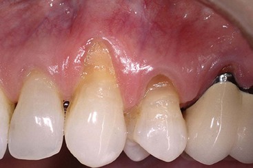


In the APT method, an incision is made in a papilla adjacent to a tooth with recession while the adjacent papilla is tunneled. The next papilla is incised, and the following papilla tunneled. Intrasulcular incisions are made facial to each tooth and interproximally at each tunneled papilla. The papilla in the anatomic midline is always tunneled to reduce tension and retraction of the recipient pouch. At each incised papilla, a V-incision (or ∧ in the mandibular arch) is made to form a new surgical papilla tip approximately 3 mm from the anatomic papilla tip. The portion of the anatomic papilla coronal to the surgical papilla is denuded to expose a vascular recipient bed for the surgical papilla when coronally advanced. The initial dissection is performed with a micro-periosteal elevator, extending apically past the mucogingival junction and laterally under the facial aspect of the tunneled papillae. The tunneling process is facilitated by the access provided at the incised papillae. The tunneled papillae are lifted from the interdental crest by blunt reflection with a curette. Following blunt reflection, supraperiosteal sharp dissection is used to deepen and mobilize the recipient pouch.
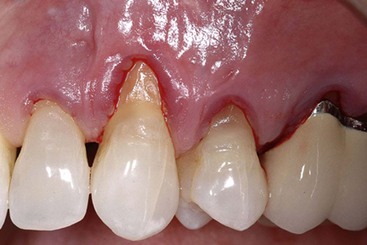
On completion of the recipient site preparation, the length of graft needed is measured and trimmed so that the graft will extend 3 mm past the last tooth with recession at each end of the prepared site. The vertical dimension of the graft should be 6 to 8 mm. The rehydrated and trimmed allograft is then placed into the surgical pouch, with the basement membrane surface facing outward, and secured coronally with 6-0 sling sutures. The graft should be well adapted to the root surface, extending to but not coronal to the cementoenamel junction (CEJ) and to the apical margin, but not over the papillary recipient beds. The pouch is then coronally advanced to completely cover the allograft and secured with 6-0 or 7-0 sling sutures.
The APT method combines the advantages of surgical access at the incised papillae with retraction resistance and wound stability at the tunneled papillae.
In the PRP method, all papillae are tunneled. Initially, intrasulcular incisions are made facial and proximal to all teeth to be treated plus an additional tooth at each end. Next, full-thickness elevation of the margin is initiated with a micro-periosteal elevator extending apically past the mucogingival junction and laterally under the facial aspect of the papillae. The pouch is extended apically and mobilized by supraperiosteal sharp dissection, and the papillae are lifted from the interdental crest as in the APT method. The allograft is rehydrated, measured, and trimmed. Placement of the allograft within the pouch may be accomplished by drawing it in with a suture or placing it through the sulcus with a curette so that it is aligned within the pouch over the exposed roots.
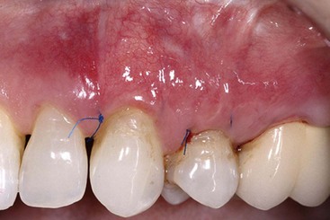
The unique feature of this method is the suturing of the allograft with a subgingival continuous subgingival double-back sling suture. Starting at the anterior end of the graft, the needle of a 6-0 suture is passed from the lingual side through the mesial embrasure and captured on the facial side. The allograft is engaged at the mesial line angle of the tooth, the needle is passed back through the mesial embrasure to the lingual side, around the lingual aspect toward the distal, through the distal embrasure back to the facial and crosses under the papilla to engage the allograft at the mesial line angle of the next tooth. This process continues until the needle has engaged the graft at the mesial line angle of the most distal tooth. At this point, the needle is passed through the mesial embrasure, around the lingual aspect, back to the facial through the distal embrasure, and the graft is engaged at the distal line angle. The needle is then passed back through the distal embrasure, around the lingual aspect, through the mesial embrasure to the facial, and under the papilla to engage the allograft at the distal line angle of the adjacent tooth. This process continues until passing through the distal embrasure of the initial tooth in which the suture is tied with the knot on the lingual side. The entire suture resides subgingivally and draws the graft and the pouch coronally. The PRP procedure is completed by coronally advancing the pouch over the allograft with a series of interrupted 6-0 or 7-0 sling sutures.
Advantages of the PRP method include enhanced retraction resistance, graft containment, and wound stability.
Postoperative care is similar for both methods and includes the following:
Developed over 15 years ago, ADM is a safe and effective biomaterial for use as a substitute for palatal connective tissue in root coverage grafting. There have been no reports of any disease transmission in medical or dental applications over this time period. ADM has proven equivalence to palatal connective tissue for root coverage procedures in randomized controlled clinical trials. It produces a thicker marginal tissue and a higher percentage of root coverage than a coronally advanced flap alone.7 It provides advantages over palatal connective tissue in that it does not require a second surgical site to obtain donor tissue and provides an unlimited amount of tissue to treat multiple teeth in one appointment. The use of AlloDerm under a coronally advanced flap extends the application of the most esthetic procedure in root coverage.
1 Aichelmann-Reidy ME, Yukna RA, Evans GH, et al. Clinical evaluation of acellular allograft dermis for the treatment of human gingival recession. J Periodontol. 2001;72(8):998-1005.
2 Cummings LC, Kaldahl WB, Allen EP. Histologic evaluation of autogenous connective tissue and acellular dermal matrix grafts in humans. J Periodontol. 2005;76(2):178-186.
3 Gapski R, Parks CA, Wang H-L. Acellular Dermal Matrix for Mucogingival Surgery: A Meta-Analysis. J Periodontol. 2005;76(11):1814-1822.
4 Novaes AB, Grisi DC, Molina GO. Comparative 6-month clinical study of a subepithelial connective tissue graft and acellular dermal matrix graft for the treatment of gingival recession. J Periodontol. 2001;72(11):1477-1484.
5 Paolantonio M, Dolci M, Esposito P, et al. Subpedicle acellular dermal matrix graft and autogenous connective tissue graft in the treatment of gingival recession: A comparative 1-year study. J Periodontol. 2002;73(11):1299-1307.
6 Tal H, Moses O, Zohar R, et al. Root coverage of advanced gingival recession: A comparative study between acellular dermal matrix allograft and subepithelial connective tissue grafts. J Periodontol. 2002;73(12):1405-1411.
7 Woodyard JG, Greenwell H, Hill M, et al. The clinical effect of acellular dermal matrix on gingival thickness and root coverage compared to coronally positioned flap alone. J Periodontol. 2004;75(1):44-56.