CHAPTER 77 SUPPLEMENT A Implant-Related Complications and Failures
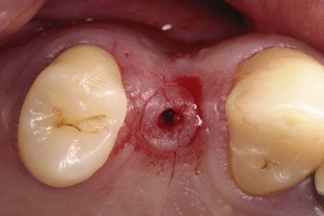
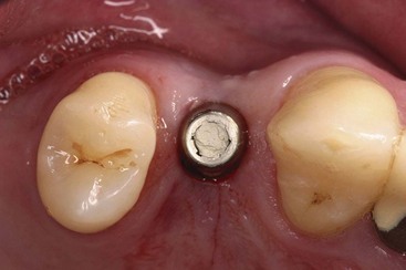
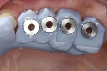
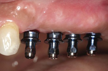
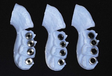
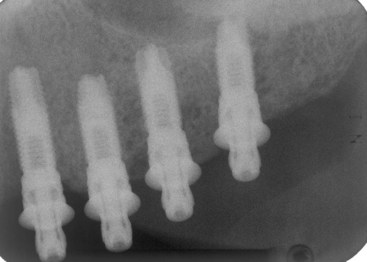
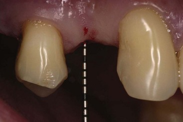
Flapless implant surgery is the placement of a dental implant without the elevation of the epithelium, connective tissue, and periosteum covering the alveolar bone. Flapless surgery is performed by removing a small circular section of tissue (Supplement A Figure 77-1), preparing the osteotomy, and placing the implant (Supplement A Figure 77-2) without flap reflection.1 Due to the lack of operator visualization of the alveolus when preparing and placing implants in a flapless procedure, there are potential complications. The most common complication associated with this technique is implant malposition. When performing flapless surgery, in order to reduce potential malposition complications, it is recommended that a three-dimensional computer tomographic (CT) or cone beam (CB) scan be taken and an anatomically correct surgical guide be used in conjunction or generated with a computer-generated surgical guide. There are several systems (i.e., Nobelguide, #i guide, Materialize I Dent) that can generate a precise surgical guide with metal sleeves of exact dimension that can direct the implant into proper position (Supplement A Figures 77-3 to 77-6). These guides are usually used in fully edentulous arches and have fixation screws that keep them in place (please reference section on CT scans). When using a flapless approach, before initiating the osteotomy, the operator should sound the soft tissue with a periodontal probe to ascertain the tissue thickness and account for this dimension when determining the final position of the implant platform in the apical−coronal dimension (Supplement A Figure 77-7). A common complication is placement of the implants apical to the crestal bone since it is difficult, even with a punch, to visualize the alveolar bony crest in relation to the platform of the implant. If this occurs, it is difficult to fully seat the abutment. Therefore, the relationship between implant and abutment connection must be verified by periapical radiographs. If bone prevents abutment seating, the implant must be backed out until it is at the crest or the bone and must be removed with a profile drill. If a fenestration occurs when placing the dental implant, full flap reflection is required, and GBR procedures should be performed as a means of augmenting the alveolous and covering the fenestration.