CHAPTER 15 Microbiology and Treatment of Endodontic Infections
Apical periodontitis is essentially an inflammatory disease of microbial etiology primarily caused by infection of the root canal system.233 Although chemical and physical factors can induce periradicular inflammation, a large body of scientific evidence indicates that endodontic infection is essential to the progression and perpetuation of the different forms of apical periodontitis.23,110,160,287 Endodontic infection develops in root canals devoid of host defenses, either as a consequence of pulp necrosis (as a sequel to caries, trauma, periodontal disease, or iatrogenic operative procedures) or pulp removal for treatment.
Although fungi and most recently archaea and viruses have been found in association with endodontic infections,217,264,273,305 bacteria are the major microorganisms implicated in the pathogenesis of apical periodontitis. In advanced stages of the endodontic infectious process, bacterial organizations resembling biofilms can be observed adhered to the canal walls.161,167,257 Therefore, there is a current trend to include apical periodontitis in the roll of biofilm-induced oral diseases. Bacteria colonizing the root canal system enter in contact with the periradicular tissues via apical/lateral foramens or root perforations. As a consequence of the encounter between bacteria and host defenses, inflammatory changes take place in the periradicular tissues and give rise to the development of apical periodontitis. Depending on several bacterial and host-related factors, endodontic infections can lead to acute or chronic apical periodontitis.
The ultimate goal of the endodontic treatment is either to prevent the development of apical periodontitis or, in cases where the disease is already present, to create adequate conditions for periradicular tissue healing. Because apical periodontitis is an infectious disease, the rationale for endodontic treatment is unarguably to eradicate the occurring infection and/or prevent microorganisms from infecting or reinfecting the root canal or the periradicular tissues. The cardinal principle of any healthcare profession is the thorough understanding of disease etiology and pathogenesis, which provides a framework for effective treatment. In this context, understanding the microbiologic aspects of apical periodontitis is the basis for endodontic practice of high quality and founded on solid scientific insight. This chapter focuses on diverse aspects of endodontic microbiology, including pathogenetic, taxonomic, morphologic, and ecological issues. Special emphasis is also placed on the underlying principles for sound antimicrobial treatment of endodontic infections.
The first recorded observation of bacteria in the root canal dates back to the 17th century and the Dutch amateur microscope builder Antony van Leeuwenhoek (1632-1723). He reported that the root canals of a decayed tooth “were stuffed with a soft matter” and that “the whole stuff” seemed to him to be alive,54 but the role of Leeuwenhoek’s “animalcules” in disease causation was unsuspected at that time. It took almost 200 years before his observation was confirmed and a cause-and-effect relationship between bacteria and apical periodontitis was suggested.
This occurred specifically in 1894, when Willoughby Dayton Miller, an American dentist working at the laboratory of Robert Koch in Berlin, Germany, published a milestone study reporting on the association between bacteria and apical periodontitis after the analysis of samples collected from root canals.155 By means of bacterioscopy of the canal samples, he found bacterial cells in the three basic morphologies known at the time: cocci, bacilli, and spirilla (or spirochetes) (Fig. 15-1). Morphologically, the endodontic microbiota was clearly different in the coronal, middle, and apical parts of the root canal. Spirochetes were found in high frequencies in abscessed cases, and an etiologic role was suspected for these bacteria. Most of the bacteria Miller had seen under light microscopy could not be cultivated using the technology available at his time. Those bacteria were conceivably anaerobic bacteria, which were only successfully cultivated about 50 to 100 years later with the advent of anaerobic culture techniques. However, it is now widely recognized that a large number of bacterial species living in diverse environments still remain to be cultivated by current technology,6,200 and the root canal is no exception (discussed later in this chapter). Based on his findings, Miller raised the hypothesis that bacteria were the causative agents of apical periodontitis.
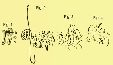
FIG. 15-1 Drawings from Miller’s classic paper showing different bacterial forms in a root canal sample observed by microscopy.
Approximately 70 years after Miller’s classic study, his assumptions were confirmed by an elegant study from Kakehashi et al.110 These authors investigated the response of the dental pulps of conventional and germ-free rats after exposure to the oral cavities. Histologic evaluation was performed and revealed that whereas pulp necrosis and apical periodontitis lesions developed in all conventional rats, the pulps of germ-free rats not only remained vital but also repaired themselves by hard-tissue formation. Dentinlike tissue sealed the exposure area and isolated the pulps again from the oral cavity.
The important role of bacteria in the etiology of apical periodontitis was further confirmed by Sundqvist’s classic study.287 This author applied advanced anaerobic culturing techniques to the evaluation of bacteria occurring in the root canals of teeth whose pulps became necrotic after trauma. Bacteria were found only in the root canals of teeth exhibiting radiographic evidence of apical periodontitis, confirming the infectious etiology of this disease. Anaerobic bacteria accounted for more than 90% of the isolates. Findings from Sundqvist’s study also served to demonstrate that in the absence of infection, the necrotic pulp tissue itself and stagnant tissue fluid in the root canal cannot induce and perpetuate apical periodontitis lesions.
Möller et al.160 also provided strong evidence about the microbial causation of apical periodontitis. Their study using monkeys’ teeth demonstrated that only devitalized pulps that were infected induced apical periodontitis lesions, whereas devitalized and noninfected pulps showed an absence of significant pathologic changes in the periradicular tissues. In addition to corroborating the importance of microorganisms for the development of apical periodontitis, this study also confirmed that necrotic pulp tissue per se is unable to induce and maintain an apical periodontitis lesion.
Under normal conditions, the pulpodentin complex is sterile and isolated from oral microbiota by overlying enamel and cementum. In the event that the integrity of these natural layers is breached (e.g., as a result of caries, trauma-induced fractures and cracks, restorative procedures, scaling and root planning, attrition, abrasion) or naturally absent (e.g., because of gaps in the cemental coating at the cervical root surface), the pulpodentin complex is exposed to the oral environment and then challenged by microorganisms present in caries lesions, in saliva bathing the exposed area, or in dental plaque formed onto the exposed area. Microorganisms from subgingival biofilms associated with periodontal disease may also have access to the pulp via dentinal tubules at the cervical region of the tooth and lateral and apical foramens. Microorganisms may also have access to the root canal any time during or after professional endodontic intervention.
Whenever dentin is exposed, the pulp is put at risk of infection as a consequence of the permeability of normal dentin dictated by its tubular structure (Fig. 15-2). Dentinal tubules traverse the entire width of the dentin and have a conical conformation, with the largest diameter located near the pulp (mean, 2.5 µm) and the smallest diameter in the periphery, near the enamel or cementum (mean, 0.9 µm).75 The smallest tubule diameter is entirely compatible with the cell diameter of most oral bacterial species, which usually ranges from 0.2 to 0.7 µm. One might well assume that once exposed, dentin offers an unimpeded access pathway for bacteria to reach the pulp via tubules. This is not necessarily the case. It has been demonstrated that bacterial invasion of dentinal tubules occurs more rapidly in nonvital teeth than in vital ones.164 In vital teeth, outward movement of dentinal fluid and the tubular contents (including odontoblast processes, collagen fibrils, and the sheathlike lamina limitans that lines the tubules) influence dentinal permeability and can conceivably delay intratubular invasion by bacteria. Because of the presence of tubular contents, the functional or physiologic diameter of the tubules is only 5% to 10% of the anatomic diameter seen by microscopy.152 Other factors such as dentinal sclerosis beneath a carious lesion, tertiary dentin, smear layer, and intratubular deposition of fibrinogen also reduce dentin permeability and thereby limit or even impede bacterial progression to the pulp via dentinal tubules.183 Host defense molecules, such as antibodies and components of the complement system, may also be present in the dentinal fluid of vital teeth and can assist in the protection against deep bacterial invasion of dentin.3,172,173 So long as the pulp is vital, dentinal exposure does not represent a significant route of pulpal infection, except when dentin thickness is considerably reduced and dentin permeability is significantly increased.
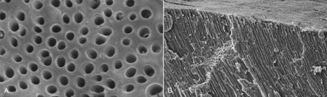
FIG. 15-2 Scanning electron micrographs of dentin showing tubules in cross-sectional (A), and longitudinal (B), views (A-B, ×850 and ×130, respectively).
Most of the bacteria in the carious process are nonmotile; they invade dentin by repeated cell division which pushes cells into tubules. Bacterial cells may also be forced into tubules by hydrostatic pressures developed on dentin during mastication.153 Bacteria inside tubules under a deep carious lesion can reach the pulp even before frank pulpal exposure.101 As mentioned, it has been assumed that the pulp will not be infected if it is still vital, and the few bacteria that reach the pulp can be of no importance, since the vital pulp can eliminate such a transient infection and rapidly clear or remove bacterial products. This efficient clearance mechanism tends to prevent injurious agents from reaching a high enough concentration to induce significant inflammatory reactions.182 On the other hand, if the vitality of the pulp is compromised and the defense mechanisms are impaired, even very few bacteria may initiate infection.
Bacteria have been isolated from traumatized teeth with necrotic pulps and apparently intact crowns.287,318 It has been suggested that bacteria from the gingival crevice or periodontal pockets might reach the root canals of teeth whose pulps became necrotic after trauma through severed blood vessels of the periodontium—a phenomenon called anachoresis.86 However, this theory has never been supported by scientific evidence. In fact, trauma can induce exposure of dentin by fracturing the crown or inducing the formation of enamel cracks. Macro- and microcracks in enamel can be present in most teeth (not only traumatized ones) and do not necessarily end at the enamel-dentin junction but extend deep into the dentin.142 A large number of dentinal tubules can be exposed to the oral environment by a single crack. These cracks can be clogged with dental plaque and provide portals of entry for bacteria. If the pulp remains vital after trauma, bacterial penetration into tubules is counteracted by the dentinal fluid and tubular contents, as discussed earlier, and pulpal health is not usually jeopardized. But if the pulp becomes necrotic as a consequence of trauma, it loses the ability to protect itself against bacterial invasion, and regardless of dentin thickness, dentinal tubules then will become true avenues through which bacteria can reach and colonize the necrotic pulp.
Direct exposure of the dental pulp to the oral cavity is the most obvious route of endodontic infection. Caries is the most common cause of pulp exposure, but bacteria may also reach the pulp via direct pulp exposure as a result of iatrogenic restorative procedures or trauma. The exposed pulp tissue enters in direct contact with oral bacteria from caries lesions, saliva, and/or plaque accumulated onto the exposed surface. Almost invariably, exposed pulps will undergo inflammation and necrosis and become infected. The time elapsed between pulp exposure and infection of the entire canal is unpredictable, but it is usually a slow process.45
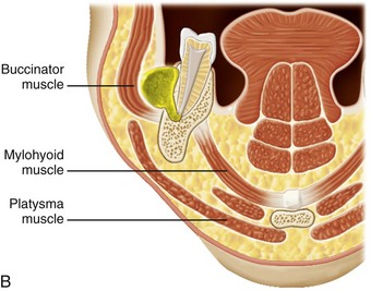
The egress of microorganisms and their products from infected root canals through apical, lateral, or furcation foramina, dentinal tubules, and iatrogenic root perforations can directly affect the surrounding periodontal tissues and give rise to pathologic changes in these tissues. However, there is no consensus as to whether the opposite is true, that is, whether subgingival biofilms associated with periodontal disease can directly cause pulpal disease. Conceptually, microorganisms in subgingival plaque biofilms associated with periodontal disease could reach the pulp by the same pathways intracanal microorganisms reach the periodontium and could thereby exert harmful effects on the pulp. However, it has been demonstrated that although degenerative and inflammatory changes of different degrees may occur in the pulp of teeth with associated marginal periodontitis, pulpal necrosis as a consequence of periodontal disease only develops if the periodontal pocket reaches the apical foramen, leading to irreversible damage to the main blood vessels that penetrate through this foramen to irrigate the pulp127 (Fig. 15-3). After the pulp becomes necrotic, periodontal bacteria can reach the root canal system via exposed dentinal tubules at the cervical area of the root or via lateral and apical foramina to establish an endodontic infectious process.
FIG. 15-3 Periodontal disease (A) mostly affects the pulp vitality when the subgingival biofilm reaches the apical foramen (B).
It has been claimed that microorganisms can reach the pulp by anachoresis. Theoretically, microorganisms are transported in the blood or lymph to an area of tissue damage, where they leave the vessel, enter the damaged tissue, and establish an infection77,203; there is no clear evidence showing that this process can represent a route for root canal infection. It has been revealed that bacteria could not be recovered from unfilled root canals when the bloodstream was experimentally infected, unless the root canals were overinstrumented during the period of bacteremia, with resulting injury to periodontal blood vessels and blood seepage into the canal.51 Another argument against anachoresis as a route of pulpal infection comes from the study by Möller et al.,160 who induced pulpal necrosis in monkeys’ teeth and reported that all cases of aseptic necrosis remained bacteria-free after 6 to 7 months of observation. Although anachoresis has been suggested to be the mechanism through which traumatized teeth with seemingly intact crowns become infected,86 current evidence indicates the main pathway of pulpal infection in these cases is dentinal exposure due to enamel cracks.141,142
Whatever the route of bacterial access to the root canal, necrosis of pulp tissue is a prerequisite for the establishment of primary endodontic infections. To reiterate: if the pulp is vital, it can protect itself against bacterial invasion and colonization. If the pulp becomes necrotic due to caries, trauma, operative procedures, or periodontal disease, it can be very easily infected. This is because host defenses do not function in the necrotic pulp tissue, and those in the periradicular tissues do not reach deep into the root canal space.
Another situation in which the root canal system is devoid of host defenses relates to cases where the pulp was removed for treatment. Microbial penetration in the canal can occur during treatment, between appointments, or even after root canal filling. The main causes of microbial introduction into the canal during treatment include remnants of dental biofilm, calculus or caries on the tooth crown; leaking rubber dam; contamination of endodontic instruments (e.g., after touch with the fingers); and contamination of irrigant solutions or other solutions of intracanal use (e.g., saline solution, distilled water, citric acid). Microorganisms can also enter the root canal system between appointments by leakage through the temporary restorative material; breakdown, fracture, or loss of the temporary restoration; fracture of the tooth structure; and in teeth left open for drainage. Microorganisms can penetrate the root canal system even after completion of the root canal filling by leakage through the temporary or permanent restorative material; breakdown, fracture, or loss of the temporary/permanent restoration; fracture of the tooth structure; recurrent decay, exposing the root canal filling material; or delay in placement of permanent restorations.242
The ability of a microorganism to cause disease is regarded as pathogenicity. Virulence denotes the degree of pathogenicity of a microorganism, and virulence factors are the microbial products, structural components, or strategies that contribute to pathogenicity. Bacterial virulence factors comprise structural cellular components and released products. Bacterial strategies that contribute to pathogenicity include the ability to coaggregate and form biofilms, which confers protection against host defenses and antimicrobial agents. Some microorganisms routinely cause disease in a given host and are called primary pathogens. Other microorganisms cause disease only when host defenses are impaired and are called opportunistic pathogens. Bacteria that make up the normal microbiota are present as harmless commensals and live in balance with the host. One of the greatest beneficial effects of human microbiota is probably the tendency to protect the host from exogenous infections by excluding other microorganisms. Nevertheless, in certain situations, the balance may be disturbed by a decrease in the normal level of resistance, and then the commensal bacteria are usually the first to take advantage. Most bacteria involved with endodontic infections are normal inhabitants of the oral microbiota that exploit changes in the balance of the host/bacteria relationship, becoming opportunistic pathogens and causing endogenous infections.
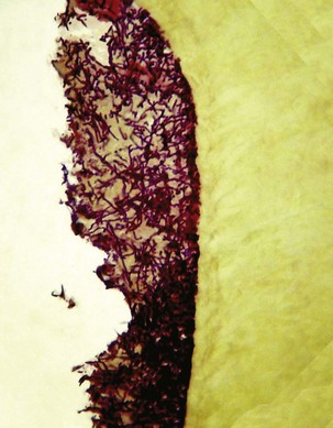
Bacteria involved with the pathogenesis of primary apical periodontitis may have participated in the early stages of pulp inflammation and necrosis, or they may have gained entry into the root canal space any time after pulpal necrosis. In the former situation, involved bacteria are usually those present in the advanced front of caries lesions and from saliva bathing the affected area. Bacteria in caries lesions form authentic biofilms adhered to dentin (Fig. 15-4). Diffusion of bacterial products through dentinal tubules induces pulpal inflammation long before the tissue is exposed. After pulp exposure, the surface of the tissue can also be colonized and covered by bacteria present in the caries biofilm. The exposed pulp tissue is in direct contact with bacteria and their products and responds with severe inflammation. Some tissue invasion by bacteria may also occur. Bacteria in the battlefront have to survive the attack from the host defenses and at the same time acquire nutrients to keep themselves alive. In this bacteria/pulp clash, the latter invariably is “defeated” and becomes necrotic, so bacteria move forward and “occupy the territory,” that is, colonize the necrotic tissue. These events advance through tissue compartments, coalesce, and move towards the apical part of the canal until virtually the entire root canal is necrotic and infected (Fig. 15-5). At this stage, involved bacteria can be regarded as the early root canal colonizers or pioneer species.
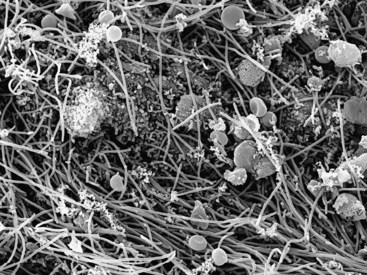
FIG. 15-4 Scanning electron micrograph showing a bacterial biofilm covering dentin in a deep carious lesion. Note the presence of different bacterial morphotypes (×3500).
(From Torabinejad M, Walton RE: Endodontics: principles and practice, ed 4, Saunders/Elsevier, St. Louis, 2009.)
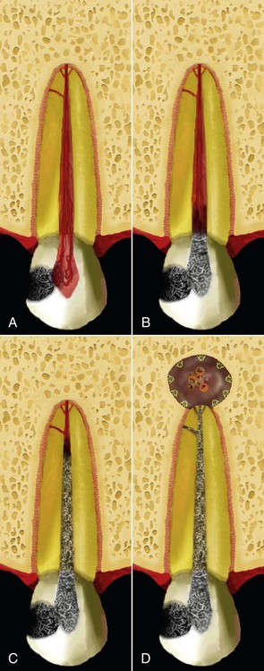
FIG. 15-5 Dynamics of pulp inflammation, necrosis, and infection from caries exposure (A) to apical periodontitis formation (D).
Bacteria colonizing the necrotic root canal induce damage to the periradicular tissues and give rise to inflammatory changes. In fact, periradicular inflammation can be observed even before the entire root canal is necrotic.7,161,279,320 Therefore, the early colonizers play an important role in the initiation of the apical periodontitis disease process. The environmental conditions in the root canal are modified by pioneer species and the disease process and may then be conducive to the establishment of bacterial groups different from the early colonizers. Once the pulp is necrotic, species other than those that participated in the initial infectious process may also have access to the canal via coronal exposure or exposed dentinal tubules, and a shift in the microbiota is observed. Rearrangement in the proportions of the pioneer species and latecomers occurs, and as the environment changes, some early colonizers are expected to no longer participate in the consortium in advanced disease. With the passage of time, the endodontic microbiota becomes more and more structurally and spatially organized.
Some virulence attributes required for pathogens to thrive in other sites may be of no value for bacteria that reach the root canal after necrosis—for instance, the ability to evade host defenses. This is because latecomers face no significant opposition from host defenses, which are no longer active in the canal after necrosis. Although colonization may appear an easy task for late colonizers, other environmental factors (e.g., interaction with pioneer species, oxygen tension, nutrient availability) will determine whether new species entering the canal will succeed in establishing themselves and join the early colonizers to make up a dynamic mixed community in the root canal. Ultimately, the root canals of teeth evincing radiographically detectable apical periodontitis lesions harbor both early colonizers that managed to stay in the canals and late colonizers that managed to adapt to the new but propitious environmental conditions.248
Bacteria exert their pathogenicity by wreaking havoc on the host tissues through direct and/or indirect mechanisms. Bacterial virulence factors that cause direct tissue harm include those that damage host cells and/or the intercellular matrix of the connective tissue. These factors usually involve secreted products, including enzymes, exotoxins, heat-shock proteins and metabolic end products.231 Furthermore, bacterial structural components, including lipopolysaccharide (LPS), peptidoglycan, lipoteichoic acid, fimbriae, flagella, outer membrane proteins and vesicles, lipoproteins, DNA, and exopolysaccharides can act as modulins by stimulating the development of host immune reactions capable not only of defending the host against infection but also of causing severe tissue destruction98,248,301 (Fig. 15-6). For instance, inflammatory and noninflammatory host cells can be stimulated by bacterial components to release chemical mediators such as cytokines and prostaglandins, which are involved in the induction of bone resorption characteristically observed in chronic apical periodontitis lesions.278 Another example of indirect damage caused by bacteria is the formation of purulent exudate in acute apical abscesses. Host defense mechanisms against bacteria emanating from the root canal appear to be the most important factor involved in formation of purulent exudate associated with abscesses. Formation of oxygen-derived free radicals, such as superoxide and hydrogen peroxide, alongside the release of lysosomal enzymes by polymorphonuclear leukocytes, gives rise to destruction of the connective extracellular matrix, leading to pus formation.300 Although bacterial products may certainly be involved in the pathogenesis of diseases related to endodontic infections, bacterial indirect destructive effects seem to be more significant in the tissue damage associated with acute and chronic apical periodontitis lesions.
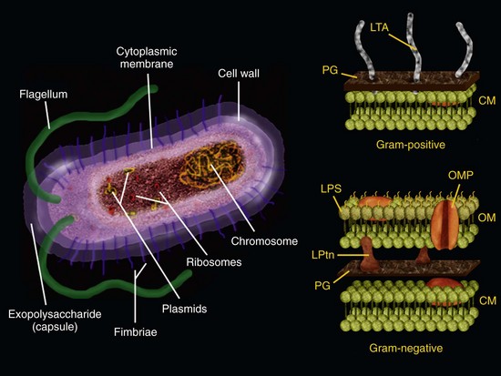
FIG. 15-6 Bacterial cell and its structural components that can act as virulence factors. Right, a detailed scheme of the bacterial cell walls from gram-positive and gram-negative bacteria. CM, Cytoplasmic membrane; LPS, lipopolysaccharide (endotoxin); LPtns, lipoproteins; LTA, lipoteichoic acid; OM, outer membrane; OMP, outer membrane protein; PG, peptidoglycan.
Depending on several factors, apical periodontitis can be chronic or acute. A chronic disease is usually associated with low virulence of the bacterial consortium involved, but this represents a persistent source of aggression to the tissues. Persistence in chronic infections is usually related to community organization in biofilms and the inaccessibility to host defenses because of the anatomic location of the infection. In chronic infections, the juxtaposition of bacterial biofilms to susceptible tissues that are not adapted to their presence triggers harmful inflammatory responses.42 Thus, bacteria in the necrotic root canal cause chronic infection by forming biofilms on the canal walls and maintaining a close contact with the apical periodontal ligament, which reacts in a damaging way because periodontal ligament and bone cells are never adapted to the presence of bacteria. An acute infection in turn is usually caused by a high virulent bacterial consortium. High virulence may be a result of synergism between species. Acute infections are usually related to bacterial cells in a planktonic state, at high numbers, with tissue invasion ability, counteracted by a diminished host resistance. It has been demonstrated for some pathogens that genes coding for many virulence factors are much more expressed in planktonic cells than in sessile (biofilm) cells, suggesting that planktonic cells are more likely to participate in acute infections.73
Apical periodontitis is a multifactorial disease that is resultant of the interplay of many host and bacterial factors. Few if any of the putative endodontic pathogens are individually capable of inducing all of the events involved in the pathogenesis of the different forms of apical periodontitis. Probably, the process requires an integrated and orchestrated interaction of the selected members of the mixed endodontic microbiota and their respective virulence attributes. Although LPS is undoubtedly the most studied and quoted virulence factor, it sounds simplistic to ascribe to this molecule all responsibility for apical periodontitis causation. This statement is further reinforced by the fact that some cases of primary infections and many cases of secondary/persistent infections harbor exclusively gram-positive bacteria. Therefore, the involvement of other factors must not be overlooked. In fact, the pathogenesis of different forms of apical periodontitis and even the same form in different individuals is unlikely to follow a stereotyped course with regard to the bacterial mediators involved.248
Individual microorganisms proliferating in a habitat give rise to populations. Such populations often occur as microcolonies in the environment. Populations interact with one another to form a community. Thus, community refers to a unified assemblage of populations that coexist and interact at a given habitat. The community and habitat are part of a larger system called an ecosystem, which can be defined as a functional self-supporting system that includes the microbial community and its environment. To sum up, the following hierarchy becomes apparent: ecosystem, community, population, and the single individual (cell).
Populations perform functions that contribute to the overall community and maintain the ecological balance of the ecosystem. Each population occupies a functional role (niche) within the community. There are a limited number of niches within the community for which populations must compete. More competent populations occupy the niches and displace those less competent. As discussed later, highly structured and spatially organized microbial communities may exhibit properties that are greater than the sum of the component populations. In reality, complex microbial communities have been shown to be endowed with the ability to confront and withstand the challenges imposed by the environment by creating a mosaic of microenvironments that enable the survival and growth of the community members.146
Historically, microbiologists have faced periods of “reductionism” and “holism.”121 Reductionism is based on the idea that the whole can be understood by examining smaller and smaller pieces of it, that is, all complex systems can be completely understood in terms of their individual components. Through reductionist approaches, individual species are isolated from complex mixed communities and metabolically and genetically studied so as to allow understanding of the community by examining every single constituent. However, it has become quite apparent for the microbiota associated with many human infectious diseases that the whole is very often greater than the simple sum of its parts. This concept has prompted microbiologists to adopt a holistic approach to understand the community behavior associated with pathogenesis of many infectious diseases known to have a polymicrobial etiology. Holism holds that any component cannot be thoroughly understood except in their relation to the whole. The holistic theory has been largely employed in ecology: the interplay of the different parts composing the ecosystem will ultimately determine its properties.
Traditionally, the study of the etiology of the main human diseases (including oral diseases) has been based on reductionism. More recently, it has been recognized that the biofilm (dental plaque) associated with caries and periodontal diseases represents a sophisticated community that exert functions essential for the biofilm architecture and physiology, with consequent pathogenetic implications. Recent evidence indicates that apical periodontitis can also be due to collaborative activities of a biofilm community.
Community profiling studies143,209,261 revealed that bacterial composition of the endodontic microbiota differs consistently between individuals. This indicates that apical periodontitis has a heterogeneous etiology, where multiple bacterial combinations can play a role in disease causation. Moreover, community structure differs significantly between different disease forms (e.g., chronic apical periodontitis versus acute apical abscess),219,261 suggesting existence of a pattern associated with each form. Differences are still observed between individuals suffering from the same disease, disclosing an interindividual variability even more pronounced when different geographic locations are studied. There is seemingly a geography-related pattern, where individuals from a given location tend to cluster together based on similarities of the microbial community profiles.
The high interindividual variability observed for samples from the same clinical disease indicates that different compositions of bacterial communities can result in similar disease outcomes. Identification of community members can reveal the presence of some species or groups of species that may be important for the causation of some forms of disease. It is reasonable to realize that different magnitudes of disease, based on intensity of signs and symptoms, may be related to the species composition of the community. Indeed, it has been demonstrated that, despite individual uniqueness, bacterial communities associated with acute abscesses (severe pain and swelling) are more similar among them in comparison to bacterial communities associated with chronic apical periodontitis (no pain).219,261 Although there may be no specific species related to some forms of disease, specificity seems to be related to community level, and certain communities (species composition) are specifically associated with some forms of apical periodontitis. From the perspective of the single-pathogen concept, apical periodontitis can be considered as having no specific microbial etiology. However, based on the community-as-a-pathogen concept, it is possible to infer that some communities are more related to certain forms of the disease.219,261
The community-forming ability can be regarded as essential for microbial survival in virtually all environments. Indeed, the vast majority of microorganisms in nature invariably grow and function as members of metabolically integrated communities, or biofilms.42,147 Biofilm can be defined as a sessile multicellular microbial community characterized by cells that are firmly attached to a surface and enmeshed in a self-produced matrix of extracellular polymeric substance (EPS), usually polysaccharide42,55 (Fig. 15-7). The ability to form biofilms has been regarded as a virulence factor,90 and biofilm infections account for an estimated 65% to 80% of bacterial infections that affect humans in the developed world.41 Given its importance in varied aspects, there has been a high level of interest in the study of biofilm properties, not only in medical microbiology but also in different sectors of industrial and environmental microbiology.
Biofilms are not merely passive assemblages of bacterial cells that are stuck to surfaces; they are structurally and dynamically organized complex biologic systems. Bacterial cells in biofilms form microcolonies (±15% by volume) that are embedded and nonrandomly distributed in the EPS matrix (±85% by volume) and separated by water channels.44,55,276,281 Microcolonies are usually shaped as “towers” or “mushrooms.” Dental biofilms can reach up to 300 or more cell layers in thickness.276 Individual microcolonies may consist of a single bacterial species but more frequently are composed of several different species in a mixed community.
As the biofilm matures on the surface, extracellular polysaccharides are continually synthesized to form an extracellular matrix that eventually may constitute as much as 85% of the volume of the biofilm.41 Although the matrix is primarily composed of polysaccharides, it can also contain proteins and nucleic acids.90 The matrix is not only important physically as part of the scaffold that determines the biofilm structure, but it is also biologically active and can retain nutrients, water, and essential enzymes within the biofilm.5 The matrix can also protect the biofilm community from exogenous threats and may participate in adherence to the surface.
Community members form distinct populations or microcolonies separated by open water channels that traverse the biofilm matrix and create primitive circulatory systems.43 Fluid in these channels carries substrate, end products of bacterial metabolism and signal molecules involved in bacterial interactions.27 Thus, vital nutrients and communication molecules can diffuse, and wastes can be washed out through these channels.
Microcolonies that form in the biofilm arise from surface colonization by planktonic (unattached) bacterial cells. During the early stages of biofilm formation, bacteria bind to many host proteins and coaggregate with other bacteria. These interactions lead to changes in growth rate, gene expression, and protein production. It has been demonstrated by proteomic techniques or DNA arrays that genes expressed by cells in biofilms differed by 20% to 70% from those expressed by the same cells growing in planktonic culture.20,174,222 Thus, bacteria in biofilms adopt a radically different phenotype compared to their planktonic counterparts. Within biofilms, sophisticated systems of cell-cell communication (quorum sensing) are also used by some bacteria to coordinate gene expression. Phenotypic heterogeneity in biofilms is also observed as a result of exposure of microcolonies to a variety of gradients (e.g., oxygen tension, pH, osmolarity, type and amounts of nutrients, cell density), which contribute to form diverse microenvironments throughout the biofilm structure.
As a community of bacteria, biofilms have a collective physiology, responding in concert to environmental challenges. Many naturally occurring biofilms have a highly diverse microbiota. The component species form populations (microcolonies) that are not randomly distributed but are spatially and functionally organized throughout the mixed community.
The properties displayed by a mixed biofilm community are mostly dictated by the interactions between populations, which create novel physiologic functions that cannot be observed with individual components. Populations are strategically positioned for optimal metabolic interaction, and the resultant architecture favors the ecological role of the community in the ecosystem. The biofilm community lifestyle affords a number of advantages to colonizing bacteria, including those discussed in the sections that follow.148
The metabolism of early colonizers alters the local environment, setting the stage for attachment and growth of latecomers (including more fastidious species). This may include increased availability of some types of nutrients, removal of harmful metabolic end products, and the development of appropriate physicochemical conditions (e.g., reduced oxygen tension and redox potential). As a consequence, the diversity of the microbiota increases over time because of bacterial succession.
Nutrients are limited in most ecosystems, so the communities that can fix and process their resources most efficiently will thrive and be perpetuated. Bacteria living in mixed communities take part in a number of nutritional interrelationships, and food webs develop. For instance, products of the metabolism of one species may become the main source of nutrients for other species. Also in food webs, the concerted action of interacting species is required for the sequential breakdown of complex host-derived substrates to simpler products, which can favor less competent species. Many complex host macromolecules, especially glycoproteins, can only be degraded efficiently by mixed consortia of oral bacteria. Byproducts of the degradation of complex nutrients are trapped in the biofilm matrix and shared with other community members.
Neighboring cells of a different species can produce enzymes such as beta-lactamases, catalase, and proteinases that are retained in the biofilm matrix and protect other bacteria against antimicrobials and host defenses. The same is true for metabolites and bacteriocins, which can inhibit competing species. The biofilm matrix also confers physical protection against phagocytosis.
Cell-cell associations and the biofilm architecture facilitate horizontal gene transfer in the community. Potential mechanisms mediating genetic exchange in biofilms include conjugation, transformation, and transduction. The main consequences of genetic exchanges among community members are related to the acquisition and dissemination of virulence and antibiotic-resistance genes.
To cause disease, bacteria must adhere to host surfaces, obtain nutrients from the host, and multiply, invade tissues, overcome or evade the host defenses, and induce tissue damage. A diverse range of virulence traits are required for these particular stages of the disease process, and it is highly likely that each will require the concerted action of bacteria in a community. Similarly, it is possible that certain species can have more than one role in disease, and different species can perform similar functions. This helps explain why communities with different bacterial composition can be found in different individuals with similar disease. In mixed communities, a broad spectrum of relationships may arise between the component species, ranging from no effect or reduced pathogenicity to additive or synergistic pathogenic effects. Endodontic abscesses are examples of polymicrobial infections whereby bacterial species that individually have low virulence and are unable to cause disease can do so when in association with others as part of a mixed consortium (pathogenic synergism).30
From a clinical standpoint, biofilm increased resistance to antimicrobial agents is of special concern. Bacteria arranged in biofilms are more resistant to antibiotics than the same cells grown in planktonic state. The antibiotic concentration required to kill bacteria in the biofilm is about 100 to 1000 times higher than that needed to kill the same species in planktonic state.145 There are several possible mechanisms involved with biofilm resistance to antimicrobials.
The agent may adsorb to and inhibit the bacteria at the biofilm surface, leaving cells deeply located in the biofilm relatively unaffected. The matrix in biofilms can also bind and retain neutralizing enzymes at concentrations that could inactivate the antimicrobial agent.280
Many antibiotics can freely penetrate the biofilm matrix, but cells are often still protected. The occurrence of starved bacteria entering the stationary phase in biofilms seems to be a significant factor in the resistance of biofilm populations to antimicrobials. Bacteria grow slowly under conditions of low availability of nutrients in an established biofilm and as a consequence are much less susceptible than faster-dividing cells. Most antibiotics require at least some degree of cellular activity to be effective. Therefore, bacterial cells in stationary phase might represent a general mechanism of antibiotic resistance in the biofilm.90
Increased tolerance of some biofilms to antibiotics may be largely due to the presence of a subpopulation of specialized survivor cells known as persisters.113 It remains unclear whether these bacteria actually represent a distinct phenotype or are simply the most resistant cells within a population.90
Quorum sensing is a term currently used for systems based on cell-cell communication that regulate gene expression in a cell density–dependent manner.180 Quorum-sensing systems capacitate bacteria to behave collectively as a group. This phenomenon has been described in both gram-positive and gram-negative bacterial species.10,57,317 Quorum sensing involves the production, release, and subsequent detection of diffusible signaling molecules called autoinducers. Two predominant types of autoinducers, N-acyl-l-homoserine lactones (AHLs) and posttranslationally modified peptides, are used by gram-negative and gram-positive bacteria, respectively. For example, the human pathogens Pseudomonas aeruginosa and Staphylococcus aureus employ AHLs and peptides, respectively, to control the expression of multiple virulence genes in concert with cell population density. Several other small-molecule quorum-sensing autoinducers have recently been discovered, including Pseudomonas quinolone signal (PQS) and AI-2.35
As members of a bacterial population producing and releasing autoinducers multiply, the extracellular concentration of these signaling molecules also increases. When autoinducers reach a crucial threshold level, the group responds with a population-wide alteration in gene expression, activating or repressing target genes.181 Since alterations in gene expression are linked to the presence of autoinducers, bacteria can perform specific functions only when living in groups. Such behavior provides some advantages to a bacterial population, affording adaptability to and protection against threatening environments.
Quorum-sensing systems are known to regulate virulence, competence for DNA uptake, entry into stationary phase, and biofilm formation.35,46,184 With regard to virulence, quorum sensing allows the community to regulate expression of virulence genes in accordance with cell density. Thus, some opportunistic pathogens express virulence factors in response to sensing their own cell density. Using quorum sensing, bacteria can amass a high cell density before virulence factors are expressed, and in so doing, the bacteria are able to make a consolidated attack to overcome the host defenses and establish infection.116 Teleologically, it appears bacteria were able to strategically wait for a critical number of cells to be reached and then start inducing damage, not giving the host sufficient time to mount an effective defense response. In reality, with regard to energy yield, it would be difficult for bacteria to produce some virulence factors before a critical community density is reached. For instance, production of proteinases by a few cells are not expected to have significant impact on the host environment, representing “wasted energy” for the growing population.180
The process of biofilm formation, including surface attachment, proliferation, matrix production, and detachment, is also partially controlled by quorum sensing.74 Moreover, bacteria can monitor crowding as a measure of competition for nutrients. At high cell density, there may be too much competition to utilize available nutrients for growth, and starvation is anticipated. If bacteria are already starved and crowded, as they often are when growing as a biofilm, it may be advantageous to enter stationary phase. Entry into stationary phase dramatically alters patterns of gene expression to allow extended cell survival in the absence of nutrients.128 Control of cell density in biofilms is also important to prevent water circulatory channels, essential for biofilm physiology, from being clogged and then inactivated by cells under excessive growth.
Several oral bacteria have been demonstrated to produce quorum-sensing signaling molecules,71,194,321 indicating that bacteria in endodontic biofilms may also communicate with one another through this system.
The endodontic microbiota has been traditionally investigated by microbiologic culture methods. Culture is the process of propagating microorganisms in the laboratory by providing them with the required nutrients and proper physicochemical conditions, including temperature, moisture, atmosphere, salt concentration, and pH.272 Essentially, culture analyses involve the following steps: sample collection and transport, dispersion, dilution, cultivation, isolation, and identification. Endodontic samples are collected and transported to the laboratory in a viability-preserving, nonsupportive, anaerobic medium. They are then dispersed by sonication or vortex mixing, diluted, distributed onto various types of agar media, and cultivated under aerobic or anaerobic conditions. After a suitable period of incubation, individual colonies are subcultivated and identified on the basis of multiple phenotype-based aspects, including colony and cellular morphology, gram-staining pattern, oxygen tolerance, comprehensive biochemical characterization, and metabolic end-product analysis by gas-liquid chromatography. The outer cellular membrane protein profile as examined by gel electrophoresis, fluorescence under ultraviolet light, and susceptibility tests to selected antibiotics can be needed for identification of some species.61 Marketed packaged kits that test for preformed enzymes have also been used for rapid identification of several species.
Culture analyses of endodontic infections have provided a substantial body of information about the etiology of apical periodontitis, composition of the endodontic microbiota in different clinical conditions, effects of treatment procedures in microbial elimination, susceptibilities of endodontic microorganisms to antibiotics, and so on. Advantages and limitations of culture methods are listed in Box 15-1. As one can tell, however, some important limitations of culture methods make a comprehensive analysis of the endodontic microbiota difficult to achieve.
The difficulties in culturing or identifying many microbial species are of special concern. Unfortunately, not all microorganisms can be cultivated under artificial conditions, and this is simply because the nutritional and physiologic needs of most microorganisms are still unknown. Investigations of many aquatic and terrestrial environments using culture-independent methods have revealed that the cultivable members of these systems represent less than 1% of the total extant population.6,314 Furthermore, 50% to 80% of bacterial species composing the microbiota associated with diverse human sites, including the oral cavity, represent unknown and still uncultivated bacteria.2,58,120,185,282
That a given species has not been cultivated does not imply that this species will remain indefinitely impossible to cultivate. A myriad of obligate anaerobic bacteria were uncultivable 100 years ago, but further developments in anaerobic culturing techniques have to a large extent helped to solve this problem. It must be assumed that no single method or culture medium is suitable for isolating the vast diversity of microorganisms present in most environments.83 While we remain relatively unaware of the requirements for many bacteria to grow, identification methods not based on cultivability are required.
In some situations, even the successful cultivation of a given microorganism does not necessarily mean that this microorganism can be successfully identified. Culture-dependent identification is based on phenotypic traits observed in reference strains, with predictable biochemical and physical properties under optimal growth conditions. However, many phenotype-related factors can lead to difficulties in identification and even to misidentification.19,25,295 As a consequence of all these factors, phenotype-based identification does not always allow an unequivocal identification.
To sidestep the limitations of culture, tools and procedures based on molecular biology have become available and have been substantially improved to achieve a more realistic description of the microbial world without the need for cultivation (Fig. 15-8). Molecular biology techniques have been widely used to directly identify microorganisms in samples without the need for cultivation. In addition, molecular technology has also been applied to reliably identify cultivated bacteria, including strains with ambiguous or aberrant phenotypic behavior, rare isolates, poorly described or uncharacterized bacteria, and newly named species.*
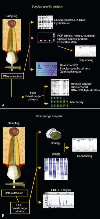
FIG. 15-8 Molecular biology methods used (or with potential to be used) in the study of endodontic infections. The choice for a particular technique will depend on the type of analysis to be performed. Some methods can be used for detection of target microbial species or groups (A) and others for a broader analysis of the microbiota (B).
Molecular approaches for microbial identification rely on certain genes that contain revealing information about the microbial identity. Of the several genes that have been chosen as targets for bacterial identification, the 16S rRNA gene (or 16S rDNA) has been the most widely used because it is universally distributed among bacteria, is long enough to be highly informative and short enough to be easily sequenced, possesses conserved and variable regions, and affords reliability for inferring phylogenetic relationships.319 Similarly, the 18S rRNA gene of fungi and other eukaryotes has also been extensively used for identification of these organisms.
Data from 16S rRNA gene sequences can be used for accurate and rapid identification of known and unknown bacterial species, using techniques that do not require cultivation. The 16S RNA gene of virtually all bacterial species in a given environment, including still uncultivated and uncharacterized bacteria, can be amplified by polymerase chain reaction (PCR) using broad-range (or universal) primers that are complementary to conserved regions of this gene. Sequencing of the variable regions flanked by the broad-range primers will provide information for accurate bacterial identification. Primers or probes that are complementary to variable regions can also be designed to detect specific target species directly in clinical samples (see Fig. 15-4).
Many molecular methods for the study of microorganisms exist; the choice of a particular approach depends on the questions being addressed (see Fig. 15-5). Broad-range PCR followed by cloning and sequencing can be used to unravel the breadth of microbial diversity in a given environment. Microbial community structures can be analyzed via fingerprinting techniques, such as denaturing gradient gel electrophoresis (DGGE) and terminal restriction fragment length polymorphism (T-RFLP). Fluorescence in situ hybridization (FISH) can measure abundance of target species and provide information on their spatial distribution in tissues. Among other applications, DNA-DNA hybridization arrays (checkerboard techniques, DNA microarrays), species-specific single PCR, nested PCR, multiplex PCR, and quantitative real-time PCR can be used to survey large numbers of clinical samples for the presence of target species. Variations in PCR technology can also be used to type microbial strains.
As with any other method, molecular methods have their own advantages and limitations (Box 15-2).
BOX 15-2 Advantages and Limitations of Molecular Biology Methods
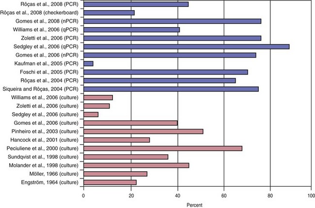
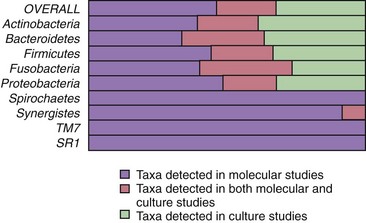
Traditionally, endodontic infections have been studied by means of culture approaches. Such studies identified a set of species thought to play an important role in the pathogenesis of apical periodontitis.288 More recently, not only have findings from culture-based methods been confirmed, but they have also been significantly supplemented with those from culture-independent molecular biology techniques.236 Molecular methods have confirmed and strengthened the association of many cultivable bacterial species with apical periodontitis and have also revealed new suspected endodontic pathogens.250 The list of candidate pathogens has expanded to include culture-difficult species or as-yet-uncultivated bacteria that had never been found in endodontic infections by culturing approaches. The results from molecular studies impact remarkably on the knowledge of bacterial diversity in endodontic infections. More than 400 different bacterial species have already been detected in different types of endodontic infections. Of these, about 45% were exclusively reported by molecular biology studies, compared to 32% detected by culture studies alone. Twenty-three percent of the total bacterial species richness has been detected by application of both culture and molecular studies (Fig. 15-9). As a consequence, it becomes quite apparent that the endodontic microbiota has been refined and redefined by molecular methods.250
Endodontic infections can be classified according to anatomic location (intraradicular or extraradicular infection). Intraradicular infection is caused by microorganisms colonizing the root canal system and can be subdivided into three categories according to the time microorganisms entered the root canal system: primary infection, caused by microorganisms that initially invade and colonize necrotic pulp tissue (initial or “virgin” infection); secondary infection, caused by microorganisms not present in the primary infection but introduced in the root canal at some time after professional intervention (i.e., secondary to intervention); and persistent infection, caused by microorganisms that were members of a primary or secondary infection and in some way resisted intracanal antimicrobial procedures and were able to endure periods of nutrient deprivation in treated canals. Both persistent and secondary infections are for the most part clinically indistinguishable, except for cases where signs and/or symptoms of infection arise in a previously noninfected tooth—a typical example of secondary infection. Extraradicular infection is characterized by microbial invasion of the inflamed periradicular tissues and is a sequel to the intraradicular infection. Admittedly, extraradicular infections can be dependent on or independent of the intraradicular infection.
Microbiota is a collective term for microorganisms and should replace terms such as flora and microflora, which perpetuate an outdated classification of microorganisms as plants.52 Diversity refers to the number of different species present (richness) and their relative abundance (evenness) in a given ecosystem.105
The oral cavity harbors one of the highest accumulations of microorganisms in the body. Even though viruses, archaea, fungi, and protozoa can be found as constituents of the oral microbiota, bacteria are by far the most dominant inhabitants of the oral cavity. There are an estimated 10 billion bacterial cells in the oral cavity,156 and culture-independent studies (microscopy and molecular biology technologies) have shown that over 50% to 60% of the oral microbiota still remains to be cultivated and fully characterized.2,275 A high diversity of bacterial species has been revealed in the oral cavity by culturing approaches, but application of molecular biology methods to the analysis of the bacterial diversity has revealed a still broader and more diverse spectrum of extant oral bacteria. Over 700 bacterial taxa have been found in the human oral cavity, and phylogenetic studies have revealed that these taxa fall into 13 separate phyla: Firmicutes, Bacteroidetes, Actinobacteria, Fusobacteria, Proteobacteria, Spirochaetes, Synergistes, TM7, Sulphur River 1 (SR1), Chloroflexi, Cyanobacteria, Deinococcus, and Acidobacteria.1,112,133,185,186
Modern anaerobic culture and sophisticated molecular biology techniques have disclosed important aspects of the endodontic microbiota in the different types of infection. Of particular interest, they have allowed the assessment of the microbial diversity in endodontic infections, collectively revealing that more than 400 different microbial species/phylotypes have been found in infected root canals, usually in combinations involving many species in primary infections and a few ones in secondary/persistent infections. Endodontic bacteria fall into 9 of the 13 phyla that have oral representatives* (Fig. 15-10). Fungi and archaea have been only occasionally found in endodontic infections.
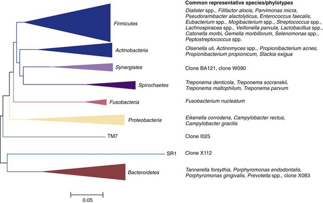
FIG. 15-10 Bacterial phyla with their respective endodontic representatives. Right, example species or phylotypes for each phylum.
Endodontic infections develop in a previously sterile place that does not contain a normal microbiota. Any species found has the potential to be an endodontic pathogen or at least play a role in the ecology of the endodontic microbial community. Culture and molecular studies reveal only prevalence of species; consequently, only association can be inferred. Causation is usually surmised on the basis of both frequency of detection and potential pathogenicity (in animal models or association with other human diseases), and several species have emerged as candidate or putative endodontic pathogens. The following sections discuss specific aspects of each type of endodontic infection.
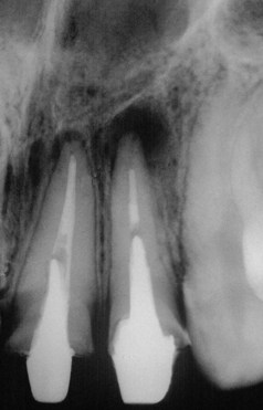
Primary intraradicular infection is infection of the necrotic pulp tissue (Fig. 15-11). Participating microorganisms can have been involved in earlier stages of pulp invasion (usually via caries) that culminated in inflammation and further necrosis, or they can be latecomers that took advantage of the environmental conditions in the root canal after pulp necrosis.
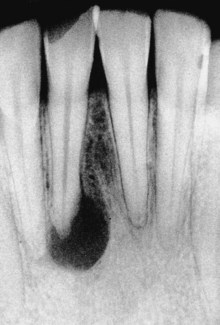
FIG. 15-11 Apical periodontitis due to primary intraradicular infection. Pulp is necrotic and lesion size is usually directly proportional to complexity of the microbiota involved.
Primary infections are characterized by a mixed community conspicuously dominated by anaerobic bacteria. The number of bacterial cells may vary from 103 to 108 per root canal.220,265,287,306 Molecular studies have disclosed a mean of 10 to 20 species/phylotypes per infected canal.162,208,250,261 Canals of teeth with sinus tracts exhibit a mean number of 17 species.208 The size of the apical periodontitis lesion has been shown to be proportional to the number of bacterial species and cells in the root canal.208,260,287 A molecular study208 demonstrated that the number of taxa per canal was clearly in direct proportion to the lesion size: small lesions (<5 mm) harbored about 12 taxa, lesions from 5 to less than 10 mm harbored 16 taxa, and lesions over 10 mm harbored about 20 species. Some canals associated with large lesions may harbor even more than 40 taxa208; the larger the lesion, the higher the bacterial diversity and density in the canal.
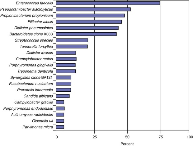
The most prevalent named bacterial species detected in primary infections, including abscessed cases, belong to diverse genera of gram-negative (Fusobacterium, Dialister, Porphyromonas, Prevotella, Tannerella, Treponema, Campylobacter, and Veillonella) and gram-positive (Parvimonas, Filifactor, Pseudoramibacter, Olsenella, Actinomyces, Peptostreptococcus, Streptococcus, Propionibacterium, and Eubacterium) bacteria.* Bacterial prevalence in primary infections may vary from study to study as a function of several factors, such as sensitivity and specificity of the detection and identification methods, sampling technique, geographic location, and accuracy or divergence in clinical diagnosis and disease classification. Even so, the species most frequently detected are expected to be the same in most well-conducted studies. Figs. 15-12 to 15-14 display the most frequently detected species associated with chronic apical periodontitis, acute apical periodontitis, and acute apical abscesses, respectively, as revealed by studies from the authors’ group using a highly sensitive molecular biology technique.
FIG. 15-12 Prevalence of bacteria detected in primary infections of teeth with chronic apical periodontitis. Data from the authors’ studies using a taxon-specific nested-polymerase chain reaction protocol.
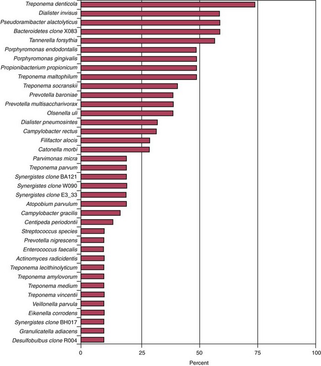
FIG. 15-13 Prevalence of bacteria detected in primary infections of teeth with acute apical periodontitis. Data from the authors’ studies using a taxon-specific nested-polymerase chain reaction protocol.
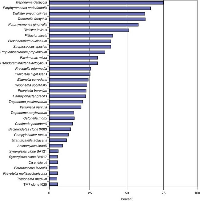
FIG. 15-14 Prevalence of bacteria detected in primary infections of teeth with acute apical abscesses. Data from the authors’ studies using a taxon-specific nested-polymerase chain reaction protocol.
About 40% to 55% of the endodontic microbiota in primary infections is composed of species still uncultivated.162,219 A molecular study revealed that 55% and 40% of the taxa detected in association with chronic apical periodontitis and acute apical abscesses, respectively, were uncultivated phylotypes.219 As for their abundance in these infections, as-yet-uncultivated phylotypes corresponded to 38% and 30% of the clones sequenced from samples of chronic apical periodontitis and abscesses, respectively.219 Molecular studies investigating the breadth of bacterial diversity in infected root canals have disclosed the occurrence of uncultivated phylotypes belonging to several genera, including Synergistes, Dialister, Prevotella, Solobacterium, Olsenella, Fusobacterium, Eubacterium, Megasphaera, Veillonella, and Selenomonas, as well as phylotypes related to the family Lachnospiraceae or the TM7 phylum.† Some uncultivated phylotypes can even be among the most prevalent bacteria in primary intraradicular infections, and others may be associated with pain.219 Two of the most prevalent such phylotypes found in endodontic infections include Synergistes clone BA121 and Bacteroidetes clone X083.207,208,251,253,255 Detection of these phylotypes in samples from endodontic infections suggests that they can be previously unrecognized bacteria that play a role in the pathogenesis of different forms of apical periodontitis. The fact that they have not yet been cultivated and phenotypically characterized does not mean that they are not important.
Acute apical periodontitis and acute apical abscesses are typical examples of symptomatic endodontic infections. In these cases, the infection is located in the canal, but it has also reached the periradicular tissues and, in abscessed cases, can spread to other anatomic spaces. Acute apical abscesses are caused by bacteria that egress the infected root canal and invade the periradicular tissues to establish an extraradicular infection and evoke purulent inflammation. Clinically, the disease leads to pain and/or swelling and has the potential to diffuse to sinuses and other facial spaces of the head and neck to form a cellulitis or other complications (Fig. 15-15). The microbiota involved in endodontic abscesses is mixed and dominated by anaerobic bacteria47,115,122,219,261 (see Fig. 15-14). Direct comparisons using molecular technology reveal an average of 12 to 18 taxa per abscess case, compared to 7 to 12 taxa present in root canals from chronic cases.219,261 Uncultivated phylotypes constitute approximately 40% of the taxa found in abscesses and collectively represent more than 30% of the 16S rRNA gene sequences retrieved in clone libraries.219
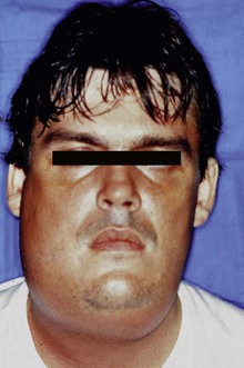
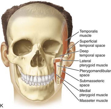
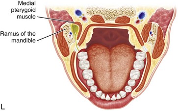
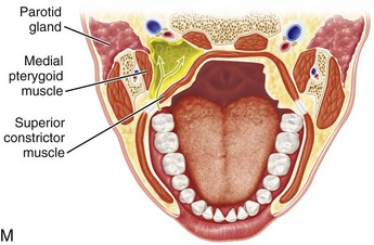
FIG. 15-15 Acute apical abscess. The infection has spread to other anatomic spaces to form cellulitis.
Whereas microbial causation of apical periodontitis is well established, there is no strong evidence disclosing specific involvement of a single species with any particular sign or symptom of apical periodontitis. Some gram-negative anaerobic bacteria have been suggested to be involved with symptomatic lesions,* but the same species may also be present in somewhat similar frequencies in asymptomatic cases,† so factors other than the mere presence of a given putative pathogenic species may play a role in the etiology of symptomatic endodontic infections.232,237 These factors could include differences in virulence ability among strains of the same species; bacterial interactions resulting in additive or synergistic effects among species in mixed infections; number of bacterial cells (load); environmental cues regulating expression of virulence factors; host resistance; and concomitant herpesvirus infection. Association of some or all of these factors (instead of an isolated event) is likely to determine the occurrence and intensity of symptoms.232,237
†References 15, 69, 89, 109, 262, and 263.
Cross-sectional studies suggest that bacterial succession occurs before symptoms arise.261 At a given moment during the endodontic infectious process, the microbiota may reach a certain degree of pathogenicity that elicits acute inflammation at the periradicular tissues, with consequent development of pain and sometimes swelling. Molecular studies using DGGE or T-RFLP analysis revealed that the structure of the endodontic bacterial communities in symptomatic teeth is significantly different from that of asymptomatic teeth.261 Differences are represented by different dominant species in the communities and larger numbers of species in symptomatic cases. A shift in the structure of the microbial community is likely to occur before appearance of symptoms. Such a shift is probably due to the arrival of new pathogenic species or variations and rearrangements in the number of members of the bacterial community. Differences in the type and load of dominant species and the resulting bacterial interactions may affect virulence of the whole bacterial community. This is in agreement with the community-as-a-pathogen concept discussed earlier.
Findings from laboratories in different countries are often quite different regarding the prevalence of the species involved in endodontic infections. Although these differences may be attributed to variations in identification methodologies, a geographic influence in the composition of the root canal microbiota has been suspected. Studies using molecular biology techniques directly compared the endodontic microbiota of patients residing in different geographic locations and suggested that significant differences in the prevalence of some important species can actually exist.14,204,241 In a more holistic approach, analysis of the bacterial community profiles of the microbiota associated with acute apical abscesses from the United States and Brazilian patients also revealed a geography-related pattern, with several species being exclusive for each location and others shared by the two locations, showing great differences in prevalence.143 The factors that can lead to differences in the composition of the endodontic microbiota and the impact of these differences on therapy, particularly in abscessed cases requiring systemic antibiotic therapy, remain elusive.
Knowledge of microbial location and organization within the root canal system assumes special importance to understanding the disease process and establishing effective antimicrobial therapeutic strategies. Most of the knowledge of the structure of the endodontic microbiota comes from morphologic studies,161,167,227,257 but these do not usually provide information about bacterial identity and numbers. Consequently, it is not possible to delineate the role of the visualized bacteria in the disease process. Because every bacterial cell observed in the root canal system might be an endodontic pathogen, findings from morphologic studies should be used to understand the topography of the root canal infection and establish therapeutic measures in an attempt to completely eradicate the infection or at least reduce the bacterial load to thresholds compatible with periradicular tissue healing.
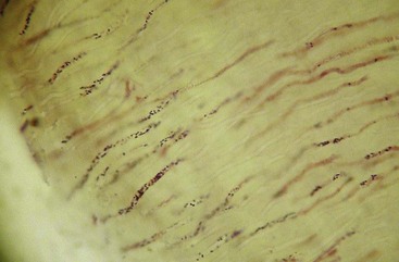
Again, mounting evidence suggests that apical periodontitis, like caries and periodontal diseases, is also a biofilm-induced disease. Morphologic studies have shown that the root canal microbiota in primary infections is dominated by bacterial morphotypes that include cocci, rods, filaments, and spirilla (spirochetes) (Fig. 15-16), and fungal cells are sporadically found227,257 (Fig. 15-17). Bacteria colonizing the root canal system usually grow in sessile biofilms adhered to the dentinal walls and also as coaggregates (cells with distinct morphologies), aggregates (cells with the same morphology), and planktonic cells suspended in the fluid phase of the main canal161,167,257 (Fig. 15-18). Lateral canals and isthmuses connecting main canals may be clogged with bacterial cells primarily organized in biofilms166,202 (Figs. 15-19 and 15-20).
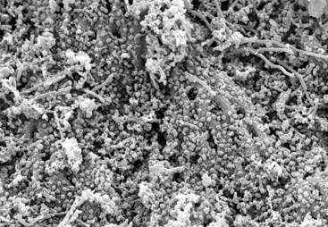
FIG. 15-16 Mixed bacterial population colonizing the root canal wall. Cocci are the predominant forms, but rods, filaments, and spirochetes are also observed. In some areas, coccoid cells are relatively apart from each other (×2200).
(From Siqueira JF Jr, Rôças IN, Lopes HP: Patterns of microbial colonization in primary root canal infections. Oral Surg Oral Med Oral Pathol Oral Radiol Endod 93:174, 2002.)
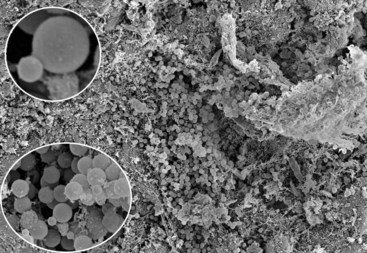
FIG. 15-17 Heavy colonization of yeast cells in the root canal of an extracted tooth with primary infection associated with apical periodontitis (×300). Note that some cells are in the stage of budding. A daughter cell is growing on the surface of the mother cell (insets) (insets: bottom ×2700, top ×3500).
(From Siqueira JF Jr, Sen BH: Fungi in endodontic infections. Oral Surg Oral Med Oral Pathol Oral Radiol Endod 97:632, 2004.)
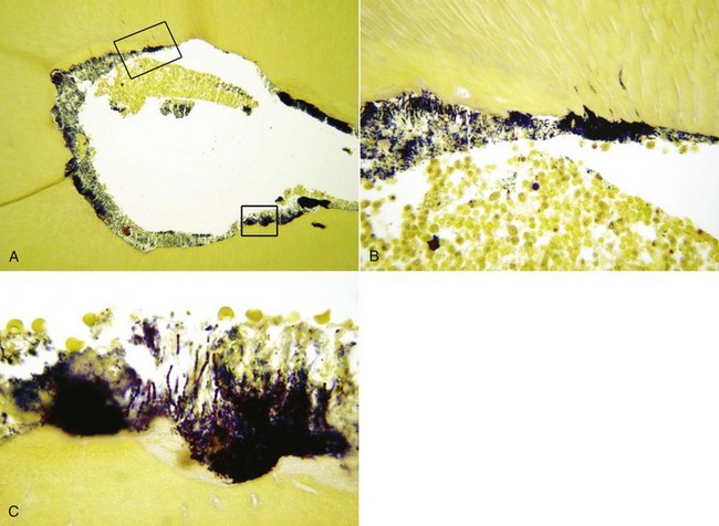
FIG. 15-18 Biofilm on the walls of a mesial root canal from a mandibular first molar (×100). The tooth was symptomatic, and an apical periodontitis lesion was present. Sections in B and C correspond to higher magnifications of the larger and smaller insets, respectively. Note the accumulation of polymorphonuclear neutrophils in the canal near the biofilm. (B, ×400; C, ×1000). Section stained with a Taylor-modified Brown and Brenn technique.
(Courtesy Dr. Domenico Ricucci.)
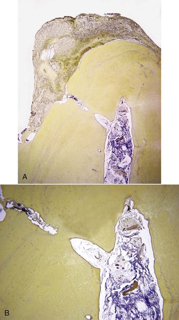
FIG. 15-19 A, Bacterial biofilm in the necrotic canal and in an apical ramification contiguous to inflamed periradicular tissues (×25). B, Higher magnification of A (×100). Sections stained with a Taylor-modified Brown and Brenn technique.
(Courtesy Dr. Domenico Ricucci.)
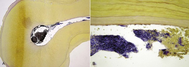
FIG. 15-20 Transverse sections of a maxillary second molar with fused mesial and palatal roots. A, Heavy bacterial infection of the canal, spreading to an isthmus (×25). B, Higher magnification of the isthmus clogged with bacteria (×400). Section stained with a Taylor-modified Brown and Brenn technique.
(Courtesy Dr. Domenico Ricucci.)
Bacterial cells from endodontic biofilms are very often seen penetrating the dentinal tubules (Fig. 15-21). The diameter of dentinal tubules is large enough to permit penetration of most oral bacteria. It has been reported that dentinal tubule infection can occur in about 70% to 80% of the teeth evincing apical periodontitis lesions.149,193 A shallow penetration is more common; bacterial cells can be observed reaching approximately 300 µm in some teeth257 (Fig. 15-22). Dividing cells can be frequently observed within tubules in in situ investigations257 (see Fig. 15-22), indicating that bacteria can derive nutrients within tubules, probably from degrading odontoblastic processes, denatured collagen, bacterial cells that die during the course of infection, and intracanal fluids that enter the tubules by capillarity.
FIG. 15-21 Heavy infection of the root canal walls, mainly by cocci, but some small rods are also seen. Cocci are penetrating into dentinal tubules (×3500).
(From Siqueira JF Jr, Rôças IN, Lopes HP: Patterns of microbial colonization in primary root canal infections. Oral Surg Oral Med Oral Pathol Oral Radiol Endod 93:174, 2002.)
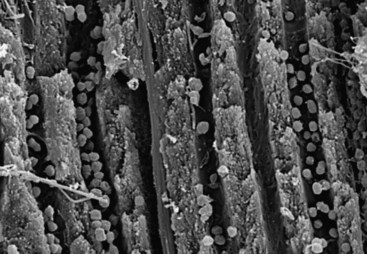
FIG. 15-22 Cocci in dentinal tubules approximately 300 µm from the main root canal (×5000).
(From Siqueira JF Jr, Rôças IN, Lopes HP: Patterns of microbial colonization in primary root canal infections. Oral Surg Oral Med Oral Pathol Oral Radiol Endod 93:174, 2002.)
Several putative endodontic pathogens have been shown to be capable of penetrating dentinal tubules in vitro, including Porphyromonas endodontalis, Porphyromonas gingivalis, Fusobacterium nucleatum, Actinomyces israelii, Propionibacterium acnes, Enterococcus faecalis, Candida albicans, and streptococci.142,191,239,258,310 In their clinical study, Peters et al.193 isolated and identified bacteria present in root dentin at different depths, and the most common isolates belonged to the genera Prevotella, Porphyromonas, Fusobacterium, Veillonella, Peptostreptococcus, Eubacterium, Actinomyces, lactobacilli, and streptococci. Using immunohistologic analysis, Matsuo et al.149 observed the intratubular occurrence of F. nucleatum, Pseudoramibacter alactolyticus, Eubacterium nodatum, Lactobacillus casei, and Parvimonas micra inside dentinal tubules from the canal walls of extracted infected teeth with apical periodontitis.
Whereas bacteria present as planktonic cells in the main root canal may be easily accessed and eliminated by instruments and substances used during treatment, those organized in biofilms attached to the canal walls or located into isthmuses, lateral canals, and dentinal tubules are definitely more difficult to reach and may require special therapeutic strategies to be eradicated.
A root canal with necrotic pulp provides a space for bacterial colonization and affords bacteria a moist, warm, nutritious, and anaerobic environment, which is by and large protected from the host defenses because of lack of active blood circulation in the necrotic pulp tissue. Also, the root canal walls are nonshedding surfaces conducive to persistent colonization and formation of complex communities. The necrotic root canal might be considered a fertile environment for bacterial growth, and colonization not a difficult task for virtually all oral bacterial species. Although a large number of bacterial species (about 100 to 200) can be found in the oral cavity of a particular individual,187 only a limited assortment of these species (about 10 to 40) is consistently selected out for growth and survival within a root canal containing necrotic pulp tissue from the same individual. This indicates that ecological determinants operate in the necrotic canal and dictate which species will succeed in colonizing this previously sterile environment. The major ecological factors that determine the composition of the root canal microbiota include oxygen tension, type and amount of available nutrients, and bacterial interactions. Other factors such as temperature, pH, and receptors for adhesins may also be involved.
The root canal infection is a dynamic process, and different bacterial species apparently dominate at different stages. Shifts in the composition of the microbiota are largely due to changes in environmental conditions, particularly in regard to oxygen tension and nutrient availability. In the very initial phases of the pulpal infectious process, facultative bacteria predominate.63 After a few days or weeks, oxygen is depleted within the root canal as a result of pulp necrosis and consumption by facultative bacteria. Further oxygen supply is interrupted with loss of blood circulation in the necrotic pulp. An anaerobic milieu develops and is highly conducive to survival and growth of obligate anaerobic bacteria. With the passage of time, anaerobic conditions become even more pronounced, particularly in the apical third of the root canal; as a consequence, anaerobes will dominate the microbiota, outnumbering facultative bacteria (Fig. 15-23).
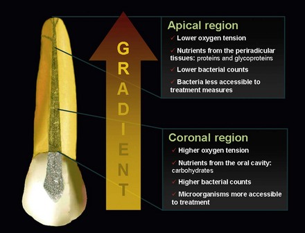
FIG. 15-23 Ecological conditions in different areas of the root canal. A gradient of oxygen tension and nutrients (type and availability) is formed. Consequently, the microbiota residing in different parts can also differ in diversity, density, and accessibility to treatment procedures.
The main sources of nutrients for bacteria colonizing the root canal system include: (1) the necrotic pulp tissue; (2) proteins and glycoproteins from tissue fluids and exudate that seep into the root canal system via apical and lateral foramens; (3) components of saliva that may coronally penetrate into the root canal; and (4) products of the metabolism of other bacteria. Because the largest amount of nutrients is available in the main canal, the most voluminous part of the root canal system, most of the infecting microbiota (particularly fastidious anaerobic species) is expected to be located in this region. Bacterial species that can best utilize and compete for nutrients in the root canal system will succeed in colonization.
In addition to being influenced by variations in oxygen levels, shifts in the microbiota colonizing the root canal system can also be dependent upon the dynamics of nutrient utilization. Saccharolytic species dominate the very early stages of the infectious process but are soon outnumbered by asaccharolytic species, which will dominate later stages.289 Even though the necrotic pulp tissue can be regarded as a finite source of nutrients to bacteria, given the small volume of tissue that is gradually degraded, induction of periradicular inflammation guarantees a sustainable source of nutrients, particularly in the form of proteins and glycoproteins present in the exudate that seep into the canal. At this stage of the infectious process, bacteria that have a proteolytic capacity or establish a cooperative interaction with those that can utilize this substrate in the metabolism start to dominate. Therefore, as the infectious process reaches the stage of induction of periradicular inflammation, proteins become the principal nutrient source, particularly in the apical part of the canal, favoring the establishment of anaerobic species that utilize peptides and/or amino acids in their metabolism (see Fig. 15-23).
Because primary endodontic infections are usually characterized by mixed communities, different bacterial species are in close proximity with one another, and interactions become inevitable. Thus, establishment of certain species in the root canal is also influenced by interactions with other species. In this regard, early colonizers play an important role in dictating which species will live along with them in the community. Bacterial interactions can be positive or negative. Positive interactions enhance the survival capacity of the interacting bacteria and enable different species to coexist in habitats where neither could exist alone. For instance, interbacterial nutritional interactions are important ecological determinants that result in higher metabolic efficiency of the whole community. Nutritional interactions are mainly represented by food webs, including utilization of metabolic end products from one species by another and bacterial cooperation for the breakdown of complex host-derived substrates. Fig. 15-24 displays a complex array of interbacterial nutritional interactions that can take place in infected root canals, where the growth of some species can be dependent upon products of metabolism of other species. Moreover, one species can provide growth conditions favorable to another—for example, by reducing oxygen tension in the environment and favoring the establishment of anaerobes, or by releasing some proteinases that can provide protection from host defenses. Negative interactions in turn act as feedback mechanisms that limit population densities. Examples include competition (for nutrients and space) and amensalism (when one species produces a substance that inhibits another species).
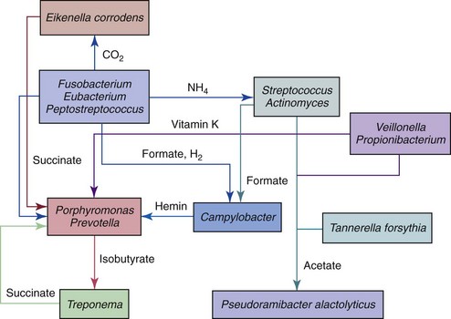
FIG. 15-24 Interbacterial nutritional interactions that can take place in infected root canals where growth of some species can be dependent upon products of metabolism of other species.
Many species adhere directly to host surfaces, other species adhere to bacteria already attached to the surface. The latter is called coaggregation and is a highly specific phenomenon with regard to the partners involved.117 Coaggregations differ from aggregations and agglutinations in that the latter two interactions occur between genetically identical cells.118 A given pair of species can attach to each other by means of specific receptor-adhesin interactions, which are usually lectinlike interactions (attachment of a specific protein on the surface of one species to a specific carbohydrate on the surface of the other). Coaggregation can favor colonization of host surfaces and also facilitate metabolic interactions between the partners. Coaggregation has been demonstrated for several pairs of bacterial taxa found in endodontic infections.114
Fungi are eukaryotic microorganisms that may colonize the oral cavity, especially Candida species, but they have been only occasionally detected in primary intraradicular infections.60,126,159,259 One molecular study16 has reported the occurrence of C. albicans in 21% of samples from primarily infected canals.
Archaea comprise a highly diverse group of prokaryotes distinct from bacteria. Members of this domain have been traditionally recognized as extremophiles, but some of these microorganisms have also been found to flourish in nonextreme environments, including the human body.59 To date, no member of the Archaea domain has been described as a human pathogen. Although one study failed to detect archaea in samples from primary endodontic infections,254 other studies detected a Methanobrevibacter oralis–like phylotype in some primarily infected canals.305,307
Viruses are not cells but inanimate particles composed of a nucleic acid molecule (DNA or RNA) and a protein coat. On their own, they have no metabolism and need to infect living cells to use the cell’s machinery to replicate the viral genome. Because viruses require viable host cells to infect and replicate themselves, they cannot thrive in the root canal with necrotic pulp. Viruses have been reported to occur in the root canal only in noninflamed vital pulps of patients infected with the human immunodeficiency virus.78 Human cytomegalovirus (HCMV) and Epstein-Barr virus (EBV) have been detected in apical periodontitis lesions,215 where living host cells are present. It has been hypothesized that HCMV and EBV may be implicated in the pathogenesis of apical periodontitis as a direct result of virus infection and replication or as a result of virally induced impairment of local host defenses, which might give rise to overgrowth of pathogenic bacteria in the very apical part of the root canal.273 Bacterial challenge emanating from the canals may cause an influx of virus-infected cells into the periradicular tissues. Reactivation of HCMV and/or EBV by tissue injury induced by bacteria may evoke impairment of host immune response in the periradicular microenvironment, changing the potential of local defense cells to mount an adequate response against infectious agents. Herpesvirus-infected inflammatory cells are stimulated to release proinflammatory cytokines,157,313 and evidence of HCMV and/or EBV infection has been more frequently observed in symptomatic apical periodontitis lesions,214,216 large lesions,215,216 and lesions from HIV-positive patients.217 However, the role of herpesviruses in the pathogenesis of apical periodontitis has still to be elucidated.
As defined earlier in this chapter, persistent intraradicular infections are caused by microorganisms that resisted intracanal antimicrobial procedures and survived in the treated canal. Involved microorganisms are remnants of a primary or secondary infection. The latter in turn is caused by microorganisms that at some time entered the root canal system secondary to professional intervention. The moment can be during treatment, between appointments, or even after root canal filling. In any circumstance, if penetrating microorganisms manage to adapt themselves to the new environment, surviving and flourishing, a secondary infection is established. Species involved can be oral microorganisms or not, depending on the source of secondary infection.
Persistent and secondary infections are for the most part clinically indistinguishable. Exceptions include infectious complications (such as an apical abscess) arising after the treatment of noninfected vital pulps or cases in which apical periodontitis was absent at the time of treatment but present on the follow-up radiograph. Both situations are typical examples of secondary infections. Both persistent and secondary infections can be responsible for several clinical problems, including persistent exudation, persistent symptoms, interappointment flare-ups, and failure of the endodontic treatment, characterized by a posttreatment apical periodontitis lesion.
Persistent or secondary intraradicular infections are the major causes of endodontic treatment failure (Fig. 15-25). This statement is supported by two strong evidence-based arguments. First, it has been demonstrated that there is an increased risk of adverse treatment outcome when bacteria are present in the canal at the time of filling.64,267,308 Second, most (if not all) root canal–treated teeth evincing persistent apical periodontitis lesions have been demonstrated to harbor an intraradicular infection.* Based on these arguments, studies investigating bacteria remaining in the root canals at the filling stage serve the purpose of disclosing species that have the potential to influence the treatment outcome (outcome into perspective). On the other hand, studies dealing with the microbiota of root canal–treated teeth evincing apical periodontitis serve to show the association of species with treatment failure, since the microorganisms detected are likely to be participating in the etiology of persistent disease (outcome already established).
FIG. 15-25 Posttreatment apical periodontitis lesions in root canal–treated teeth. In poorly treated canals, the microbiota is similar to primary infections. In cases apparently well treated, fewer species are found. Regardless of treatment quality, persistent or secondary intraradicular infections are the main causative agents of endodontic treatment failure.
Diligent antimicrobial treatment may still fail to completely eliminate bacteria from the infected root canal system. This is because persisting bacteria are either resistant or inaccessible to treatment procedures. Whatever the cause of persistence, bacterial diversity and density are substantially reduced after treatment. Root canal samples positive for bacterial growth after chemomechanical procedures followed or not by intracanal medication have been shown to harbor 1 to 5 bacterial species per case, and the number of bacterial cells usually varies from 102 to 105 per sample.*
*References 34, 220, 245, 246, 267, and 306.
No single species has been significantly found to persist after treatment procedures. Gram-negative bacteria, which are common members of primary infections, are usually eliminated. Exceptions include some anaerobic rods, such as F. nucleatum, Prevotella species, and Campylobacter rectus, which are among the species found in postinstrumentation or postmedication samples.† Most studies on this subject have clearly revealed that when bacteria resist treatment procedures, gram-positive bacteria are more frequently present. Gram-positive facultatives or anaerobes often detected in these samples include streptococci, P. micra, Actinomyces species, Propionibacterium species, P. alactolyticus, lactobacilli, E. faecalis, and Olsenella uli.‡ This gives support to the notion that gram-positive bacteria can be more resistant to antimicrobial treatment measures and have the ability to adapt to the harsh environmental conditions in instrumented and medicated root canals. About 40% of the taxa found in posttreatment samples are as-yet-uncultivated bacteria,220 indicating that along with other reasons (such as inaccessibility to sampling or low bacterial levels), attainment of negative cultures after treatment procedures does not necessarily imply sterility.
‡References 34, 36-38, 40, 80, 190, 192, 220, 240, 245, 246, 267, and 293.
Bacteria persisting in the root canal after chemomechanical procedures or intracanal medication will not always maintain an infectious process. This statement is supported by evidence that some apical periodontitis lesions healed even after bacteria were found in the canal at the filling stage.64,267 There are some possible explanations:
Bacteria that resist intracanal procedures and are present in the canal at the time of filling can influence the outcome of the endodontic treatment provided they:
In this context, it should not be forgotten that host resistance to infection is also an important and probably decisive counteracting factor.
The microbiota in root canal–treated teeth with apical periodontitis also exhibits decreased diversity in comparison to primary infections. Canals apparently well treated harbor one to five species, but the number of species in canals with inadequate treatment can reach up to 10 to 30 species, which is very similar to untreated canals (see Fig. 15-25).197,209,221,252,290 The number of bacterial cells in treated teeth varies from 103 to 107 per canal.190,225
Several culture and molecular biology studies have revealed that E. faecalis is the most frequent species in root canal–treated teeth, with prevalence values reaching up to 90% of cases* (Fig. 15-26). Root canal–treated teeth are about nine times more likely to harbor E. faecalis than cases of primary infections.211 This suggests that this species can be inhibited by other members of a mixed bacterial consortium commonly present in primary infections, and that the bleak environmental conditions within filled root canals do not prevent its survival. The fact that E. faecalis has been commonly recovered from cases treated in multiple visits and/or in teeth left open for drainage266 suggests that this species may be a secondary invader that succeeds in colonizing the canal, resists treatment, and causes a secondary infection that then becomes persistent.
FIG. 15-26 Prevalence of Enterococcus faecalis in samples from root canal–treated teeth with apical periodontitis. Data from culture (orange bars) and molecular (blue bars) studies below.
For a given microorganism to survive in treated canals, it has to resist intracanal disinfection procedures and adapt to the harsh environmental conditions caused by treatment. The ability of E. faecalis to penetrate dentinal tubules, sometimes to a deep extent88,239 (Fig. 15-27), can enable it to escape the action of endodontic instruments and irrigants used during chemomechanical preparation.88,238 Moreover, its ability to form biofilms in root canals can be important for its resistance to and persistence after intracanal antimicrobial procedures.53 Enterococcus faecalis is also resistant to calcium hydroxide32; such an ability to resist high pH values seems to be related to a functioning proton pump that drives protons into the cell to acidify the cytoplasm.62 Unlike most putative endodontic pathogens frequently found in primary infections, E. faecalis may colonize root canals in single infections,290 and such relative independence of living without deriving nutrients from other bacteria can be extremely important for its establishment in treated root canals. Finally, environmental cues can regulate gene expression in E. faecalis, affording this bacterium the ability to adapt to varying (and adverse) conditions.107 Indeed, E. faecalis can enter a so-called viable but non-cultivable (VBNC) state,136 which is a survival mechanism adopted by several bacteria when exposed to unfavorable environmental conditions.137 In VBNC state, bacteria lose the ability to grow in culture media but maintain viability and pathogenicity and sometimes can resume division when optimal environmental conditions are restored. Studies have demonstrated that E. faecalis has the ability to survive in environments with scarcity of nutrients and then flourish when the nutrient source is reestablished.66 It also has the capacity to recover from a prolonged starvation state in obturated canals,226 suggesting that viable cells of this species entombed at the time of root filling may provide a long-term nidus for subsequent infection.
FIG. 15-27 Dentinal tubule infection by Enterococcus faecalis in dog’s teeth after experimental infection. Notice that cells invaded the entire extent of some tubules up to the cementum (Brown and Brenn staining, ×1000).
Taken together, all these properties help explain the significantly high prevalence of E. faecalis in root canal–treated teeth. Although association of this species with posttreatment disease is suggested by epidemiologic studies and supported by the species attributes that allow it to survive under unfavorable environmental conditions, causation is not proven. In fact, the status of E. faecalis as the main causative agent of endodontic failures has been questioned by the following findings from studies carried out in independent laboratories:
Other bacteria found in root canal–treated teeth with apical periodontitis include streptococci and some fastidious anaerobic bacterial species: P. alactolyticus, Propionibacterium propionicum, Filifactor alocis, Dialister pneumosintes, Dialister invisus, Tannerella forsythia, P. micra, Prevotella intermedia, and Treponema denticola* (Fig. 15-28). Uncultivated phylotypes correspond to 55% of the taxa detected in treated canals, and collectively they can also be in high proportions, corresponding to about half of the 16S rRNA gene sequences retrieved in clone libraries.221 Some still uncultivated phylotypes, such as Bacteroidetes clone X083 and Synergistes clone BA121, can be among the most prevalent taxa in treated canals205,221 (see Fig. 15-28). As-yet-uncultivated bacteria can also dominate the community, helping explain why culture studies failed to detect bacteria in some treated root canals.
FIG. 15-28 Prevalence of microorganisms detected in root canal–treated teeth with posttreatment disease. Data from the authors’ studies using a taxon-specific polymerase chain reaction assay.
The bacterial community profiles in treated cases vary from individual to individual, indicating that distinct bacterial combinations can play a role in treatment failure.209,221 All these findings strongly suggest that the microbiota of root canal–treated teeth with apical periodontitis is more complex than previously anticipated by culture studies.
Fungi are only occasionally found in primary infections, but Candida species have been detected in root canal–treated teeth in up to 18% of the cases.† Fungi gain access to root canals via contamination during endodontic therapy (secondary infection) or they overgrow after inefficient intracanal antimicrobial procedures that cause an imbalance in the primary endodontic microbiota.264 Candida albicans is by far the most commonly detected fungal species in root canal–treated teeth. This species has several properties that can be involved in persistence following treatment, including its ability to colonize and invade dentin228,229,258 and resistance to calcium hydroxide.309,311
Apical periodontitis lesions are formed in response to intraradicular infection and by and large constitute an effective barrier against spread of the infection to the alveolar bone and other body sites. In most situations, apical periodontitis inflammatory lesions succeed in preventing microorganisms from invading the periradicular tissues. Nevertheless, in some specific circumstances, microorganisms can overcome this defense barrier and establish an extraradicular infection. The most common form of extraradicular infection is the acute apical abscess, characterized by purulent inflammation in the periradicular tissues in response to a massive egress of virulent bacteria from the root canal. There is, however, another form of extraradicular infection which, unlike the acute abscess, is usually characterized by absence of overt symptoms. This condition entails the establishment of microorganisms in the periradicular tissues, either by adherence to the apical external root surface in the form of biofilm structures169,297 or by formation of cohesive actinomycotic colonies within the body of the inflammatory lesion.92 These extraradicular microorganisms have been discussed as one of the etiologies of persistence of apical periodontitis lesions in spite of diligent root canal treatment.234,299
Certain situations permit intraradicular bacteria to reach the periradicular tissues and establish an extraradicular infection.235 Some may be:
FIG. 15-29 Scanning electron micrograph showing extensive bacterial colonization in the very apical part of the canal, near and at the apical foramen (A-B, ×550 and ×850, respectively). C, Higher magnification of left inset in B showing a bacterial biofilm adhered to the very apical canal walls (×3700). D, Higher magnification of right inset in B showing a fully developed “corn cob” in tissue meshwork adjacent to the apical foramen (×4000).
(Modified from Siqueira JF Jr, Lopes HP: Bacteria on the apical root surfaces of untreated teeth with periradicular lesions: a scanning electron microscopy study. Int Endod J 34:216, 2001.)
Conceivably, the extraradicular infection can be dependent on or independent of the intraradicular infection.234 The acute apical abscess is the main example of dependent extraradicular infection. Independent extraradicular infections would be those that are no longer fostered by the intraradicular infection and can persist even after successful eradication of the latter. So far, it has been suggested that the main bacterial species implicated in independent extraradicular infections are Actinomyces species and P. propionicum, in a pathologic entity named apical (or periapical, periradicular) actinomycosis33,92,270,291 (Fig. 15-30). These bacteria form cohesive colonies that may be collectively resistant to phagocytosis.65 Existence of apical actinomycosis as a self-sustained pathologic entity, no longer nurtured by the intraradicular infection, and its involvement as an exclusive cause of treatment failure is speculative and remains to be proven.202,247
FIG. 15-30 Bacterial aggregate in an epithelialized periradicular lesion, suggestive of actinomycosis. This case presented a severe flare-up in the past, but the tooth was asymptomatic at the time of extraction. A, Section not passing through the canal; note cyst cavity and bacterial aggregation on the left (×25). B, Detail of area with colony (×100). C, Higher magnification of colony, which is surrounded by neutrophils (×400). Sections stained with a Taylor-modified Brown and Brenn technique.
(Courtesy Dr. Domenico Ricucci.)
Except for apical actinomycosis and cases evincing sinus tracts, it is still controversial whether chronic apical periodontitis lesions can harbor bacteria for very long beyond initial tissue invasion.11 Studies using culture-dependent283,298,315 or culture-independent molecular biology methods, such as checkerboard hybridization76,285 and FISH,284 have reported the extraradicular occurrence of a complex microbiota associated with apical periodontitis lesions that do not respond favorably to the root canal treatment. Anaerobic bacteria have been reported to be the dominant microorganisms in several of those lesions.283,285 Apart from a discussion about whether contamination can be effectively prevented during surgical sampling of periradicular lesions, these studies did not evaluate the bacteriologic conditions of the apical part of the root canal. This makes it difficult to ascertain whether extraradicular infections were dependent on or independent of an intraradicular infection.
The incidence of extraradicular infections in untreated teeth is conceivably low,167,243 which is congruent with the high success rate of nonsurgical root canal treatment.269 Even in root canal–treated teeth with recalcitrant lesions in which a higher incidence of extraradicular bacteria has been reported, a high rate of healing following retreatment269 indicates that the major cause of posttreatment disease is located within the root canal system, characterizing a persistent or secondary intraradicular infection. This has been confirmed by studies investigating the microbiologic conditions of root canals associated with persistent apical periodontitis.158,197,209,252,290 Based on such data, it is reasonable to assume that most extraradicular infections observed in association with root canal–treated teeth must have been fostered by an intraradicular infection.
Focal infection consists of an infectious disorder caused by microorganisms or their products that have disseminated from a distant body site (focus of infection).179 Microorganisms from an infected oral site might spontaneously or after dental procedures gain entry into the blood circulation and cause disease in remote sites. The relationship of oral infections and systemic diseases is not a new issue; in 400 bc, Hippocrates reported that a patient was cured of arthritis after tooth extraction.
In a paper published in 1891, Miller described several human diseases, including non-oral diseases, which had been traced to the action of oral microorganisms.154 He did not necessarily recommend extracting teeth considered to be a focus of infection, and indeed suggested treatment of infected root canals. Conservative dentistry suffered a severe attack at the beginning of the 20th century, after spread of the concept of oral sepsis into the medical literature by the English physician William Hunter in 1900.103 Hunter believed that oral sepsis was responsible for several infectious diseases in the body, including osteomyelitis, gastritis, middle-ear suppurations, tonsillitis, endocarditis, meningitis, nephritis, and other septic conditions.104 In 1900, he wrote:
The more I study it [oral sepsis as a cause of medical diseases] the more impressed I am, at once with its importance, and with the extraordinary neglect with which it is treated alike by physicians and surgeons.… I confess I think it urgent, in the interests of the many sufferers from gastritis as well as in the interests of those suffering from pyogenic conditions generally, that some similar steps be taken with regard to the mouth—the chief channel of access, in my judgement, of all pyogenic infections.103
In 1911, he stressed the oral sepsis concept still more emphatically and attacked American dentists:
It is an all-important matter of sepsis and antisepsis that concerns every branch of the medical profession, and concerns very closely the public health of the community.… My clinical experience satisfies me that if oral sepsis could be successfully excluded, the other channels by which medical sepsis gains entrance into the body might almost be ignored.… No one has probably had more reason than I have had to admire the sheer ingenuity and mechanical skill constantly displayed by the dental surgeon. Such are the fruits of this baneful so-called “conservative dentistry.” … The title that would best describe the dentistry here referred to would be that of “septic dentistry.” Conservative it is, but only in one sense. It conserves the sepsis which it produces by the gold work it places over and around the teeth, by the satisfaction which it gives the patient, by the pride which the dentist responsible for it feels in his “high-class American” work, and by his inability or unwillingness to recognise the septic effects which it produces.104
As a result of the focal infection theory, the avoidance of conservative dentistry in favor of tooth extractions was what followed in dentistry for years. Fortunately, improvements in dental treatment (including root canal therapy), accompanied by advances in epidemiologic and biologic research giving scientific support to treatment modalities, significantly contributed to rescuing the fate of the dental profession. In 1952, an editorial in the Journal of the American Medical Association heralded the apparent end of the focal infection theory as it was acknowledged at that time:
After exerting a tremendous influence on the practice of medicine for a generation, the theory of focal infection in the past 10 or 15 years has fallen in part into disfavor. This has been due partly to the following observations that seem to discredit it: 1) Many patients with diseases presumably caused by foci of infection have not been relieved of their symptoms by removal of the foci; 2) many patients with these same systemic diseases have no evident focus of infection; 3) foci of infection are, according to some statistical studies, as common in apparently healthy persons as in those with disease.274
Nevertheless, the last 2 decades have witnessed a revived interest in the focal infection theory, thanks to reports from epidemiologic studies suggesting the involvement of oral microorganisms with systemic diseases.150,292 Although a plethora of focal infections having the oral cavity as their suspected source have been described, the most documented examples are bacterial endocarditis,22 brain abscess,132 and orthopedic joint infections.179 Oral bacteria have also been implicated in aspiration pneumonia,224 preterm low birth weight,170 and coronary heart disease.18 Infected root canals and apical periodontitis lesions have long been suggested to be potential foci of infection. Once again the focal infection concept is knocking on our doors. Fortunately, this time it can be evaluated and discussed on a scientific basis and has to go through the scrutiny of scientific methods.
To cause a focal disease, bacteria must travel from the focus of infection to a distant body site. This occurs via bacteremia, a condition in which viable bacteria are present in the blood circulation. There is no evidence showing that bacteremia spontaneously arises from infected root canals associated with a chronic apical periodontitis lesion. On the other hand, bacteremia can occur in cases of acute apical abscesses and during the treatment of infected root canals or periradicular surgery.* It has been shown that it is far more probable that bacteremia occurs if root canal procedures are performed beyond the apical foramen than when maintained within the root canal.21 Bender et al.21 reported that the incidence of bacteremia was zero if the instrumentation remained within the canal and 15% if it extended beyond the apical foramen. Even so, the bacteremia following endodontic procedures was demonstrated to last no longer than 10 minutes, as a result of competent clearance of bacteria from within the circulation. Using improved anaerobic cultivation methods, Baumgartner et al.12,13 revealed that nonsurgical root canal treatment resulted in a lower incidence of bacteremia (3%, as a result of overinstrumentation) than surgical flap reflection (83%), periradicular curettage (33%), or tooth extraction (100%). In a study that performed intentional instrumentation beyond the apex, a 34% to 54% incidence of bacteremia was reported.108
However, it has been demonstrated that bacteremia can occur even if instrumentation is maintained within the root canal system. Debelian et al.49 investigated the incidence of bacteremia following endodontic treatment of teeth with apical periodontitis lesions. In the treatment of half of the patients, the first three reamers (sizes 15, 20, and 25) were used to a level 2 mm beyond the root apex, while in the other half, instrumentation ended inside the root canal, 1 mm short of the apex. Bacteremia was observed in 54% of the cases where instrumentation through the foramen occurred and in 31% of the cases where instrumentation was confined to the canals. No statistically significant difference was found when the frequency of bacteremias in the two groups was compared. This may be explained by the fact that all instrumentation techniques induce apical extrusion of debris, some more than others, even when instrumentation is confined to the interior of the canal.4,102,140,201 If debris is infected, bacteria are propelled into the periradicular tissues and then can enter the circulation. In a study where instrumentation was accomplished short of the apical foramen and patency files were not utilized,223 culture analysis revealed detectable bacteremia in 30% of the patients who had no positive preoperative control blood sample.
There is no doubt that root canal procedures can induce bacteremia. The questions now are at what magnitude (number of bacterial cells in the blood) bacteremia occurs, how long it persists, and whether endodontic bacteria can cause disease at distant sites. It is very difficult to prove that a given oral microorganism is the causative agent of a focal infection. Although it has been demonstrated that the microbial species present in the blood of patients undergoing root canal treatment are of the same clonal types as those present in their root canals,48 such findings only mean root canal treatment can cause bacteremia, not that microorganisms from the root canal cause disease in remote sites of the body. For bacteria present in the bloodstream to reach other body sites and induce disease, they must (1) survive the host defenses in the blood vessels as well as in the distant anatomic site, (2) encounter predisposing conditions in the distant site for their attachment and further colonization, (3) reach sufficient numbers to induce disease, and (4) possess an array of virulence factors that can inflict direct or indirect damage to the host tissues.
Establishing a causal link between oral disease and disorders in remote parts of the body depends on some important factors. Perhaps one of the most important is determining whether the microorganism associated with the medical disorder is genotypically the same as the suspected oral microorganism. In addition, the onset of the medical disease should follow the onset of the oral disease. In cases where a dental procedure is suspected of causing bacteremia, the onset of the medical disease should be within the incubation period.271
It is apparent from well-conducted studies that oral bacteria are rarely a cause of systemic disease.179 Periodontal pathogens seldom cause endocarditis, with about 100 reported cases due to Aggregatibacter actinomycetemcomitans, two due to Prevotella oralis, one due to Prevotella bivia, one due to black-pigmented anaerobic bacteria, and five due to Veillonella species.175,189 Except for A. actinomycetemcomitans, which has been infrequently found in endodontic infections,256 the other species have been isolated from infected root canals or apical abscesses. Obligate anaerobic bacteria from the oral cavity do not appear to survive well in other body locations, and viridans group streptococci, considered the principal oral culprits in endocarditis, are not primary pathogens but rather opportunistic bacteria that usually require altered biologic tissues to induce disease.175
Culture-independent techniques for bacterial identification have revealed previously unsuspected degrees of diversity in the oral microbiota.119,185 Since many oral (and endodontic) bacteria remain to be cultivated and then fully characterized, their pathogenicity and involvement in causation of disease have yet to be elucidated. A study using molecular technology revealed a large amount of bacterial DNA in blood specimens from healthy individuals,168 and many of the DNA sequences detected were from uncultivated and unknown bacteria. The presence of bacterial DNA in the blood has important implications for a possible role of previously uncharacterized bacterial species in some diseases, sometimes distant from the focus of infection. It would appear that to date, no molecular study has been performed to detect the occurrence of uncultivated bacterial phylotypes from the oral cavity in bacteremias following endodontic procedures. In addition, no study has evaluated the presence of those phylotypes in remote diseases in the body. Therefore, future research is warranted to ascertain whether uncultivated bacteria from the oral cavity can be involved in focal diseases.
Although there is no definitive evidence that bacteria from infected root canals can cause systemic diseases after bacteremia, there is a potential risk in certain patients, so it would be prudent to avoid situations that could predispose to bacteremias, such as overinstrumentation. Overinstrumentation induces damage to the periradicular tissues, affecting cells, extracellular matrix, and vessels; when it occurs during preparation of infected root canals, large numbers of bacteria can also be carried into the periradicular tissues. Bacteria introduced into periradicular tissues can enter injured vessels, and a bacteremia ensues. It has been postulated that lymphatics and not blood vessels (where the pressure gradient is outward and not inward after trauma) may be the primary means of entry of oral bacteria into the blood.179 In addition to a higher risk of bacteremia, as alluded to earlier, bacteria pushed into the periradicular tissues during overinstrumentation may cause postoperative pain or even the failure of root canal treatment.235 For all these reasons, overinstrumentation should be avoided.
The focal infection theory has remained controversial owing to the lack of indisputable evidence regarding the causal relationship between endodontic infections and other medical conditions. A cross-sectional study72 revealed that there was no association between coronary heart disease and root canal–treated teeth or teeth with apical periodontitis. Nonsurgical endodontics is perhaps the least likely dental treatment procedure to produce a significant bacteremia in either incidence or magnitude.179
One should bear in mind that bacteremia can occur naturally as a result of normal daily activities, including toothbrushing and mastication.8,87,139 Because of the 1000 to 8000 times greater chance of any bacteremia originating from normal daily activities, determining whether bacteremia has emanated from an endodontic intervention or a time before or after it is impossible.179 Whatever the origin, bacteremias are usually transient. Detection of bacteria after dental procedures is significant after 10 minutes, and some reports indicate that positive blood cultures can be detected for as long as 30 minutes after the procedure.9,96,138,198,199 However, bacteremia caused by some pathogenic species has been observed to persist for at least 60 minutes after brushing and extraction without antibiotic prophylaxis.139
In the absence of clear evidence regarding the effects of bacteremia in some compromised patients, and before elucidation of the speculative involvement of uncultivated bacteria, empirical consensus indicates that antibiotic prophylaxis should be performed in patients at risk of developing infective endocarditis. Antibiotic prophylaxis should also be considered for immunosuppressed patients, individuals with indwelling catheters, or patients with orthopedic prosthetic devices. Such individuals may receive some benefit from antibiotic prophylaxis, although there is unclear evidence in this regard.175
The ultimate goal of endodontic treatment is either to prevent the development of apical periodontitis or, in cases where disease is present, to create adequate conditions for periradicular tissue healing. Taking into account the microbial etiology of apical periodontitis, the logical goals of the endodontic treatment are then to eliminate or at least substantially reduce the microbial population within the root canal system (through disinfection means) and to prevent microorganisms from infecting or reinfecting the root canal or the periradicular tissues (through aseptic means and a tight coronal seal provided by both the root canal filling and the permanent coronal restoration). The success of the endodontic treatment will depend on how effective the clinician is in accomplishing these goals.
Inasmuch as apical periodontitis has a polymicrobial etiology, bacterial community profiles significantly vary from one individual to another,219 and differences are even more pronounced when samples are taken from different geographical locations,143,204 endodontic infections should be treated using a broad-spectrum, nonspecific antimicrobial strategy with the potential to reach the most likely members of the endodontic bacterial community.
The privileged anatomic localization of the root canal system places bacteria entrenched in it beyond the reach of the host’s defenses. Endodontic infections can only be treated by means of professional intervention using both chemical and mechanical procedures. The main steps of endodontic treatment involved with infection control are represented by the chemomechanical preparation and the interappointment medication. In this regard, the chemomechanical preparation is of paramount importance for root canal disinfection, since instruments and irrigants act primarily on the main canal, which is the most voluminous area of the system and consequently harbors the largest numbers of bacterial cells. Bacterial elimination from the root canal is carried out by means of the mechanical action of instruments, the flow and backflow of the irrigant solution, and the antibacterial effects of irrigants. Several irrigants have been proposed over the years, but sodium hypochlorite (NaOCl) remains the most widely used, although studies have revealed that instrumentation and irrigation with NaOCl per se do not suffice to predictably render root canals free of cultivable bacteria. About 40% to 60% of canals are still positive for bacterial presence after chemomechanical preparation using different NaOCl concentrations.* Chlorhexidine has been proposed as an alternative, but clinical studies showed that it is not superior to NaOCl with regard to antibacterial effectiveness.265,306
Because residual bacteria can adversely affect treatment outcome,64,267 the use of an interappointment medication has been recommended to complement the antibacterial effects of chemomechanical procedures and eliminate persisting bacteria.151,245,268 Calcium hydroxide is arguably the most used intracanal medication. Addition of other antimicrobial mixing substances can enhance the effectiveness of calcium hydroxide.244 Studies have shown that intracanal medication with a calcium hydroxide paste may be necessary to supplement the antibacterial effects of chemomechanical procedures and predictably render root canals free of cultivable bacteria before filling.230,240,245,246,268
If bacteria from the infected root canal gain entry into the periradicular tissues and the immune system is unable to suppress the invasion, an otherwise healthy patient eventually shows signs and symptoms of an acute apical abscess, which can in turn evolve to cellulitis (see Fig. 15-15). Clinically, the patient experiences swelling and mild to severe pain. Depending on the relationship of the apices of the involved tooth to the muscular attachments, the swelling may be localized to the vestibule or extend into a fascial space. The patient may also have systemic manifestations, such as fever, chills, lymphadenopathy, headache, and nausea. Because the reaction to the infection may occur very quickly, the involved tooth may or may not show radiographic evidence of a widened periodontal ligament space. In most cases, the tooth elicits a positive response to percussion, and the periradicular area is tender to palpation. The tooth is serving as a focus of infection because it leads to periradicular infection and secondary (i.e., metastatic) spread to the fascial spaces of the head and neck, resulting in cellulitis and systemic signs and symptoms of infection.
In such cases, treatment involves incision for drainage and root canal treatment of the involved tooth to remove the source of the infection. Antibiotic therapy may be indicated with compromised host resistance, the presence of systemic symptoms, or fascial space involvement. Fascial space infections of odontogenic origin are infections that have spread into the fascial spaces from the periradicular area of a tooth, the focus of infection. They are not examples of the theory of focal infection, which describes the dissemination of bacteria or their products from a distant focus of infection.
Fascial spaces are potential anatomic areas that exist between the fascia and underlying organs and other tissues. During infection, these spaces are formed as a result of the spread of purulent exudate. The spread of infections of odontogenic origin into the fascial spaces of the head and neck is determined by the location of the root end of the involved tooth in relation to its overlying buccal or lingual cortical plate and the relationship of the apex to the attachment of a muscle (Fig. 15-31, A). For example, if the source of the infection is a mandibular molar and the apices of the molar lie closer to the lingual cortical plate and above the attachment of the mylohyoid muscle of the floor of the mouth, the purulent exudate breaks through the lingual cortical plate into the sublingual space. If the apices lie below the attachment of the mylohyoid muscle, the infection spreads into the submandibular space.
FIG. 15-31 A, Spread of odontogenic infections. B, Mandibular buccal vestibule (posterior tooth). C, Mandibular buccal vestibule (anterior tooth). D, Mental space. E, Submental space. F, Sublingual space. G, Submandibular space. H, Maxillary buccal vestibule. I, Buccal space. J, Submasseteric space. K, Temporal space. L, Pterygomandibular space. M, Parapharyngeal spaces. N, Cervical spaces. O, Canine (infraorbital) and periorbital space.
As described by Hohl et al.,100 the fascial spaces of the head and neck can be categorized into four anatomic groups:
Swellings of and below the mandible include six anatomic areas or fascial spaces:
The mandibular buccal vestibule is the anatomic area between the buccal cortical plate, the overlying alveolar mucosa, and the buccinator muscle (posterior) or the mentalis muscle (anterior) (see Figs. 15-31, B and C). In this case, the source of the infection is a mandibular posterior or anterior tooth in which the purulent exudate breaks through the buccal cortical plate, and the apex or apices of the involved tooth lie above the attachment of the buccinator or mentalis muscle, respectively.
The space of the body of the mandible is the potential anatomic area between the buccal or lingual cortical plate and its overlying periosteum. The source of infection is a mandibular tooth in which the purulent exudate has broken through the overlying cortical plate but not yet perforated the overlying periosteum. Involvement of this space can also occur as a result of a postsurgical infection.
The mental space (Fig. 15-31, D) is the potential bilateral anatomic area of the chin that lies between the mentalis muscle superiorly and the platysma muscle inferiorly. The source of the infection is an anterior tooth in which the purulent exudate breaks through the buccal cortical plate, and the apex of the tooth lies below the attachment of the mentalis muscle.
The submental space (Fig. 15-31, E) is the potential anatomic area between the mylohyoid muscle superiorly and the platysma muscle inferiorly. The source of the infection is an anterior tooth in which the purulent exudate breaks through the lingual cortical plate, and the apex of the tooth lies below the attachment of the mylohyoid muscle.
The sublingual space (Fig. 15-31, F) is the potential anatomic area between the oral mucosa of the floor of the mouth superiorly and the mylohyoid muscle inferiorly. The lateral boundaries of the space are the lingual surfaces of the mandible. The source of infection is any mandibular tooth in which the purulent exudate breaks through the lingual cortical plate, and the apex or apices of the tooth lie above the attachment of the mylohyoid muscle.
The submandibular space (Fig. 15-31, G) is the potential space between the mylohyoid muscle superiorly and the platysma muscle inferiorly. The source of infection is a posterior tooth, usually a molar, in which the purulent exudate breaks through the lingual cortical plate, and the apices of the tooth lie below the attachment of the mylohyoid muscle. If the submental, sublingual, and submandibular spaces are involved at the same time, a diagnosis of Ludwig’s angina is made. This life-threatening cellulitis can advance into the pharyngeal and cervical spaces, resulting in airway obstruction.
Swellings of the lateral face and cheek include four anatomic areas or fascial spaces:
Anatomically, the buccal vestibular space (Fig. 15-31, H) is the area between the buccal cortical plate, the overlying mucosa, and the buccinator muscle. The superior extent of the space is the attachment of the buccinator muscle to the zygomatic process. The source of infection is a maxillary posterior tooth in which the purulent exudate breaks through the buccal cortical plate, and the apex of the tooth lies below the attachment of the buccinator muscle.
The buccal space (Fig. 15-31, I) is the potential space between the lateral surface of the buccinator muscle and the medial surface of the skin of the cheek. The superior extent of the space is the attachment of the buccinator muscle to the zygomatic arch, whereas the inferior and posterior boundaries are the attachment of the buccinator to the inferior border of the mandible and the anterior margin of the masseter muscle, respectively. The source of the infection can be either a posterior mandibular or maxillary tooth in which the purulent exudate breaks through the buccal cortical plate, and the apex or apices of the tooth lie above the attachment of the buccinator muscle (i.e., maxilla) or below the attachment of the buccinator muscle (i.e., mandible).
As the name implies, the submasseteric space (Fig. 15-31, J) is the potential space between the lateral surface of the ramus of the mandible and the medial surface of the masseter muscle. The source of the infection is usually an impacted third molar in which the purulent exudate breaks through the lingual cortical plate, and the apices of the tooth lie very close to or within the space.
The temporal space (Fig. 15-31, K) is divided into two compartments by the temporalis muscle. The deep temporal space is the potential space between the lateral surface of the skull and the medial surface of the temporalis muscle; the superficial temporal space lies between the temporalis muscle and its overlying fascia. The deep or superficial temporal spaces are involved indirectly if an infection spreads superiorly from the inferior pterygomandibular or submasseteric spaces, respectively.
Swellings of the pharyngeal and cervical areas include the following fascial spaces:
The pterygomandibular space (Fig. 15-31, L) is the potential space between the lateral surface of the medial pterygoid muscle and the medial surface of the ramus of the mandible. The superior extent of the space is the lateral pterygoid muscle. The source of the infection is mandibular second or third molars in which the purulent exudate drains directly into the space. In addition, contaminated inferior alveolar nerve injections can lead to infection of the space.
The parapharyngeal spaces comprise the lateral pharyngeal and retropharyngeal spaces (Fig. 15-31, M). The lateral pharyngeal space is bilateral and lies between the lateral surface of the medial pterygoid muscle and the posterior surface of the superior constrictor muscle. The superior and inferior margins of the space are the base of the skull and the hyoid bone, respectively, and the posterior margin is the carotid space, or sheath, which contains the common carotid artery, internal jugular vein, and the vagus nerve. Anatomically, the retropharyngeal space lies between the anterior surface of the prevertebral fascia and the posterior surface of the superior constrictor muscle and extends inferiorly into the retroesophageal space, which extends into the posterior compartment of the mediastinum. The pharyngeal spaces usually become involved as a result of secondary spread of infection from other fascial spaces or directly from a peritonsillar abscess.
The cervical spaces comprise the pretracheal, retrovisceral, danger, and prevertebral spaces (Fig. 15-31, N). The pretracheal space is the potential space surrounding the trachea. It extends from the thyroid cartilage inferiorly into the superior portion of the anterior compartment of the mediastinum to the level of the aortic arch. Because of its anatomic location, odontogenic infections do not spread to the pretracheal space. The retrovisceral space comprises the retropharyngeal space superiorly and the retroesophageal space inferiorly. The space extends from the base of the skull into the posterior compartment of the mediastinum to a level between vertebrae C6 and T4. The danger space (i.e., space 4)85 is the potential space between the alar and prevertebral fascia. Because this space is composed of loose connective tissue, it is considered an actual anatomic space extending from the base of the skull into the posterior compartment of the mediastinum to a level corresponding to the diaphragm. The prevertebral space is the potential space surrounding the vertebral column. As such, it extends from vertebra C1 to the coccyx. A retrospective study showed that 71% of cases in which the mediastinum was involved arose from the spread of infection from the retrovisceral space (21% from the carotid space and 8% from the pretracheal space).130
Swellings of the midface consist of four anatomic areas and spaces:
Odontogenic infections can spread into the areas between the palate and its overlying periosteum and mucosa and the base of the upper lip, which lies superior to the orbicularis oris muscle, even though these areas are not considered actual fascial spaces. The source of infection of the palate is any of the maxillary teeth in which the apex of the involved tooth lies close to the palate. The source of infection of the base of the upper lip is a maxillary central incisor in which the apex lies close to the buccal cortical plate and above the attachment of the orbicularis oris muscle.
The canine, or infraorbital, space (Fig. 15-31, O) is the potential space between the levator anguli oris muscle inferiorly and the levator labii superioris muscle superiorly. The source of infection is the maxillary canine or first premolar in which the purulent exudate breaks through the buccal cortical plate, and the apex of the tooth lies above the attachment of the levator anguli oris muscle.
The periorbital space (see Fig. 15-31, O) is the potential space that lies deep to the orbicularis oculi muscle. This space becomes involved through the spread of infection from the canine or buccal spaces. Infections of the midface can be very dangerous because they can result in cavernous sinus thrombosis, a life-threatening infection in which a thrombus formed in the cavernous sinus breaks free, resulting in blockage of an artery or the spread of infection. Under normal conditions, the angular and ophthalmic veins and the pterygoid plexus of veins flow into the facial and external jugular veins. If an infection has spread into the midfacial area, however, edema and resultant increased pressure from the inflammatory response cause the blood to back up into the cavernous sinus. Once in the sinus, the blood stagnates and clots. The resultant infected thrombi remain in the cavernous sinus or escape into the circulation.171,323
The two most important elements of effective patient management are correct diagnosis and removal of the cause by controlling the endodontic infection. In an otherwise healthy patient, chemomechanical preparation of the infected root canals and incision for drainage of periradicular swelling usually prompt rapid improvement in clinical signs and symptoms. The majority of cases of endodontic infections can be treated effectively without the use of adjunctive antibiotics. The appropriate treatment is removal of the cause of the inflammatory condition.
Antibiotics are not recommended for irreversible pulpitis, acute apical periodontitis, draining sinus tracts, after endodontic surgery, to prevent flare-ups, or after incision for drainage of a localized swelling (without cellulitis, fever, or lymphadenopathy).70,99,165,196,312 When the ratio of risk to benefit is considered in these situations, antibiotic use may put the patient at risk for side effects of the antimicrobial agent and select for resistant microorganisms. Analgesics (not antibiotics) are indicated for the treatment of pain.
Antibiotics in conjunction with appropriate endodontic treatment are recommended for progressive or persistent infections with systemic signs and symptoms such as fever (100° F [37.8° C]), malaise, cellulitis, unexplained trismus, and progressive or persistent swelling (or both). In such cases, antibiotic therapy is indicated as an adjunct to effective débridement of the root canal system, which is the reservoir of microorganisms. In addition, aggressive incision for drainage is indicated for any infection marked by cellulitis. Incision for drainage is indicated whether the cellulitis is indurated or fluctuant. It is important to provide a pathway of drainage to prevent further spread of the abscess and/or cellulitis. An incision for drainage allows decompression of the increased tissue pressure associated with edema and provides significant pain relief. Furthermore, the incision provides a draining pathway not only for bacteria and their products but also for the inflammatory mediators associated with the spread of cellulitis.
A minimum inhibitory concentration of antibiotic may not reach the source of the infection because of decreased blood flow and because the antibiotic must diffuse through the edematous fluid and pus. Drainage of edematous fluid and purulent exudate improves circulation to the tissues associated with an abscess or cellulitis, providing better delivery of the antibiotic to the area. Placement of a drain may not be indicated for localized fluctuant swellings if complete evacuation of the purulent exudate is believed to have occurred.
For effective drainage, a stab incision is made through the periosteum in the most dependent site of the swelling. The incision must be long enough to allow blunt dissection using a curved hemostat or periosteal elevator under the periosteum for drainage of pockets of inflammatory exudate. A rubber dam drain or a Penrose drain is indicated for any patient with a progressive abscess or cellulitis to maintain an open pathway for drainage. Patients should improve rapidly after removal of the cause of the infection and drainage.
Patients with cellulitis should be followed on a daily basis to ensure that the infection is resolving. The best practical guide for determining the duration of antibiotic therapy is clinical improvement of the patient. When clinical evidence indicates that the infection is certain to resolve or is resolved, antibiotics should be administered for no longer than 1 or 2 days more.
Endodontic treatment should be completed as soon as possible after the incision for drainage. The drain usually can be removed 1 or 2 days after improvement. If no significant clinical improvement occurs, the diagnosis and treatment must be reviewed carefully. Consultation with a specialist and referral may be indicated for severe infections or persistent infections. Likewise, patients requiring extraoral drainage should be referred to a clinician trained in the technique.
A hundred years ago, infectious diseases were the major recognized causes of death in the world. The advent of antibiotics resulted in a significant decline in the incidence of life-threatening infections and heralded a new era in the therapy of infectious diseases, but the enthusiasm generated turned out to be premature. Over the years, microbial evolutionary responses to the selective pressure exerted by antibiotics have resulted in microbial species resistant to virtually every known antibiotic agent.93 The rapid emergence of resistant microbial strains comes as a consequence of the astonishing power of natural selection among microorganisms. If a given member of a microbial community possesses genes of resistance against a certain antibiotic and the community is persistently exposed to the drug, the resistant microorganism is selected to emerge and multiply in detriment of the susceptible portion of community. The emergence of multidrug-resistant strains of several bacterial species capable of causing life-threatening infections has been reported.93,144,188,296,316 Antibiotic resistance among obligately anaerobic bacteria is increasing, with resistance to penicillins, clindamycin, and cephalosporins noted at community hospitals and major medical centers.94,95
Among oral bacteria, there have been a handful of reports on emerging resistance to commonly used antibiotics. Resistance has been found in F. nucleatum strains for penicillin, amoxicillin, and metronidazole, in P. intermedia for tetracycline and amoxicillin, and in A. actinomycetemcomitans for amoxicillin and azithromycin.124,303 Macrolides (erythromycin and azithromycin) have presented decreased activity against Fusobacterium and nonpigmented Prevotella species.97,123,124 Production of beta-lactamase by oral bacteria has also been reported, with the most prominent beta-lactamase-producing bacteria belonging to the anaerobic genus Prevotella.24,31,68,91,304 Kuriyama et al.123 revealed that beta-lactamase production was detected in 36% of the black-pigmented Prevotella and 32% of the nonpigmented Prevotella species isolated from pus samples of oral abscesses. Susceptibility of Prevotella strains to several cephalosporins, erythromycin, and azithromycin has been found to correlate with amoxicillin susceptibility; amoxicillin-resistant strains can be similarly resistant to these other antibiotics.124 This finding suggests that there is little value in the use of oral cephalosporins and macrolides in managing endodontic abscesses, particularly when penicillin-resistant strains are evident. Other enzyme-producing oral anaerobic species include strains of F. nucleatum, P. acnes, Actinomyces species, and Peptostreptococcus species.31,68,91,304 Facultative bacteria such as Capnocytophaga and Neisseria species have also been detected among the beta-lactamase producers.91 Bacteria that produce beta-lactamases protect not only themselves but also other penicillin-susceptible bacteria present in a mixed community by releasing free beta-lactamase into the environment.29
Overuse and misuse of antibiotics has been considered the major cause for the emergence of multidrug-resistant strains. Improper use of antibiotics includes use in cases with no infection, erroneous choice of the agent, dosage and/or duration of therapy, and excessive use in prophylaxis.177,178 Antibiotics are used in clinical practice far more often than necessary. Antibiotic therapy is actually warranted in about 20% of the individuals who are seen for clinical infectious disease, but they are prescribed up to 80% of the time. To complicate matters further, in up to 50% of cases, recommended agents, doses, or duration of therapy are incorrect.144
The appalling rise in the frequency of multidrug resistance among leading pathogens should cause great concern and incite a commitment to act carefully and responsibly. A single erroneous use of antibiotics can be a significant contribution to the current scenario of increasing microbial resistance. Diseases that were effectively treated in the past with a given antibiotic may now require the use of another drug, usually more expensive and potentially more toxic, to achieve effective antimicrobial treatment. Unfortunately, even the new drug may not be effective.
Antibiotics are defined as naturally occurring substances of microbial origin or similar synthetic (or semisynthetic) substances that have antimicrobial activity in low concentrations and inhibit the growth of or kill selective microorganisms. The purpose of antibiotic therapy is to aid the host defenses in controlling and eliminating microorganisms that temporarily have overwhelmed the host defense mechanisms.177 Based on the earlier discussion, it becomes clear that the most important decision making in antibiotic therapy is not so much which antibiotic should be employed but whether antibiotics should be used at all.176 One should bear in mind that antibiotics are very useful drugs classically used to treat or help treat infectious disease and provide prophylaxis in carefully selected cases.
The vast majority of infections of endodontic origin are treated without the need for antibiotics. As mentioned, the absence of blood circulation in a necrotic pulp prevents antibiotics from reaching and eliminating microorganisms present in the root canal system, so the source of infection is often unaffected by systemic antibiotic therapy. Antibiotics can, however, help impede spread of the infection and development of focal infections in medically compromised patients and be a valuable adjunct for managing selected cases of endodontic infection. In addition to the indications for systemic antibiotics discussed earlier for acute abscesses and cellulitis, antibiotics are also prescribed for prophylaxis in medically compromised patients during routine endodontic therapy, in some cases of persistent exudation not resolved after revision of intracanal procedures, and replantation of avulsed teeth.
Selection of antibiotics in clinical practice is either empirical or based on the results of microbial susceptibility tests. For diseases with known microbial causes, empirical therapy may be used. This is especially applicable to infections of endodontic origin, because culture-dependent antimicrobial tests of anaerobic bacteria can take too long to provide results about their susceptibility to antibiotics (7 to 14 days). Therefore, it is preferable to opt for an antimicrobial agent whose spectrum of action includes the most commonly detected bacteria. Most of the bacterial species involved with endodontic infections, including abscesses, are susceptible to penicillins,17,106,115,124 which make them first-line drugs of choice. Since the use of antibiotics is restricted to severe infections or prophylaxis, it seems prudent to use amoxicillin, a semisynthetic penicillin with a broad spectrum of antimicrobial activity. In more serious cases, including life-threatening conditions, combining amoxicillin with clavulanic acid or metronidazole may be required to achieve optimum antimicrobial effects as a result of the extended spectrum of action to include penicillin-resistant strains.124 In patients allergic to penicillins or in cases refractory to amoxicillin therapy, clindamycin is indicated. Clindamycin has strong antimicrobial activity against oral anaerobes.122,124,125
To sum up, the risk/benefit ratio should be always weighed before prescribing antibiotics. Appropriately selected patients will benefit from systemically administered antibiotics. A restrictive and conservative use of antibiotics is highly recommended in endodontic practice, but indiscriminate use (including cases of pulpitis—no infection!) is contrary to sound clinical practice. This may cause a selective pressure and consequent overgrowth of intrinsically resistant bacteria, predisposing patients to secondary and superinfections and rendering drugs ineffective against potentially fatal medical infectious diseases. Future generations will thank us for today’s conscientious and judicious use of antibiotics.
The authors acknowledge the outstanding work of Drs. J. Craig Baumgartner and Jeffrey W. Hutter in previous editions of this text. The sections on fascial space infections and management of abscesses and cellulitis were built upon their foundation work.
1. Aas JA, Barbuto SM, Alpagot T, Olsen I, Dewhirst FE, Paster BJ. Subgingival plaque microbiota in HIV positive patients. J Clin Periodontol. 2007;34:189.
2. Aas JA, Paster BJ, Stokes LN, Olsen I, Dewhirst FE. Defining the normal bacterial flora of the oral cavity. J Clin Microbiol. 2005;43:5721.
3. Ackermans F, Klein JP, Frank RM. Ultrastructural localization of immunoglobulins in carious human dentine. Arch Oral Biol. 1981;26:879.
4. al-Omari MA, Dummer PM. Canal blockage and debris extrusion with eight preparation techniques. J Endod. 1995;21:154.
5. Allison DG. The biofilm matrix. Biofouling. 2003;19:139.
6. Amann RI, Ludwig W, Schleifer KH. Phylogenetic identification and in situ detection of individual microbial cells without cultivation. Microbiol Rev. 1995;59:143.
7. Armada-Dias L, Breda J, Provenzano JC, et al. Development of periradicular lesions in normal and diabetic rats. J Appl Oral Sci. 2006;14:371.
8. Bahrani-Mougeot FK, Paster BJ, Coleman S, Ashar J, Barbuto S, Lockhart PB. Diverse and novel oral bacterial species in blood following dental procedures. J Clin Microbiol. 2008;46:2129.
9. Baltch AL, Pressman HL, Schaffer C, et al. Bacteremia in patients undergoing oral procedures. Study following parenteral antimicrobial prophylaxis as recommended by the American Heart Association, 1977. Arch Intern Med. 1988;148:1084.
10. Bassler BL. How bacteria talk to each other: regulation of gene expression by quorum sensing. Curr Opin Microbiol. 1999;2:582.
11. Baumgartner JC. Microbiologic aspects of endodontic infections. J Calif Dent Assoc. 2004;32:459.
12. Baumgartner JC, Heggers JP, Harrison JW. The incidence of bacteremias related to endodontic procedures. I. Nonsurgical endodontics. J Endod. 1976;2:135.
13. Baumgartner JC, Heggers JP, Harrison JW. Incidence of bacteremias related to endodontic procedures. II. Surgical endodontics. J Endod. 1977;3:399.
14. Baumgartner JC, Siqueira JFJr, Xia T, Rôças IN. Geographical differences in bacteria detected in endodontic infections using polymerase chain reaction. J Endod. 2004;30:141.
15. Baumgartner JC, Watkins BJ, Bae KS, Xia T. Association of black-pigmented bacteria with endodontic infections. J Endod. 1999;25:413.
16. Baumgartner JC, Watts CM, Xia T. Occurrence of Candida albicans in infections of endodontic origin. J Endod. 2000;26:695.
17. Baumgartner JC, Xia T. Antibiotic susceptibility of bacteria associated with endodontic abscesses. J Endod. 2003;29:44.
18. Beck J, Gracia R, Heiss G, Vokonas PS, Offenbacher S. Periodontal disease and cardiovascular disease. J Periodontol. 1996;67:1123.
19. Beighton D, Hardie JM, Whiley RA. A scheme for the identification of viridans streptococci. J Med Microbiol. 1991;35:367.
20. Beloin C, Valle J, Latour-Lambert P, et al. Global impact of mature biofilm lifestyle on Escherichia coli K-12 gene expression. Mol Microbiol. 2004;51:659.
21. Bender IB, Seltzer S, Yermish M. The incidence of bacteremia in endodontic manipulation. Oral Surg Oral Med Oral Pathol. 1960;13:353.
22. Berbari E, Cockerill FRIII, Steckelberg JM. Infective endocarditis due to unusual or fastidious microorganisms. Mayo Clin Proc. 1997;72:532.
23. Bergenholtz G. Micro-organisms from necrotic pulp of traumatized teeth. Odontol Revy. 1974;25:347.
24. Bernal LA, Guillot E, Paquet C, Mouton C. Beta-lactamase-producing strains in the species Prevotella intermedia and Prevotella nigrescens. Oral Microbiol Immunol. 1998;13:36.
25. Bosshard PP, Abels S, Altwegg M, Bottger EC, Zbinden R. Comparison of conventional and molecular methods for identification of aerobic catalase-negative gram-positive cocci in the clinical laboratory. J Clin Microbiol. 2004;42:2065.
26. Bosshard PP, Abels S, Zbinden R, Bottger EC, Altwegg M. Ribosomal DNA sequencing for identification of aerobic gram-positive rods in the clinical laboratory (an 18-month evaluation). J Clin Microbiol. 2003;41:4134.
27. Bowden GH. The microbial ecology of dental caries. Microb Ecol Health Dis. 2000;12:138-148.
28. Brito LC, Teles FR, Teles RP, et al. Use of multiple-displacement amplification and checkerboard DNA-DNA hybridization to examine the microbiota of endodontic infections. J Clin Microbiol. 2007;45:3039.
29. Brook I. beta-Lactamase-producing bacteria in mixed infections. Clin Microbiol Infect. 2004;10:777.
30. Brook I. Encapsulated anaerobic bacteria in synergistic infections. Microbiol Rev. 1986;50:452.
31. Brook I, Frazier EH, Gher MEJr. Microbiology of periapical abscesses and associated maxillary sinusitis. J Periodontol. 1996;67:608.
32. Byström A, Claesson R, Sundqvist G. The antibacterial effect of camphorated paramonochlorophenol, camphorated phenol and calcium hydroxide in the treatment of infected root canals. Endod Dent Traumatol. 1985;1:170.
33. Byström A, Happonen RP, Sjogren U, Sundqvist G. Healing of periapical lesions of pulpless teeth after endodontic treatment with controlled asepsis. Endod Dent Traumatol. 1987;3:58.
34. Byström A, Sundqvist G. The antibacterial action of sodium hypochlorite and EDTA in 60 cases of endodontic therapy. Int Endod J. 1985;18:35.
35. Camilli A, Bassler BL. Bacterial small-molecule signaling pathways. Science. 2006;311:1113.
36. Chavez de Paz L, Svensater G, Dahlen G, Bergenholtz G. Streptococci from root canals in teeth with apical periodontitis receiving endodontic treatment. Oral Surg Oral Med Oral Pathol Oral Radiol Endod. 2005;100:232.
37. Chavez de Paz LE, Dahlen G, Molander A, Moller A, Bergenholtz G. Bacteria recovered from teeth with apical periodontitis after antimicrobial endodontic treatment. Int Endod J. 2003;36:500.
38. Chavez de Paz LE, Molander A, Dahlen G. Gram-positive rods prevailing in teeth with apical periodontitis undergoing root canal treatment. Int Endod J. 2004;37:579.
39. Cheung GS, Ho MW. Microbial flora of root canal-treated teeth associated with asymptomatic periapical radiolucent lesions. Oral Microbiol Immunol. 2001;16:332.
40. Chu FC, Leung WK, Tsang PC, Chow TW, Samaranayake LP. Identification of cultivable microorganisms from root canals with apical periodontitis following two-visit endodontic treatment with antibiotics/steroid or calcium hydroxide dressings. J Endod. 2006;32:17.
41. Costerton B. Microbial ecology comes of age and joins the general ecology community. Proc Natl Acad Sci U S A. 2004;101:16983.
42. Costerton JW. The biofilm primer. Berlin, Heidelberg: Springer-Verlag; 2007.
43. Costerton JW, Lewandowski Z, Caldwell DE, Korber DR, Lappin-Scott HM. Microbial biofilms. Annu Rev Microbiol. 1995;49:711.
44. Costerton JW, Stewart PS, Greenberg EP. Bacterial biofilms: a common cause of persistent infections. Science. 1999;284:1318.
45. Cvek M, Cleaton-Jones PE, Austin JC, Andreasen JO. Pulp reactions to exposure after experimental crown fractures or grinding in adult monkeys. J Endod. 1982;8:391.
46. Davies DG, Parsek MR, Pearson JP, Iglewski BH, Costerton JW, Greenberg EP. The involvement of cell-to-cell signals in the development of a bacterial biofilm. Science. 1998;280:295.
47. de Sousa EL, Ferraz CC, Gomes BP, Pinheiro ET, Teixeira FB, de Souza-Filho FJ. Bacteriological study of root canals associated with periapical abscesses. Oral Surg Oral Med Oral Pathol Oral Radiol Endod. 2003;96:332.
48. Debelian GJ, Eribe ER, Olsen I, Tronstad L. Ribotyping of bacteria from root canal and blood of patients receiving endodontic therapy. Anaerobe. 1997;3:237.
49. Debelian GJ, Olsen I, Tronstad L. Bacteremia in conjunction with endodontic therapy. Endod Dent Traumatol. 1995;11:142.
50. Debelian GJ, Olsen I, Tronstad L. Systemic diseases caused by oral microorganisms. Endod Dent Traumatol. 1994;10:57.
51. Delivanis PD, Fan VS. The localization of blood-borne bacteria in instrumented unfilled and overinstrumented canals. J Endod. 1984;10:521.
52. Dethlefsen L, Eckburg PB, Bik EM, Relman DA. Assembly of the human intestinal microbiota. Trends Ecol Evol. 2006;21:517.
53. Distel JW, Hatton JF, Gillespie MJ. Biofilm formation in medicated root canals. J Endod. 2002;28:689.
54. Dobell C. Antony van Leeuwenhoek and his “little animals”. London: Staples Press Limited; 1932.
55. Donlan RM, Costerton JW. Biofilms: survival mechanisms of clinically relevant microorganisms. Clin Microbiol Rev. 2002;15:167.
56. Drancourt M, Bollet C, Carlioz A, Martelin R, Gayral JP, Raoult D. 16S ribosomal DNA sequence analysis of a large collection of environmental and clinical unidentifiable bacterial isolates. J Clin Microbiol. 2000;38:3623.
57. Dunny GM, Leonard BA. Cell-cell communication in gram-positive bacteria. Annu Rev Microbiol. 1997;51:527.
58. Eckburg PB, Bik EM, Bernstein CN, et al. Diversity of the human intestinal microbial flora. Science. 2005;308:1635.
59. Eckburg PB, Lepp PW, Relman DA. Archaea and their potential role in human disease. Infect Immun. 2003;71:591.
60. Egan MW, Spratt DA, Ng YL, Lam JM, Moles DR, Gulabivala K. Prevalence of yeasts in saliva and root canals of teeth associated with apical periodontitis. Int Endod J. 2002;35:321.
61. Engelkirk PG, Duben-Engelkirk J, Dowell VRJr. Principles and practice of clinical anaerobic bacteriology. Belmont, CA: Star Publishing Company; 1992.
62. Evans M, Davies JK, Sundqvist G, Figdor D. Mechanisms involved in the resistance of Enterococcus faecalis to calcium hydroxide. Int Endod J. 2002;35:221.
63. Fabricius L, Dahlén G, Ohman AE, Möller AJR. Predominant indigenous oral bacteria isolated from infected root canals after varied times of closure. Scand J Dent Res. 1982;90:134.
64. Fabricius L, Dahlén G, Sundqvist G, Happonen RP, Möller AJR. Influence of residual bacteria on periapical tissue healing after chemomechanical treatment and root filling of experimentally infected monkey teeth. Eur J Oral Sci. 2006;114:278.
65. Figdor D. Microbial aetiology of endodontic treatment failure and pathogenic properties of selected species [Odontological Dissertation no.79]. Umea, Sweden: University of Umea; 2002.
66. Figdor D, Davies JK, Sundqvist G. Starvation survival, growth and recovery of Enterococcus faecalis in human serum. Oral Microbiol Immunol. 2003;18:234.
67. Foschi F, Cavrini F, Montebugnoli L, Stashenko P, Sambri V, Prati C. Detection of bacteria in endodontic samples by polymerase chain reaction assays and association with defined clinical signs in Italian patients. Oral Microbiol Immunol. 2005;20:289.
68. Fosse T, Madinier I, Hitzig C, Charbit Y. Prevalence of beta-lactamase-producing strains among 149 anaerobic gram-negative rods isolated from periodontal pockets. Oral Microbiol Immunol. 1999;14:352.
69. Fouad AF, Barry J, Caimano M, et al. PCR-based identification of bacteria associated with endodontic infections. J Clin Microbiol. 2002;40:3223.
70. Fouad AF, Rivera EM, Walton RE. Penicillin as a supplement in resolving the localized acute apical abscess. Oral Surg Oral Med Oral Pathol Oral Radiol Endod. 1996;81:590.
71. Frias J, Olle E, Alsina M. Periodontal pathogens produce quorum sensing signal molecules. Infect Immun. 2001;69:3431.
72. Frisk F, Hakeberg M, Ahlqwist M, Bengtsson C. Endodontic variables and coronary heart disease. Acta Odont Scand. 2003;61:257.
73. Furukawa S, Kuchma SL, O’Toole GA. Keeping their options open: acute versus persistent infections. J Bacteriol. 2006;188:1211.
74. Fux CA, Costerton JW, Stewart PS, Stoodley P. Survival strategies of infectious biofilms. Trends Microbiol. 2005;13:34.
75. Garberoglio R, Brännström M. Scanning electron microscopic investigation of human dentinal tubules. Arch Oral Biol. 1976;21:355.
76. Gatti JJ, Dobeck JM, Smith C, White RR, Socransky SS, Skobe Z. Bacteria of asymptomatic periradicular endodontic lesions identified by DNA-DNA hybridization. Endod Dent Traumatol. 2000;16:197.
77. Gier RE, Mitchell DF. Anachoretic effect of pulpitis. J Dent Res. 1968;47:564.
78. Glick M, Trope M, Bagasra O, Pliskin ME. Human immunodeficiency virus infection of fibroblasts of dental pulp in seropositive patients. Oral Surg Oral Med Oral Pathol. 1991;71:733.
79. Gomes BP, Lilley JD, Drucker DB. Clinical significance of dental root canal microflora. J Dent. 1996;24:47.
80. Gomes BP, Lilley JD, Drucker DB. Variations in the susceptibilities of components of the endodontic microflora to biomechanical procedures. Int Endod J. 1996;29:235.
81. Gomes BP, Pinheiro ET, Gade-Neto CR, et al. Microbiological examination of infected dental root canals. Oral Microbiol Immunol. 2004;19:71.
82. Gomes BP, Pinheiro ET, Jacinto RC, Zaia AA, Ferraz CC, Souza-Filho FJ. Microbial analysis of canals of root-filled teeth with periapical lesions using polymerase chain reaction. J Endod. 2008;34:537.
83. Green BD, Keller M. Capturing the uncultivated majority. Curr Opin Biotechnol. 2006;17:236.
84. Griffee MB, Patterson SS, Miller CH, Kafrawy AH, Newton CW. The relationship of Bacteroides melaninogenicus to symptoms associated with pulpal necrosis. Oral Surg Oral Med Oral Pathol. 1980;50:457.
85. Grodinsky M, Holyoke EA. The fasciae and fascial spaces of the head, neck, and adjacent regions. Am J Anat. 1938;63:367.
86. Grossman LI. Origin of microorganisms in traumatized, pulpless, sound teeth. J Dent Res. 1967;46:551.
87. Guntheroth WG. How important are dental procedures as a cause of infective endocarditis? Am J Cardiol. 1984;54:797.
88. Haapasalo M, Orstavik D. In vitro infection and disinfection of dentinal tubules. J Dent Res. 1987;66:1375.
89. Haapasalo M, Ranta H, Ranta K, Shah H. Black-pigmented Bacteroides spp. in human apical periodontitis. Infect Immun. 1986;53:149.
90. Hall-Stoodley L, Costerton JW, Stoodley P. Bacterial biofilms: from the natural environment to infectious diseases. Nat Rev Microbiol. 2004;2:95.
91. Handal T, Olsen I, Walker CB, Caugant DA. Beta-lactamase production and antimicrobial susceptibility of subgingival bacteria from refractory periodontitis. Oral Microbiol Immunol. 2004;19:303.
92. Happonen RP. Periapical actinomycosis: a follow-up study of 16 surgically treated cases. Endod Dent Traumatol. 1986;2:205.
93. Hayward CMM, Griffin GE. Antibiotic resistance: the current position and the molecular mechanisms involved. Br J Hosp Med. 1994;52:473.
94. Hecht DW. Prevalence of antibiotic resistance in anaerobic bacteria: worrisome developments. Clin Infect Dis. 2004;39:92.
95. Hecht DW, Vedantam G, Osmolski JR. Antibiotic resistance among anaerobes: what does it mean? Anaerobe. 1999;5:421.
96. Heimdahl A, Hall G, Hedberg M, et al. Detection and quantitation by lysis-filtration of bacteremia after different oral surgical procedures. J Clin Microbiol. 1990;28:2205.
97. Heimdahl A, von Konow L, Satoh T, Nord CE. Clinical appearance of orofacial infections of odontogenic origin in relation to microbiological findings. J Clin Microbiol. 1985;22:299.
98. Henderson B, Poole S, Wilson M. Bacterial modulins: a novel class of virulence factors which cause host tissue pathology by inducing cytokine synthesis. Microbiol Rev. 1996;60:316.
99. Henry M, Reader A, Beck M. Effect of penicillin on postoperative endodontic pain and swelling in symptomatic necrotic teeth. J Endod. 2001;27:117.
100. Hohl TH, Whitacre RJ, Hooley JR, Williams B. A self instructional guide: diagnosis and treatment of odontogenic infections. Seattle: Stoma Press, Inc; 1983.
101. Hoshino E, Ando N, Sato M, Kota K. Bacterial invasion of non-exposed dental pulp. Int Endod J. 1992;25:2.
102. Hulsmann M, Peters OA, Dummer PMH. Mechanical preparation of root canals: shaping goals, techniques and means. Endod Topics. 2005;10:30.
103. Hunter W. Oral sepsis as a cause of disease. BMJ. 1900;2:215.
104. Hunter W. The rôle of sepsis and antisepsis in medicine. Lancet. 1911;14:79.
105. Huston MA. Biological diversity. Cambridge, UK: Cambridge University Press; 1994.
106. Jacinto RC, Gomes BP, Ferraz CC, Zaia AA, Filho FJ. Microbiological analysis of infected root canals from symptomatic and asymptomatic teeth with periapical periodontitis and the antimicrobial susceptibility of some isolated anaerobic bacteria. Oral Microbiol Immunol. 2003;18:285.
107. Jett BD, Huycke MM, Gilmore MS. Virulence of enterococci. Clin Microbiol Rev. 1994;7:462.
108. Jostes JL. Anaerobic bacteremia and fungemia in patients undergoing endodontic therapy: an overview. Oral Surg Oral Med Oral Pathol Oral Radiol Endod. 1999;88:483.
109. Jung IY, Choi BK, Kum KY, et al. Molecular epidemiology and association of putative pathogens in root canal infection. J Endod. 2000;26:599.
110. Kakehashi S, Stanley HR, Fitzgerald RJ. The effects of surgical exposures of dental pulps in germ-free and conventional laboratory rats. Oral Surg Oral Med Oral Pathol. 1965;20:340.
111. Kaufman B, Spangberg L, Barry J, Fouad AF. Enterococcus spp. in endodontically treated teeth with and without periradicular lesions. J Endod. 2005;31:851.
112. Kazor CE, Mitchell PM, Lee AM, et al. Diversity of bacterial populations on the tongue dorsa of patients with halitosis and healthy patients. J Clin Microbiol. 2003;41:558.
113. Keren I, Kaldalu N, Spoering A, Wang Y, Lewis K. Persister cells and tolerance to antimicrobials. FEMS Microbiol Lett. 2004;230:13.
114. Khemaleelakul S, Baumgartner JC, Pruksakom S. Autoaggregation and coaggregation of bacteria associated with acute endodontic infections. J Endod. 2006;32:312.
115. Khemaleelakul S, Baumgartner JC, Pruksakorn S. Identification of bacteria in acute endodontic infections and their antimicrobial susceptibility. Oral Surg Oral Med Oral Pathol Oral Radiol Endod. 2002;94:746.
116. Kievit TR, Iglewski BH. Bacterial quorum sensing in pathogenic relationships. Infect Immun. 2000;68:4839.
117. Kolenbrander PE, Andersen RN, Blehert DS, Egland PG, Foster JS, Palmer RJJr. Communication among oral bacteria. Microbiol Mol Biol Rev. 2002;66:486.
118. Kolenbrander PE, Egland PG, Diaz PI, Palmer RJJr. Genome-genome interactions: bacterial communities in initial dental plaque. Trends Microbiol. 2005;13:11.
119. Kroes I, Lepp PW, Relman DA. Bacterial diversity within the human subgingival crevice. Proc Natl Acad Sci U S A. 1999;96:14547.
120. Kumar PS, Griffen AL, Moeschberger ML, Leys EJ. Identification of candidate periodontal pathogens and beneficial species by quantitative 16S clonal analysis. J Clin Microbiol. 2005;43:3944.
121. Kuramitsu HK, He X, Lux R, Anderson MH, Shi W. Interspecies interactions within oral microbial communities. Microbiol Mol Biol Rev. 2007;71:653.
122. Kuriyama T, Karasawa T, Nakagawa K, Saiki Y, Yamamoto E, Nakamura S. Bacteriologic features and antimicrobial susceptibility in isolates from orofacial odontogenic infections. Oral Surg Oral Med Oral Pathol Oral Radiol Endod. 2000;90:600.
123. Kuriyama T, Karasawa T, Nakagawa K, Yamamoto E, Nakamura S. Incidence of beta-lactamase production and antimicrobial susceptibility of anaerobic gram-negative rods isolated from pus specimens of orofacial odontogenic infections. Oral Microbiol Immunol. 2001;16:10.
124. Kuriyama T, Williams DW, Yanagisawa M, et al. Antimicrobial susceptibility of 800 anaerobic isolates from patients with dentoalveolar infection to 13 oral antibiotics. Oral Microbiol Immunol. 2007;22:285.
125. Lakhssassi N, Elhajoui N, Lodter JP, Pineill JL, Sixou M. Antimicrobial susceptibility variation of 50 anaerobic periopathogens in aggressive periodontitis: an interindividual variability study. Oral Microbiol Immunol. 2005;20:244.
126. Lana MA, Ribeiro-Sobrinho AP, Stehling R, et al. Microorganisms isolated from root canals presenting necrotic pulp and their drug susceptibility in vitro. Oral Microbiol Immunol. 2001;16:100.
127. Langeland K, Rodrigues H, Dowden W. Periodontal disease, bacteria, and pulpal histopathology. Oral Surg Oral Med Oral Pathol. 1974;37:257.
128. Lazazzera BA. Quorum sensing and starvation: signals for entry into stationary phase. Curr Opin Microbiol. 2000;3:177.
129. Le Goff A, Bunetel L, Mouton C, Bonnaure-Mallet M. Evaluation of root canal bacteria and their antimicrobial susceptibility in teeth with necrotic pulp. Oral Microbiol Immunol. 1997;12:318.
130. Levitt GW. The surgical treatment of deep neck infections. Laryngoscope. 1970;81:403.
131. Li X, Kolltveit KM, Tronstad L, Olsen I. Systemic diseases caused by oral infection. Clin Microbiol Rev. 2000;13:547.
132. Li X, Tronstad L, Olsen I. Brain abscesses caused by oral infection. Endod Dent Traumatol. 1999;15:95.
133. Lillo A, Ashley FP, Palmer RM, et al. Novel subgingival bacterial phylotypes detected using multiple universal polymerase chain reaction primer sets. Oral Microbiol Immunol. 2006;21:61.
134. Lin LM, Pascon EA, Skribner J, Gangler P, Langeland K. Clinical, radiographic, and histologic study of endodontic treatment failures. Oral Surg Oral Med Oral Pathol. 1991;71:603.
135. Lin LM, Skribner JE, Gaengler P. Factors associated with endodontic treatment failures. J Endod. 1992;18:625.
136. Lleo MM, Bonato B, Tafi MC, Signoretto C, Boaretti M, Canepari P. Resuscitation rate in different enterococcal species in the viable but nonculturable state. J Appl Microbiol. 2001;91:1095.
137. Lleo MM, Bonato B, Tafi MC, Signoretto C, Pruzzo C, Canepari P. Molecular vs culture methods for the detection of bacterial faecal indicators in groundwater for human use. Lett Appl Microbiol. 2005;40:289.
138. Lockhart PB, Brennan MT, Kent ML, Norton HJ, Weinrib DA. Impact of amoxicillin prophylaxis on the incidence, nature, and duration of bacteremia in children after intubation and dental procedures. Circulation. 2004;109:2878.
139. Lockhart PB, Brennan MT, Sasser HC, Fox PC, Paster BJ, Bahrani-Mougeot FK. Bacteremia associated with toothbrushing and dental extraction. Circulation. 2008;117:3118.
140. Lopes HP, Elias CN, Silveira GEL, Araújo-Filho WR, Siqueira JFJr. Extrusão de material do canal via forame apical. Rev Paul Odont. 1997;19:34.
141. Love RM. Bacterial penetration of the root canal of intact incisor teeth after a simulated traumatic injury. Endod Dent Traumatol. 1996;12:289.
142. Love RM, Jenkinson HF. Invasion of dentinal tubules by oral bacteria. Crit Rev Oral Biol Med. 2002;13:171.
143. Machado de Oliveira JC, Siqueira JFJr, Rôças IN, et al. Bacterial community profiles of endodontic abscesses from Brazilian and USA subjects as compared by denaturing gradient gel electrophoresis analysis. Oral Microbiol Immunol. 2007;22:14.
144. Madigan MT, Martinko JM, Parker J. Brock biology of microorganisms, ed 9. Upper Saddle River, NJ: Prentice-Hall; 2000.
145. Mah TF, O’Toole GA. Mechanisms of biofilm resistance to antimicrobial agents. Trends Microbiol. 2001;9:34.
146. Marsh PD. Are dental diseases examples of ecological catastrophes? Microbiology. 2003;149:279.
147. Marsh PD. Dental plaque as a microbial biofilm. Caries Res. 2004;38:204.
148. Marsh PD. Dental plaque: biological significance of a biofilm and community life-style. J Clin Periodontol. 2005;32(Suppl 6):7.
149. Matsuo T, Shirakami T, Ozaki K, Nakanishi T, Yumoto H, Ebisu S. An immunohistological study of the localization of bacteria invading root pulpal walls of teeth with periapical lesions. J Endod. 2003;29:194.
150. Mattila KJ, Nieminen MS, Valtonen VV, et al. Association between dental health and acute myocardial infarction. BMJ. 1989;298:779.
151. McGurkin-Smith R, Trope M, Caplan D, Sigurdsson A. Reduction of intracanal bacteria using GT rotary instrumentation, 5.25% NaOCl, EDTA, and Ca(OH)2. J Endod. 2005;31:359.
152. Michelich V, Pashley DH, Whitford GM. Dentin permeability: a comparison of functional versus anatomical tubular radii. J Dent Res. 1978;57:1019.
153. Michelich VJ, Schuster GS, Pashley DH. Bacterial penetration of human dentin in vitro. J Dent Res. 1980;59:1398.
154. Miller WD. The human mouth as a focus of infection. Dent Cosmos. 1891;33:689.
155. Miller WD. An introduction to the study of the bacterio-pathology of the dental pulp. Dent Cosmos. 1894;36:505.
156. Mims C, Nash A, Stephen J. Mims’ pathogenesis of infectious diseases, ed 5. San Diego: Academic Press; 2001.
157. Mogensen TH, Paludan SR. Molecular pathways in virus-induced cytokine production. Microbiol Mol Biol Rev. 2001;65:131.
158. Molander A, Reit C, Dahlen G, Kvist T. Microbiological status of root-filled teeth with apical periodontitis. Int Endod J. 1998;31:1.
159. Möller AJR. Microbial examination of root canals and periapical tissues of human teeth. Odontol Tidskr. 1966;74(supplement):1.
160. Möller AJR, Fabricius L, Dahlén G, Öhman AE, Heyden G. Influence on periapical tissues of indigenous oral bacteria and necrotic pulp tissue in monkeys. Scand J Dent Res. 1981;89:475.
161. Molven O, Olsen I, Kerekes K. Scanning electron microscopy of bacteria in the apical part of root canals in permanent teeth with periapical lesions. Endod Dent Traumatol. 1991;7:226.
162. Munson MA, Pitt-Ford T, Chong B, Weightman A, Wade WG. Molecular and cultural analysis of the microflora associated with endodontic infections. J Dent Res. 2002;81:761.
163. Murray CA, Saunders WP. Root canal treatment and general health: a review of the literature. Int Endod J. 2000;33:1.
164. Nagaoka S, Miyazaki Y, Liu HJ, Iwamoto Y, Kitano M, Kawagoe M. Bacterial invasion into dentinal tubules of human vital and nonvital teeth. J Endod. 1995;21:70.
165. Nagle D, Reader A, Beck M, Weaver J. Effect of systemic penicillin on pain in untreated irreversible pulpitis. Oral Surg Oral Med Oral Pathol Oral Radiol Endod. 2000;90:636.
166. Nair PN, Henry S, Cano V, Vera J. Microbial status of apical root canal system of human mandibular first molars with primary apical periodontitis after “one-visit” endodontic treatment. Oral Surg Oral Med Oral Pathol Oral Radiol Endod. 2005;99:231.
167. Nair PNR. Light and electron microscopic studies of root canal flora and periapical lesions. J Endod. 1987;13:29.
168. Nikkari S, McLaughlin IJ, Bi W, Dodge DE, Relman DA. Does blood of healthy subjects contain bacterial ribosomal DNA? J Clin Microbiol. 2001;39:1956.
169. Noiri Y, Ehara A, Kawahara T, Takemura N, Ebisu S. Participation of bacterial biofilms in refractory and chronic periapical periodontitis. J Endod. 2002;28:679.
170. Offenbacher S, Jared HL, O’Reilly PG, et al. Potential pathogenic mechanisms of periodontitis associated pregnancy complications. Ann Periodontol. 1998;3:233.
171. Ogundiya DA, Keith DA, Mirowski J. Cavernous sinus thrombosis and blindness as complications of an odontogenic infection. Oral Maxillofac Surg. 1989;47:1317.
172. Okamura K, Maeda M, Nishikawa T, Tsutsui M. Dentinal response against carious invasion: localization of antibodies in odontoblastic body and process. J Dent Res. 1980;59:1368.
173. Okamura K, Tsubakimoto K, Uobe K, Nishida K, Tsutsui M. Serum proteins and secretory component in human carious dentin. J Dent Res. 1979;58:1127.
174. Oosthuizen MC, Steyn B, Theron J, et al. Proteomic analysis reveals differential protein expression by Bacillus cereus during biofilm formation. Appl Environ Microbiol. 2002;68:2770.
175. Pallasch TJ. Antibiotic prophylaxis. Endod Topics. 2003;4:46.
176. Pallasch TJ. Antibiotics in endodontics. Dent Clin North Am. 1979;23:737.
177. Pallasch TJ. Pharmacokinetic principles of antimicrobial therapy. Periodontol 2000. 1996;10:5.
178. Pallasch TJ, Slots J. Antibiotic prophylaxis and the medically compromised patient. Periodontol 2000. 1996;10:107.
179. Pallasch TJ, Wahl MJ. Focal infection: new age or ancient history? Endod Topics. 2003;4:32.
180. Parsek MR, Greenberg EP. Relationships between community behavior and pathogenesis in Pseudomonas aeruginosa. In: Brogden KA, Roth JA, Stanton TB, Bolin CA, Minion FC, Wannemuehler MJ, editors. Virulence mechanisms of bacterial pathogens. Washington, DC: ASM Press; 2000:77.
181. Parsek MR, Greenberg EP. Sociomicrobiology: the connections between quorum sensing and biofilms. Trends Microbiol. 2005;13:27.
182. Pashley DH. Clinical considerations of microleakage. J Endod. 1990;16:70.
183. Pashley DH. Dynamics of the pulpo-dentin complex. Crit Rev Oral Biol Med. 1996;7:104.
184. Passador L, Cook JM, Gambello MJ, Rust L, Iglewski BH. Expression of Pseudomonas aeruginosa virulence genes requires cell-to-cell communication. Science. 1993;260:1127.
185. Paster BJ, Boches SK, Galvin JL, et al. Bacterial diversity in human subgingival plaque. J Bacteriol. 2001;183:3770.
186. Paster BJ, Falkler WAJr, Enwonwu CO, et al. Prevalent bacterial species and novel phylotypes in advanced noma lesions. J Clin Microbiol. 2002;40:2187.
187. Paster BJ, Olsen I, Aas JA, Dewhirst FE. The breadth of bacterial diversity in the human periodontal pocket and other oral sites. Periodontology 2000. 2006;42:80.
188. Patel R. Clinical impact of vancomycin-resistant enterococci. J Antimicrob Chemother. 2003;51(Suppl 3):iii13.
189. Paturel L, Casalta JP, Habib G, Nezri M, Raoult D. Actinobacillus actinomycetemcomitans endocarditis. Clin Microbiol Infect. 2004;10:98.
190. Peciuliene V, Reynaud AH, Balciuniene I, Haapasalo M. Isolation of yeasts and enteric bacteria in root-filled teeth with chronic apical periodontitis. Int Endod J. 2001;34:429.
191. Perez F, Calas P, de Falguerolles A, Maurette A. Migration of a Streptococcus sanguis strain through the root dentinal tubules. J Endod. 1993;19:297.
192. Peters LB, van Winkelhoff AJ, Buijs JF, Wesselink PR. Effects of instrumentation, irrigation and dressing with calcium hydroxide on infection in pulpless teeth with periapical bone lesions. Int Endod J. 2002;35:13.
193. Peters LB, Wesselink PR, Buijs JF, van Winkelhoff AJ. Viable bacteria in root dentinal tubules of teeth with apical periodontitis. J Endod. 2001;27:76.
194. Petersen FC, Pecharki D, Scheie AA. Biofilm mode of growth of Streptococcus intermedius favored by a competence-stimulating signaling peptide. J Bacteriol. 2004;186:6327.
195. Petti CA, Polage CR, Schreckenberger P. The role of 16S rRNA gene sequencing in identification of microorganisms misidentified by conventional methods. J Clin Microbiol. 2005;43:6123-6125.
196. Pickenpaugh L, Reader A, Beck M, Meyers WJ, Peterson LJ. Effect of prophylactic amoxicillin on endodontic flare-up in asymptomatic, necrotic teeth. J Endod. 2001;27:53.
197. Pinheiro ET, Gomes BP, Ferraz CC, Sousa EL, Teixeira FB, Souza-Filho FJ. Microorganisms from canals of root-filled teeth with periapical lesions. Int Endod J. 2003;36:1.
198. Rajasuo A, Nyfors S, Kanervo A, Jousimies-Somer H, Lindqvist C, Suuronen R. Bacteremia after plate removal and tooth extraction. Int J Oral Maxillofac Surg. 2004;33:356.
199. Rajasuo A, Perkki K, Nyfors S, Jousimies-Somer H, Meurman JH. Bacteremia following surgical dental extraction with an emphasis on anaerobic strains. J Dent Res. 2004;83:170.
200. Rappe MS, Giovannoni SJ. The uncultured microbial majority. Annu Rev Microbiol. 2003;57:369.
201. Reddy SA, Hicks ML. Apical extrusion of debris using two hand and two rotary instrumentation techniques. J Endod. 1998;24:180.
202. Ricucci D, Siqueira JFJr. Apical actinomycosis as a continuum of intraradicular and extraradicular infection: case report and critical review on its involvement with treatment failure. J Endod. 2008;34:1124.
203. Robinson HBG, Boling LR. The anachoretic effect in pulpitis. Bacteriologic studies. J Am Dent Assoc. 1941;28:268.
204. Rôças IN, Baumgartner JC, Xia T, Siqueira JFJr. Prevalence of selected bacterial named species and uncultivated phylotypes in endodontic abscesses from two geographic locations. J Endod. 2006;32:1135.
205. Rôças IN, Hulsmann M, Siqueira JFJr. Microorganisms in root canal-treated teeth from a German population. J Endod. 2008;34:926.
206. Rôças IN, Jung IY, Lee CY, Siqueira JFJr. Polymerase chain reaction identification of microorganisms in previously root-filled teeth in a South Korean population. J Endod. 2004;30:504.
207. Rôças IN, Siqueira JFJr. Detection of novel oral species and phylotypes in symptomatic endodontic infections including abscesses. FEMS Microbiol Lett. 2005;250:279.
208. Rôças IN, Siqueira JFJr. Root canal microbiota of teeth with chronic apical periodontitis. J Clin Microbiol. 2008;46:3599-3606.
209. Rôças IN, Siqueira JFJr, Aboim MC, Rosado AS. Denaturing gradient gel electrophoresis analysis of bacterial communities associated with failed endodontic treatment. Oral Surg Oral Med Oral Pathol Oral Radiol Endod. 2004;98:741.
210. Rôças IN, Siqueira JFJr, Andrade AFB, Uzeda M. Identification of selected putative oral pathogens in primary root canal infections associated with symptoms. Anaerobe. 2002;8:200.
211. Rôças IN, Siqueira JFJr, Santos KR. Association of Enterococcus faecalis with different forms of periradicular diseases. J Endod. 2004;30:315.
212. Rôças IN, Siqueira JFJr. Characterization of Dialister species in infected root canals. J Endod. 2006;32:1057.
213. Rolph HJ, Lennon A, Riggio MP, et al. Molecular identification of microorganisms from endodontic infections. J Clin Microbiol. 2001;39:3282.
214. Sabeti M, Simon JH, Slots J. Cytomegalovirus and Epstein-Barr virus are associated with symptomatic periapical pathosis. Oral Microbiol Immunol. 2003;18:327.
215. Sabeti M, Slots J. Herpesviral-bacterial coinfection in periapical pathosis. J Endod. 2004;30:69.
216. Sabeti M, Valles Y, Nowzari H, Simon JH, Kermani-Arab V, Slots J. Cytomegalovirus and Epstein-Barr virus DNA transcription in endodontic symptomatic lesions. Oral Microbiol Immunol. 2003;18:104.
217. Saboia-Dantas CJ, Coutrin de Toledo LF, Sampaio-Filho HR, Siqueira JFJr. Herpesviruses in asymptomatic apical periodontitis lesions: an immunohistochemical approach. Oral Microbiol Immunol. 2007;22:320.
218. Saito D, de Toledo Leonardo R, Rodrigues JLM, Tsai SM, Hofling JF, Gonçalves RB. Identification of bacteria in endodontic infections by sequence analysis of 16S rDNA clone libraries. J Med Microbiol. 2006;55:101.
219. Sakamoto M, Rôças IN, Siqueira JFJr, Benno Y. Molecular analysis of bacteria in asymptomatic and symptomatic endodontic infections. Oral Microbiol Immunol. 2006;21:112.
220. Sakamoto M, Siqueira JFJr, Rôças IN, Benno Y. Bacterial reduction and persistence after endodontic treatment procedures. Oral Microbiol Immunol. 2007;22:19.
221. Sakamoto M, Siqueira JFJr, Rôças IN, Benno Y. Molecular analysis of the root canal microbiota associated with endodontic treatment failures. Oral Microbiol Immunol. 2008;23:275-281.
222. Sauer K, Camper AK, Ehrlich GD, Costerton JW, Davies DG. Pseudomonas aeruginosa displays multiple phenotypes during development as a biofilm. J Bacteriol. 2002;184:1140.
223. Savarrio L, Mackenzie D, Riggio M, Saunders WP, Bagg J. Detection of bacteraemias during non-surgical root canal treatment. J Dent. 2005;33:293.
224. Scannapieco FA. Role of oral bacteria in respiratory infection. J Periodontol. 1999;70:793.
225. Sedgley C, Nagel A, Dahlen G, Reit C, Molander A. Real-time quantitative polymerase chain reaction and culture analyses of Enterococcus faecalis in root canals. J Endod. 2006;32:173.
226. Sedgley CM, Lennan SL, Appelbe OK. Survival of Enterococcus faecalis in root canals ex vivo. Int Endod J. 2005;38:735.
227. Sen BH, Piskin B, Demirci T. Observation of bacteria and fungi in infected root canals and dentinal tubules by SEM. Endod Dent Traumatol. 1995;11:6.
228. Sen BH, Safavi KE, Spangberg LS. Colonization of Candida albicans on cleaned human dental hard tissues. Arch Oral Biol. 1997;42:513.
229. Sen BH, Safavi KE, Spangberg LS. Growth patterns of Candida albicans in relation to radicular dentin. Oral Surg Oral Med Oral Pathol Oral Radiol Endod. 1997;84:68.
230. Shuping GB, Orstavik D, Sigurdsson A, Trope M. Reduction of intracanal bacteria using nickel-titanium rotary instrumentation and various medications. J Endod. 2000;26:751.
231. Siqueira JFJr. Endodontic infections: concepts, paradigms, and perspectives. Oral Surg Oral Med Oral Pathol Oral Radiol Endod. 2002;94:281.
232. Siqueira JFJr. Microbial causes of endodontic flare-ups. Int Endod J. 2003;36:453.
233. Siqueira JFJr. Microbiology of apical periodontitis. In: Ørstavik D, Pitt Ford T, editors. Essential endodontology. ed 2. Oxford, UK: Blackwell Munksgaard Ltd; 2008:135.
234. Siqueira JFJr. Periapical actinomycosis and infection with Propionibacterium propionicum. Endod Topics. 2003;6:78.
235. Siqueira JFJr. Reaction of periradicular tissues to root canal treatment: benefits and drawbacks. Endod Topics. 2005;10:123.
236. Siqueira JFJr. Taxonomic changes of bacteria associated with endodontic infections. J Endod. 2003;29:619.
237. Siqueira JFJr, Barnett F. Interappointment pain: mechanisms, diagnosis, and treatment. Endod Topics. 2004;7:93.
238. Siqueira JFJr, de Uzeda M. Disinfection by calcium hydroxide pastes of dentinal tubules infected with two obligate and one facultative anaerobic bacteria. J Endod. 1996;22:674.
239. Siqueira JFJr, de Uzeda M, Fonseca ME. A scanning electron microscopic evaluation of in vitro dentinal tubules penetration by selected anaerobic bacteria. J Endod. 1996;22:308.
240. Siqueira JFJr, Guimarães-Pinto T, Rôças IN. Effects of chemomechanical preparation with 2.5% sodium hypochlorite and intracanal medication with calcium hydroxide on cultivable bacteria in infected root canals. J Endod. 2007;33:800.
241. Siqueira JFJr, Jung IY, Rôças IN, Lee CY. Differences in prevalence of selected bacterial species in primary endodontic infections from two distinct geographic locations. Oral Surg Oral Med Oral Pathol Oral Radiol Endod. 2005;99:641.
242. Siqueira JFJr, Lima KC. Staphylococcus epidermidis and Staphylococcus xylosus in a secondary root canal infection with persistent symptoms: a case report. Aust Endod J. 2002;28:61.
243. Siqueira JFJr, Lopes HP. Bacteria on the apical root surfaces of untreated teeth with periradicular lesions: a scanning electron microscopy study. Int Endod J. 2001;34:216.
244. Siqueira JFJr, Lopes HP. Mechanisms of antimicrobial activity of calcium hydroxide: a critical review. Int Endod J. 1999;32:361.
245. Siqueira JFJr, Magalhães KM, Rôças IN. Bacterial reduction in infected root canals treated with 2.5% NaOCl as an irrigant and calcium hydroxide/camphorated paramonochlorophenol paste as an intracanal dressing. J Endod. 2007;33:667-672.
246. Siqueira JFJr, Paiva SS, Rôças IN. Reduction in the cultivable bacterial populations in infected root canals by a chlorhexidine-based antimicrobial protocol. J Endod. 2007;33:541.
247. Siqueira JFJr, Ricucci D. Periapikale aktinomykose. mikrobiologie, pathogenese und therapie. Endodontie. 2008;17:45.
248. Siqueira JFJr, Rôças IN. Bacterial pathogenesis and mediators in apical periodontitis. Braz Dent J. 2007;18:267.
249. Siqueira JFJr, Rôças IN. Exploiting molecular methods to explore endodontic infections: Part 1–current molecular technologies for microbiological diagnosis. J Endod. 2005;31:411.
250. Siqueira JFJr, Rôças IN. Exploiting molecular methods to explore endodontic infections: Part 2–Redefining the endodontic microbiota. J Endod. 2005;31:488.
251. Siqueira JFJr, Rôças IN. Molecular detection and identification of Synergistes phylotypes in primary endodontic infections. Oral Dis. 2007;13:398.
252. Siqueira JFJr, Rôças IN. Polymerase chain reaction-based analysis of microorganisms associated with failed endodontic treatment. Oral Surg Oral Med Oral Pathol Oral Radiol Endod. 2004;97:85.
253. Siqueira JFJr, Rôças IN. Uncultivated phylotypes and newly named species associated with primary and persistent endodontic infections. J Clin Microbiol. 2005;43:3314.
254. Siqueira JFJr, Rôças IN, Baumgartner JC, Xia T. Searching for Archaea in infections of endodontic origin. J Endod. 2005;31:719.
255. Siqueira JFJr, Rôças IN, Cunha CD, Rosado AS. Novel bacterial phylotypes in endodontic infections. J Dent Res. 2005;84:565.
256. Siqueira JFJr, Rôças IN, de Uzeda M, Colombo AP, Santos KR. Comparison of 16S rDNA-based PCR and checkerboard DNA-DNA hybridisation for detection of selected endodontic pathogens. J Med Microbiol. 2002;51:1090.
257. Siqueira JFJr, Rôças IN, Lopes HP. Patterns of microbial colonization in primary root canal infections. Oral Surg Oral Med Oral Pathol Oral Radiol Endod. 2002;93:174.
258. Siqueira JFJr, Rôças IN, Lopes HP, Elias CN, de Uzeda M. Fungal infection of the radicular dentin. J Endod. 2002;28:770.
259. Siqueira JFJr, Rôças IN, Moraes SR, Santos KR. Direct amplification of rRNA gene sequences for identification of selected oral pathogens in root canal infections. Int Endod J. 2002;35:345.
260. Siqueira JFJr, Rôças IN, Paiva SSM, Magalhães KM, Guimarães-Pinto T. Cultivable bacteria in infected root canals as identified by 16S rRNA gene sequencing. Oral Microbiol Immunol. 2007;22:266.
261. Siqueira JFJr, Rôças IN, Rosado AS. Investigation of bacterial communities associated with asymptomatic and symptomatic endodontic infections by denaturing gradient gel electrophoresis fingerprinting approach. Oral Microbiol Immunol. 2004;19:363.
262. Siqueira JFJr, Rôças IN, Souto R, de Uzeda M, Colombo AP. Checkerboard DNA-DNA hybridization analysis of endodontic infections. Oral Surg Oral Med Oral Pathol Oral Radiol Endod. 2000;89:744.
263. Siqueira JFJr, Rôças IN, Souto R, Uzeda M, Colombo AP. Microbiological evaluation of acute periradicular abscesses by DNA-DNA hybridization. Oral Surg Oral Med Oral Pathol Oral Radiol Endod. 2001;92:451.
264. Siqueira JFJr, Sen BH. Fungi in endodontic infections. Oral Surg Oral Med Oral Pathol Oral Radiol Endod. 2004;97:632.
265. Siqueira JFJr, Rôças IN, Paiva SS, Guimarães-Pinto T, Magalhães KM, Lima KC. Bacteriologic investigation of the effects of sodium hypochlorite and chlorhexidine during the endodontic treatment of teeth with apical periodontitis. Oral Surg Oral Med Oral Pathol Oral Radiol Endod. 2007;104:122.
266. Siren EK, Haapasalo MP, Ranta K, Salmi P, Kerosuo EN. Microbiological findings and clinical treatment procedures in endodontic cases selected for microbiological investigation. Int Endod J. 1997;30:91.
267. Sjögren U, Figdor D, Persson S, Sundqvist G. Influence of infection at the time of root filling on the outcome of endodontic treatment of teeth with apical periodontitis. Int Endod J. 1997;30:297.
268. Sjögren U, Figdor D, Spangberg L, Sundqvist G. The antimicrobial effect of calcium hydroxide as a short-term intracanal dressing. Int Endod J. 1991;24:119.
269. Sjögren U, Hagglund B, Sundqvist G, Wing K. Factors affecting the long-term results of endodontic treatment. J Endod. 1990;16:498.
270. Sjögren U, Happonen RP, Kahnberg KE, Sundqvist G. Survival of Arachnia propionica in periapical tissue. Int Endod J. 1988;21:277.
271. Slots J. Casual or causal relationship between periodontal infection and non-oral disease? J Dent Res. 1998;77:1764.
272. Slots J. Rapid identification of important periodontal microorganisms by cultivation. Oral Microbiol Immunol. 1986;1:48.
273. Slots J, Sabeti M, Simon JH. Herpesviruses in periapical pathosis: an etiopathogenic relationship? Oral Surg Oral Med Oral Pathol Oral Radiol Endod. 2003;96:327.
274. Smith A. Editorial. Focal infection. J Am Med Assoc. 1952;150:490.
275. Socransky SS, Gibbons RJ, Dale AC, Bortnick L, Rosenthal E, MacDonald JB. The microbiota of the gingival crevice in man. 1. Total microscopic and viable counts and counts of specific organisms. Archives of Oral Biology. 1963;8:275-280.
276. Socransky SS, Haffajee AD. Dental biofilms: difficult therapeutic targets. Periodontol 2000. 2002;28:12.
277. Song Y, Liu C, McTeague M, Finegold SM. 16S ribosomal DNA sequence-based analysis of clinically significant gram-positive anaerobic cocci. J Clin Microbiol. 2003;41:1363.
278. Stashenko P. Interrelationship of dental pulp and apical periodontitis. In: Hargreaves KM, Goodis HE, editors. Seltzer and Bender’s dental pulp. Chicago: Quintessence Publishing Co, Inc; 2002:389.
279. Stashenko P, Wang CY, Riley E, Wu Y, Ostroff G, Niederman R. Reduction of infection-stimulated periapical bone resorption by the biological response modifier PGG glucan. J Dent Res. 1995;74:323.
280. Stewart PS, Costerton JW. Antibiotic resistance of bacteria in biofilms. Lancet. 2001;358:135.
281. Stoodley P, Sauer K, Davies DG, Costerton JW. Biofilms as complex differentiated communities. Annu Rev Microbiol. 2002;56:187.
282. Suau A, Bonnet R, Sutren M, et al. Direct analysis of genes encoding 16S rRNA from complex communities reveals many novel molecular species within the human gut. Appl Environ Microbiol. 1999;65:4799.
283. Sunde PT, Olsen I, Debelian GJ, Tronstad L. Microbiota of periapical lesions refractory to endodontic therapy. J Endod. 2002;28:304.
284. Sunde PT, Olsen I, Gobel UB, et al. Fluorescence in situ hybridization (FISH) for direct visualization of bacteria in periapical lesions of asymptomatic root-filled teeth. Microbiology. 2003;149:1095.
285. Sunde PT, Tronstad L, Eribe ER, Lind PO, Olsen I. Assessment of periradicular microbiota by DNA-DNA hybridization. Endod Dent Traumatol. 2000;16:191.
286. Sundqvist G. Associations between microbial species in dental root canal infections. Oral Microbiol Immunol. 1992;7:257.
287. Sundqvist G. Bacteriological studies of necrotic dental pulps [Odontological Dissertation no.7]. Umea, Sweden: University of Umea; 1976.
288. Sundqvist G. Taxonomy, ecology, and pathogenicity of the root canal flora. Oral Surg Oral Med Oral Pathol. 1994;78:522.
289. Sundqvist G, Figdor D. Life as an endodontic pathogen. Ecological differences between the untreated and root-filled root canals. Endod Topics. 2003;6:3.
290. Sundqvist G, Figdor D, Persson S, Sjogren U. Microbiologic analysis of teeth with failed endodontic treatment and the outcome of conservative re-treatment. Oral Surg Oral Med Oral Pathol Oral Radiol Endod. 1998;85:86.
291. Sundqvist G, Reuterving CO. Isolation of Actinomyces israelii from periapical lesion. J Endod. 1980;6:602.
292. Syrjänen J, Peltola J, Valtonen V, Livanainen M, Kaste M, Huttunen JK. Dental infections in association with certain infarction in young and middle-aged men. J Intern Med. 1989;225:179.
293. Tang G, Samaranayake LP, Yip HK. Molecular evaluation of residual endodontic microorganisms after instrumentation, irrigation and medication with either calcium hydroxide or Septomixine. Oral Dis. 2004;10:389.
294. Tang YW, Ellis NM, Hopkins MK, Smith DH, Dodge DE, Persing DH. Comparison of phenotypic and genotypic techniques for identification of unusual aerobic pathogenic gram-negative bacilli. J Clin Microbiol. 1998;36:3674-3679.
295. Tanner A, Lai C-H, Maiden M. Characteristics of oral gram-negative species. In: Slots J, Taubman MA, editors. Contemporary oral microbiology and immunology. St Louis: Mosby; 1992:299.
296. Tendolkar PM, Baghdayan AS, Shankar N. Pathogenic enterococci: new developments in the 21st century. Cell Mol Life Sci. 2003;60:2622.
297. Tronstad L, Barnett F, Cervone F. Periapical bacterial plaque in teeth refractory to endodontic treatment. Endod Dent Traumatol. 1990;6:73.
298. Tronstad L, Barnett F, Riso K, Slots J. Extraradicular endodontic infections. Endod Dent Traumatol. 1987;3:86.
299. Tronstad L, Sunde PT. The evolving new understanding of endodontic infections. Endod Topics. 2003;6:57.
300. Trowbridge HO, Emling RC. Inflammation: a review of the process, ed 5. Chicago: Quintessence; 1997.
301. van Amersfoort ES, van Berkel TJC, Kuiper J. Receptors, mediators, and mechanisms involved in bacterial sepsis and septic shock. Clin Microbiol Rev. 2003;16:379.
302. van Winkelhoff AJ, Carlee AW, de Graaff J. Bacteroides endodontalis and others black-pigmented Bacteroides species in odontogenic abscesses. Infect Immun. 1985;49:494.
303. van Winkelhoff AJ, Herrera D, Oteo A, Sanz M. Antimicrobial profiles of periodontal pathogens isolated from periodontitis patients in The Netherlands and Spain. J Clin Periodontol. 2005;32:893.
304. van Winkelhoff AJ, Winkel EG, Barendregt D, Dellemijn-Kippuw N, Stijne A, van der Velden U. beta-Lactamase producing bacteria in adult periodontitis. J Clin Periodontol. 1997;24:538.
305. Vianna ME, Conrads G, Gomes BPFA, Horz HP. Identification and quantification of archaea involved in primary endodontic infections. J Clin Microbiol. 2006;44:1274-1282.
306. Vianna ME, Horz HP, Gomes BP, Conrads G. In vivo evaluation of microbial reduction after chemo-mechanical preparation of human root canals containing necrotic pulp tissue. Int Endod J. 2006;39:484-492.
307. Vickerman MM, Brossard KA, Funk DB, Jesionowski AM, Gill SR. Phylogenetic analysis of bacterial and archaeal species in symptomatic and asymptomatic endodontic infections. J Med Microbiol. 2007;56:110.
308. Waltimo T, Trope M, Haapasalo M, Orstavik D. Clinical efficacy of treatment procedures in endodontic infection control and one year follow-up of periapical healing. J Endod. 2005;31:863.
309. Waltimo TM, Orstavik D, Siren EK, Haapasalo MP. In vitro susceptibility of Candida albicans to four disinfectants and their combinations. Int Endod J. 1999;32:421.
310. Waltimo TM, Orstavik D, Siren EK, Haapasalo MP. In vitro yeast infection of human dentin. J Endod. 2000;26:207.
311. Waltimo TM, Siren EK, Orstavik D, Haapasalo MP. Susceptibility of oral Candida species to calcium hydroxide in vitro. Int Endod J. 1999;32:94.
312. Walton RE, Chiappinelli J. Prophylactic penicillin: effect on posttreatment symptoms following root canal treatment of asymptomatic periapical pathosis. J Endod. 1993;19:466.
313. Wara-Aswapati N, Boch JA, Auron PE. Activation of interleukin 1beta gene transcription by human cytomegalovirus: molecular mechanisms and relevance to periodontitis. Oral Microbiol Immunol. 2003;18:67.
314. Ward DM, Weller R, Bateson MM. 16S rRNA sequences reveal numerous uncultured microorganisms in a natural community. Nature. 1990;345:63.
315. Wayman BE, Murata SM, Almeida RJ, Fowler CB. A bacteriological and histological evaluation of 58 periapical lesions. J Endod. 1992;18:152.
316. Whitney CG, Farley MM, Hadler J, et al. Increasing prevalence of multidrug-resistant Streptococcus pneumoniae in the United States. N Engl J Med. 2000;343:1917.
317. Withers H, Swift S, Williams P. Quorum sensing as an integral component of gene regulatory networks in gram-negative bacteria. Curr Opin Microbiol. 2001;4:186.
318. Wittgow WCJr, Sabiston CBJr. Microorganisms from pulpal chambers of intact teeth with necrotic pulps. J Endod. 1975;1:168.
319. Woese CR. Bacterial evolution. Microbiol Rev. 1987;51:221.
320. Yamasaki M, Kumazawa M, Kohsaka T, Nakamura H, Kameyama Y. Pulpal and periapical tissue reactions after experimental pulpal exposure in rats. J Endod. 1994;20:13.
321. Yoshida A, Ansai T, Takehara T, Kuramitsu HK. LuxS-based signaling affects Streptococcus mutans biofilm formation. Appl Environ Microbiol. 2005;71:2372.
322. Yoshida M, Fukushima H, Yamamoto K, Ogawa K, Toda T, Sagawa H. Correlation between clinical symptoms and microorganisms isolated from root canals of teeth with periapical pathosis. J Endod. 1987;13:24.
323. Yun MW, Hwang CF, Lui CC. Cavernous sinus thrombus following odontogenic and cervicofacial infection. Eur Arch Otorhinolaryngol. 1991;248:422.
324. Zoletti GO, Siqueira JFJr, Santos KR. Identification of Enterococcus faecalis in root-filled teeth with or without periradicular lesions by culture-dependent and -independent approaches. J Endod. 2006;32:722.
* References 26, 56, 195, 249, 277, and 294.
* References 162, 208, 218-220, 253, 260, and 307.
* References 15, 28, 67, 69, 79, 81, 89, 115, 126, 129, 162, 208, 218, 219, 250, 260, 262, 263, 286, 287, and 307.
* References 79, 84, 210, 219, 287, 302, and 322.
* References 134, 135, 197, 206, 221, 252, and 290.
† References 34, 80, 192, 220, 265, and 267.
* References 158, 197, 206, 211, 225, 252, 290, and 324.
* References 82, 158, 197, 205, 209, 221, 252, 253, and 290.
† References 39, 60, 158, 159, 190, 197, 252, and 290.