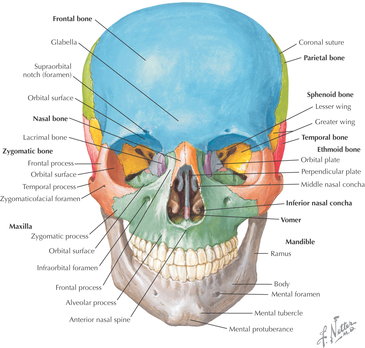
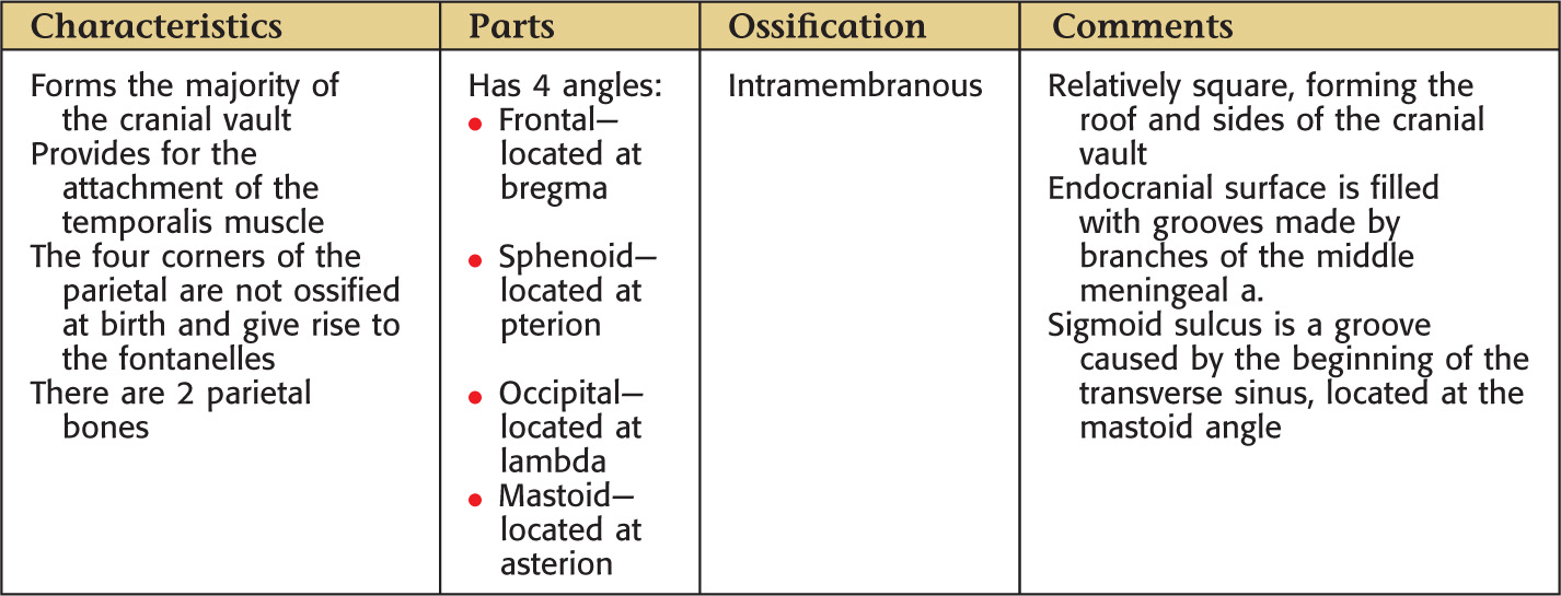
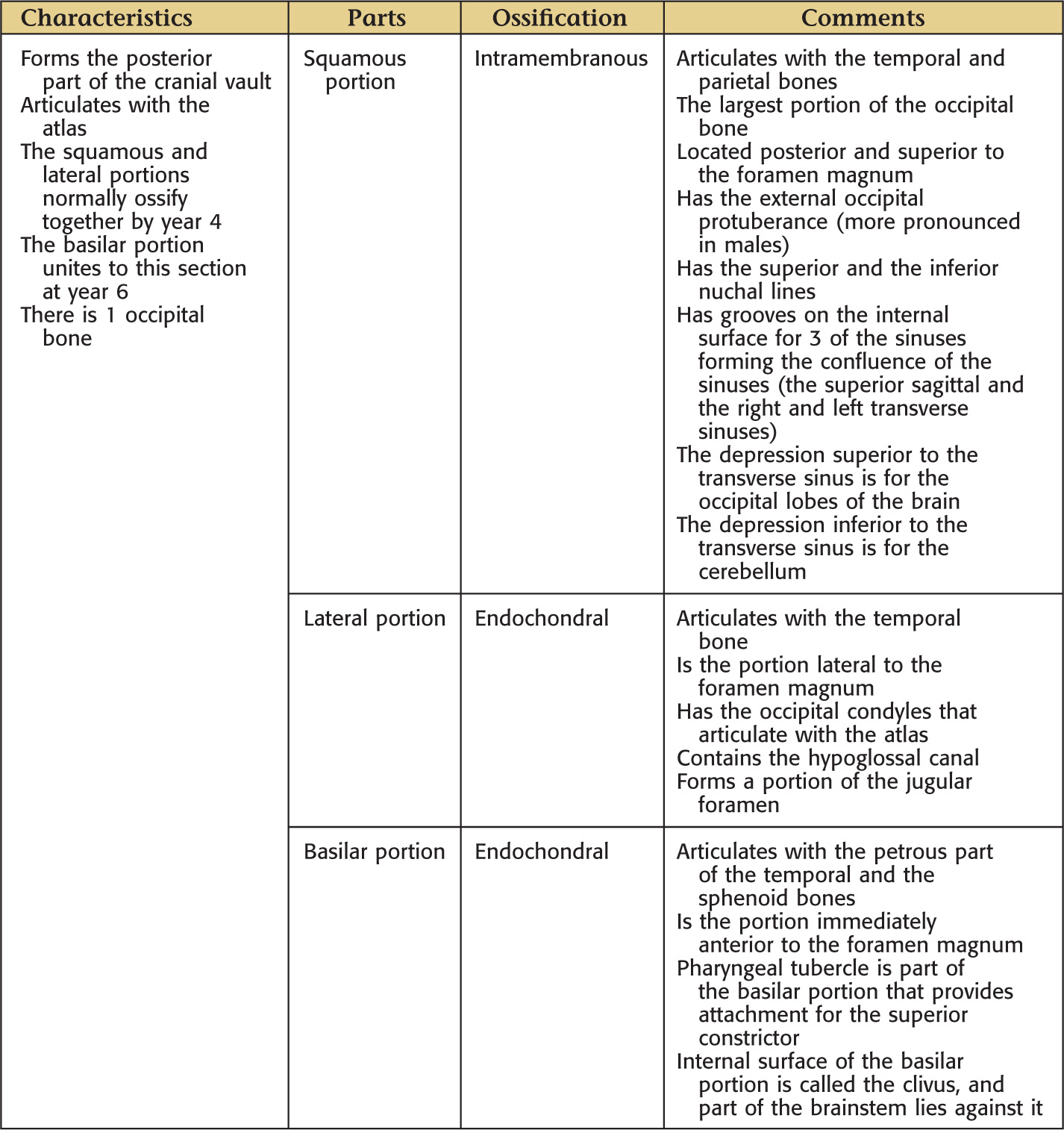
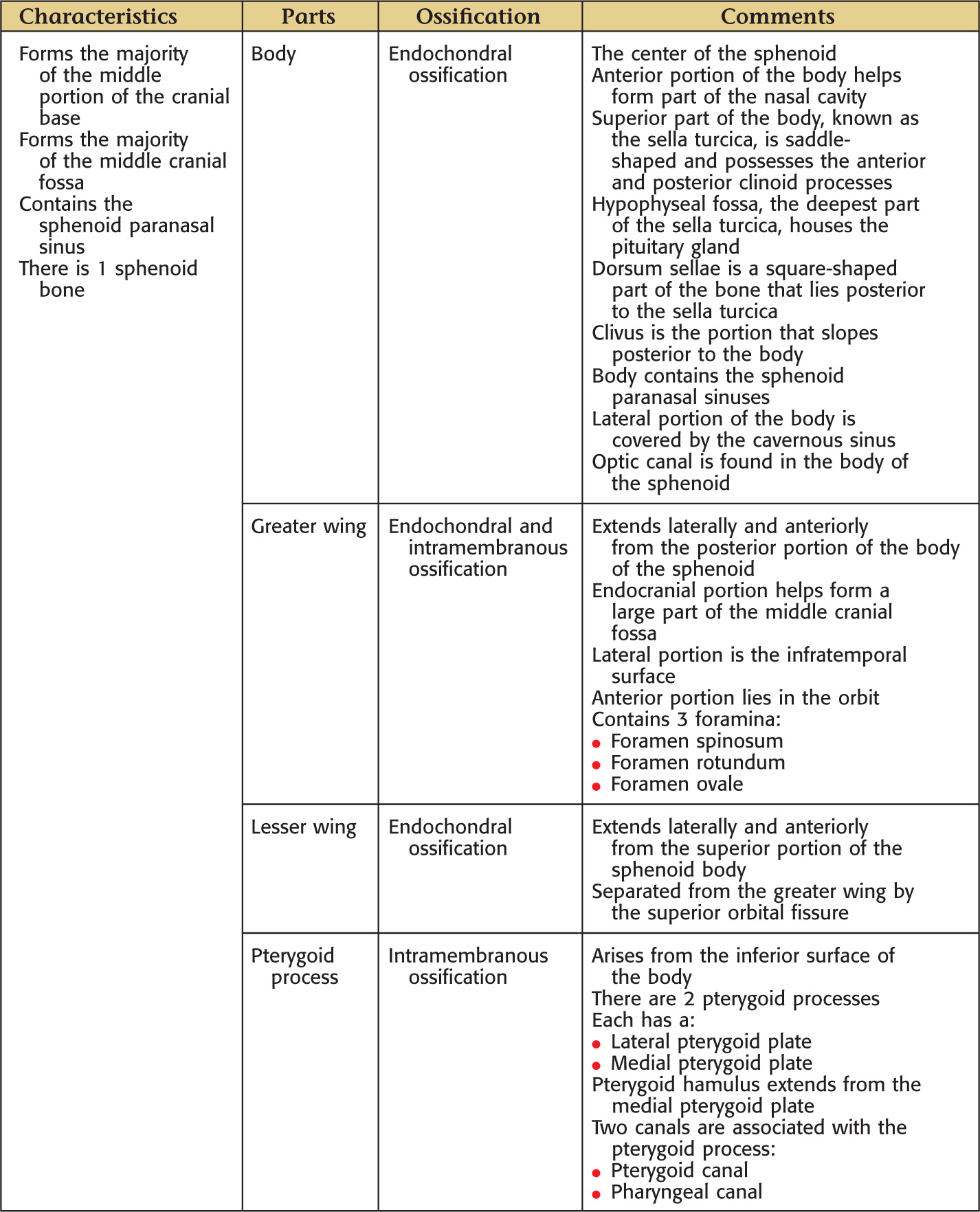
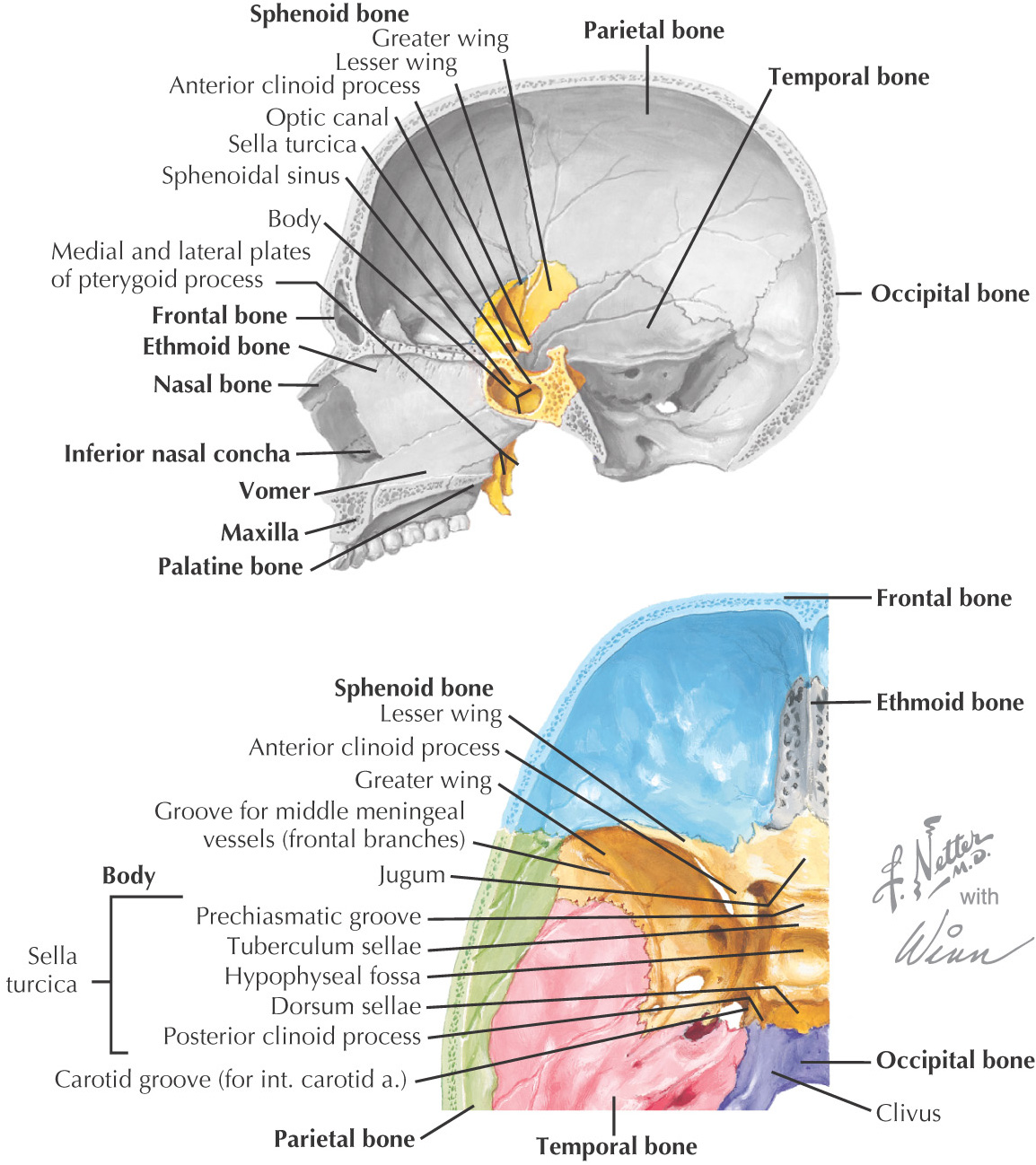
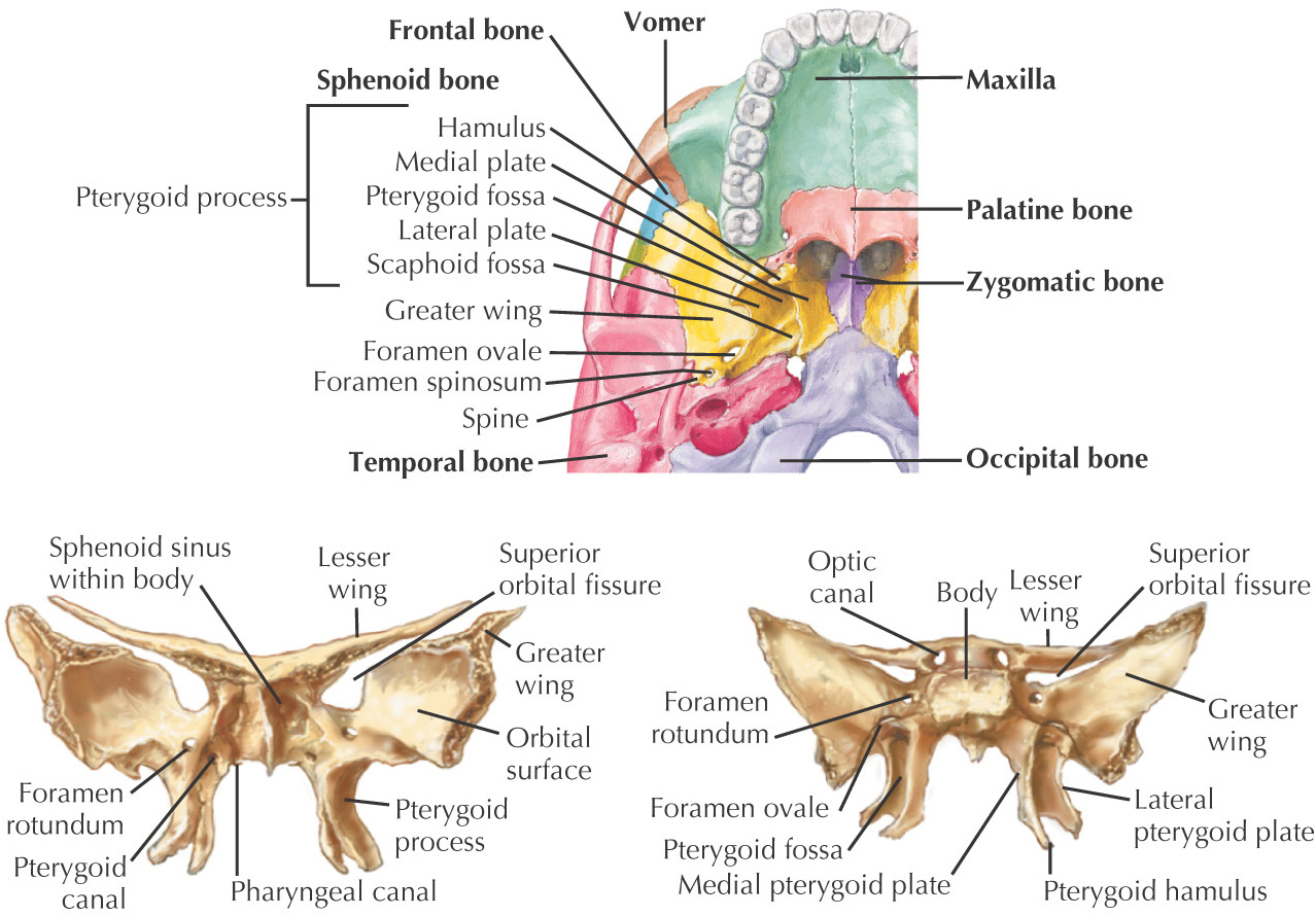
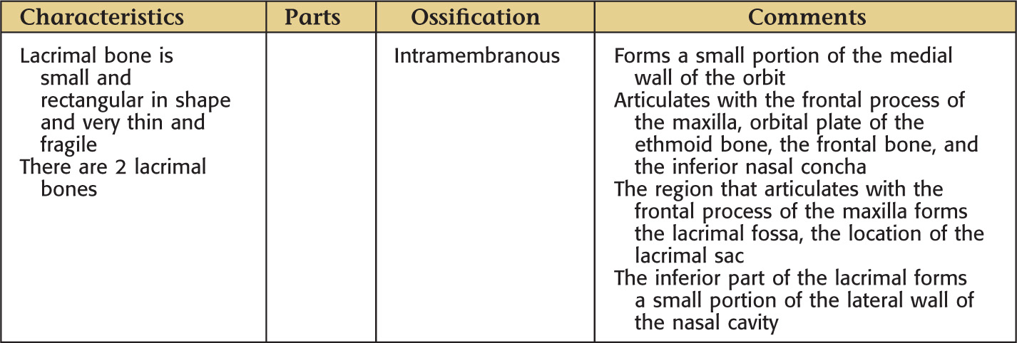
Most complicated bony structure in the human body
The complete bony framework of the head; includes the mandible
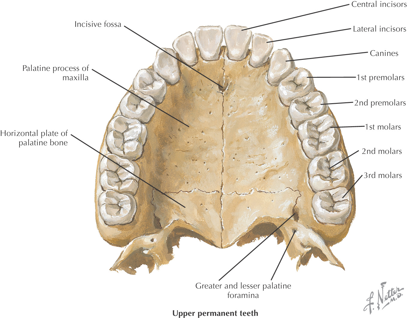
28 individual bones make up the skull:
• 11 are paired
• 6 are single
Wormian bones, or sutural bones, are irregularly shaped small bones found along sutures that occur naturally
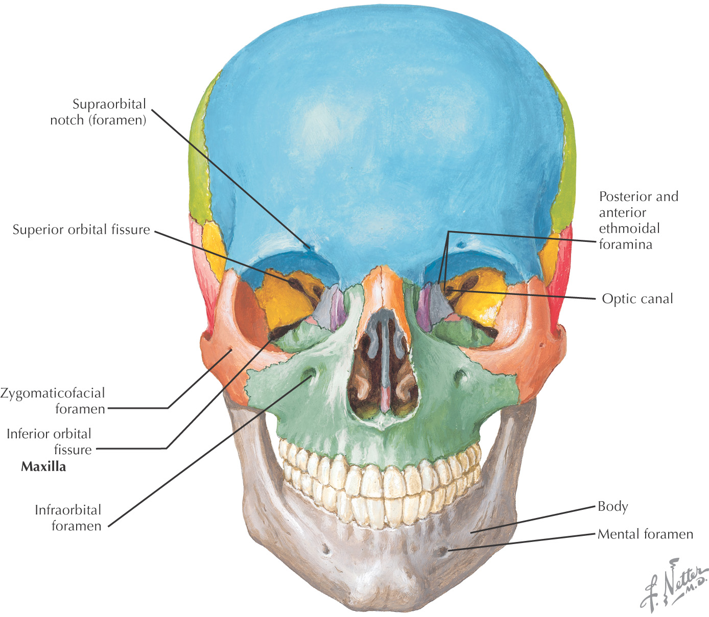
Most important function: to protect the brain
Also protects the 5 organs of special sense:
Two major ways to divide the bones of the skull:
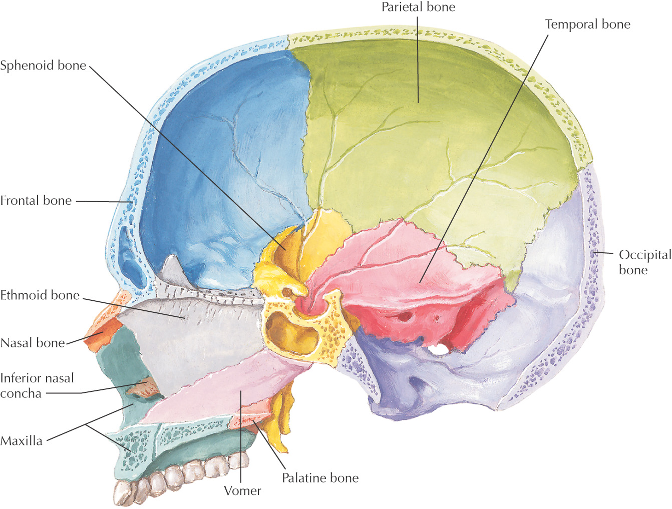
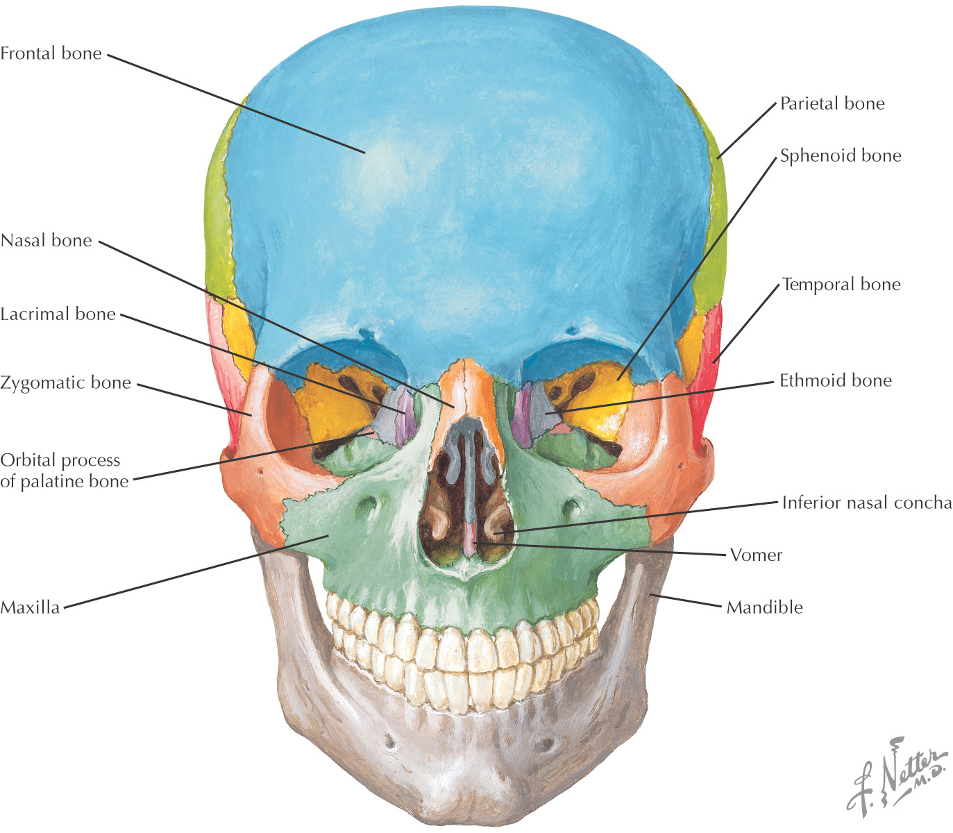
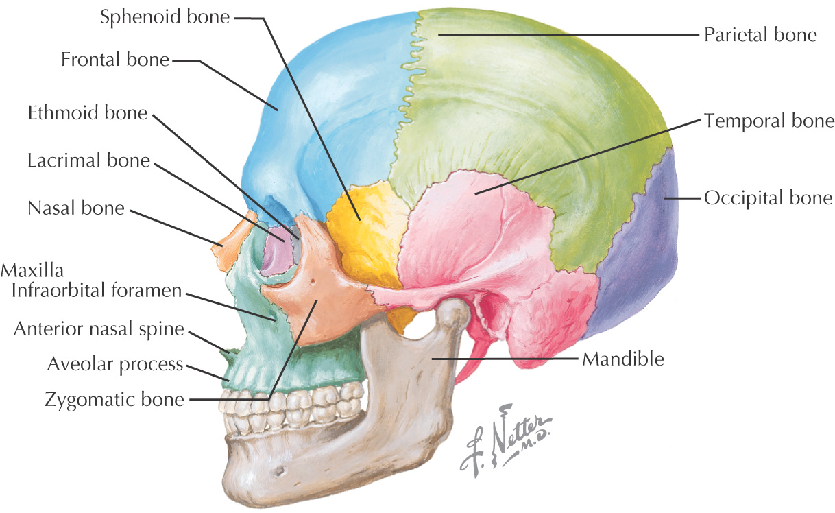
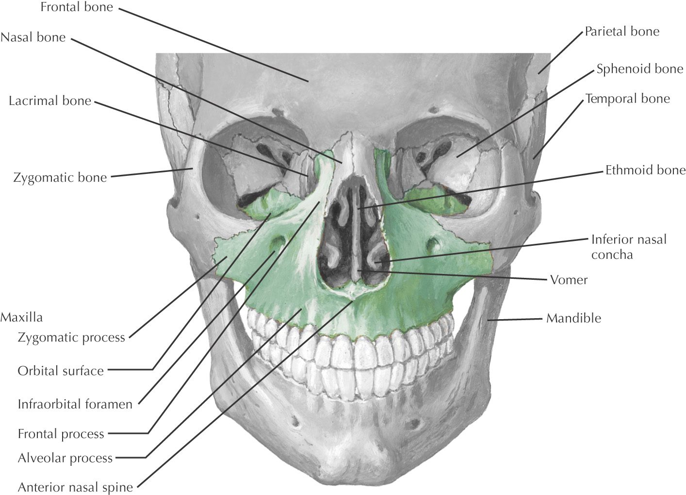
Regionally, the skull is divided into the mandible (lower jaw) and cranium (skull without the mandible)
Cranium is further divided into:
• Cranial vault–upper portion of the skull
• Cranial base–inferior portion of the skull
• Cranial cavity–interior of the skull
• Facial skeleton–bones that make up the face
• Acoustic skeleton–ear ossicles
Developmentally, the skull is divided into:
• Viscerocranium–the portion of the skull related to the digestive and respiratory systems
• Neurocranium–the portion of the skull that protects the brain and the 5 organs of special sense
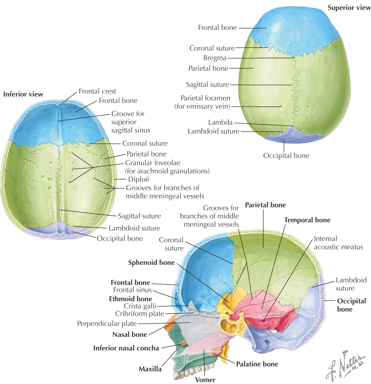
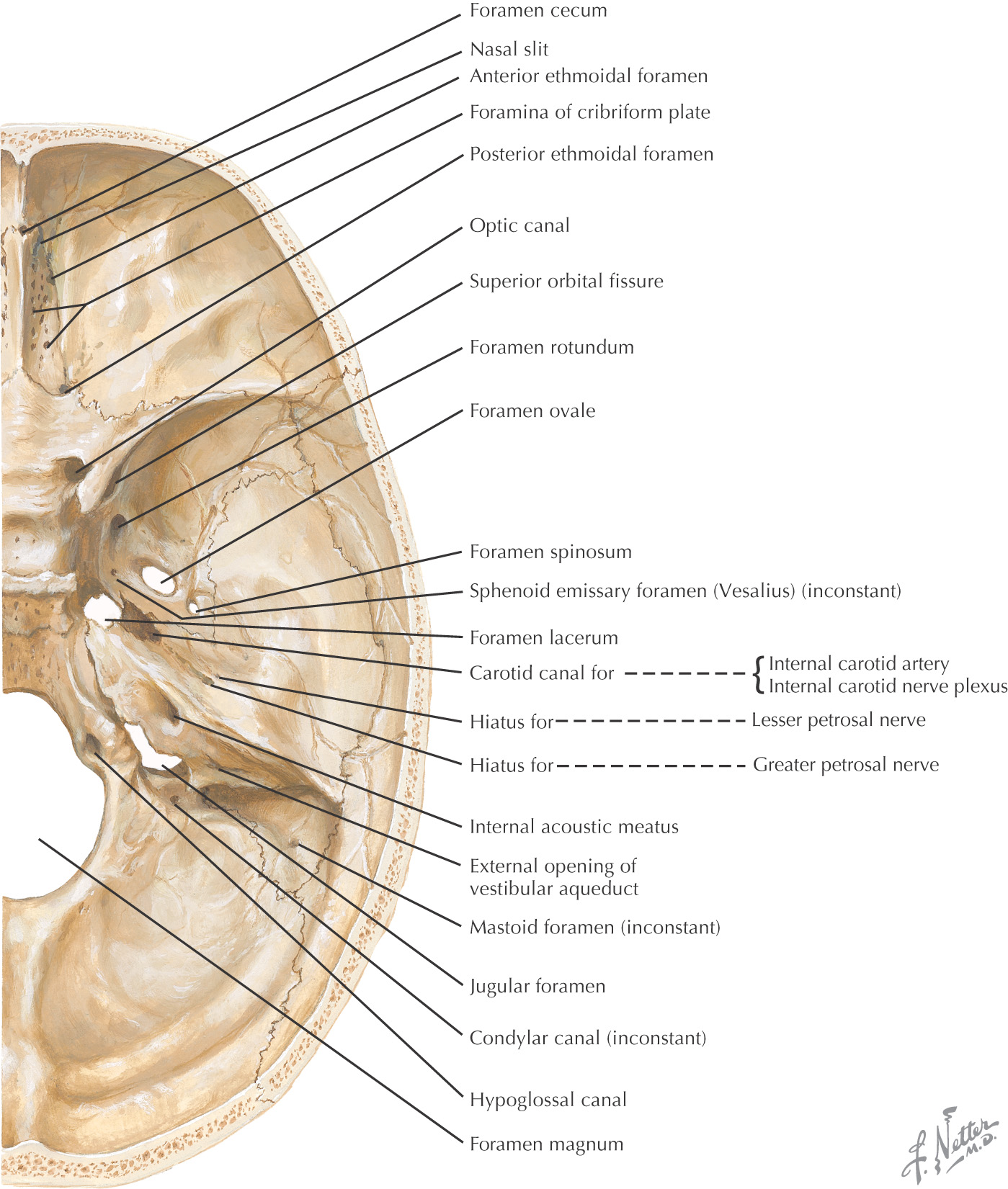
Cranial cavity divisions:
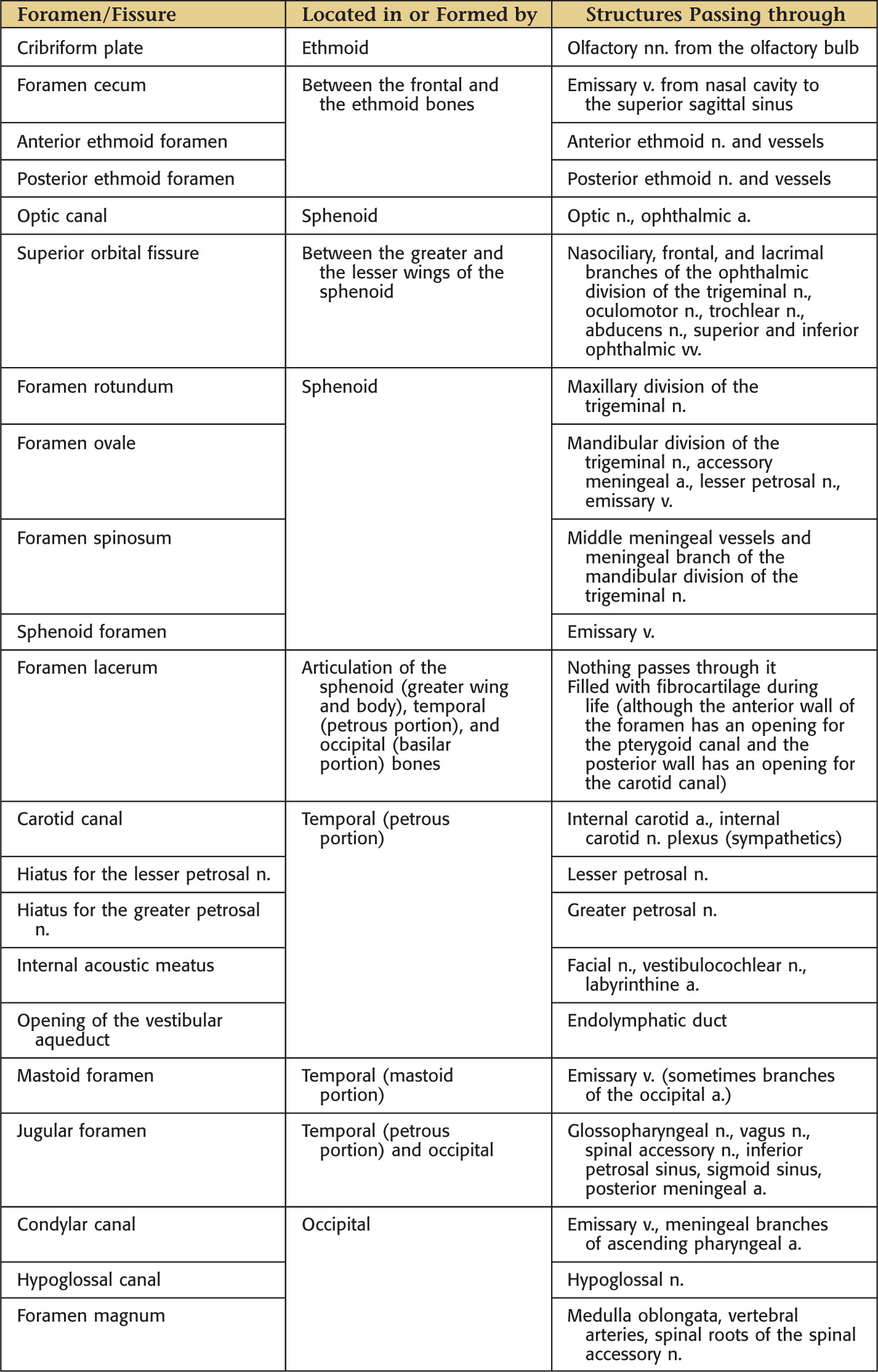
• Anterior cranial fossa–contains the frontal lobe of the brain
• Middle cranial fossa–contains the temporal lobe of the brain
• Posterior cranial fossa–contains the cerebellum
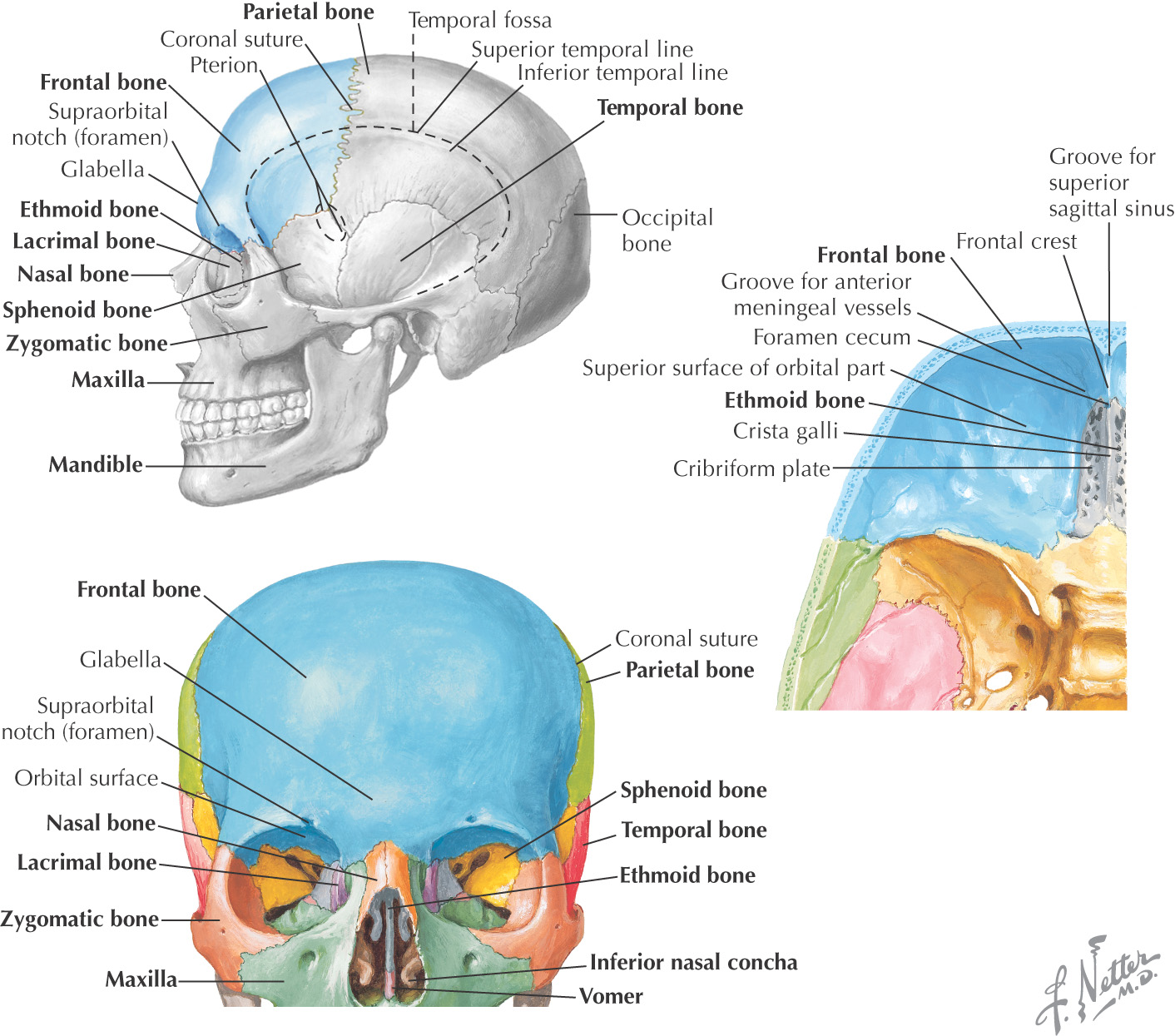
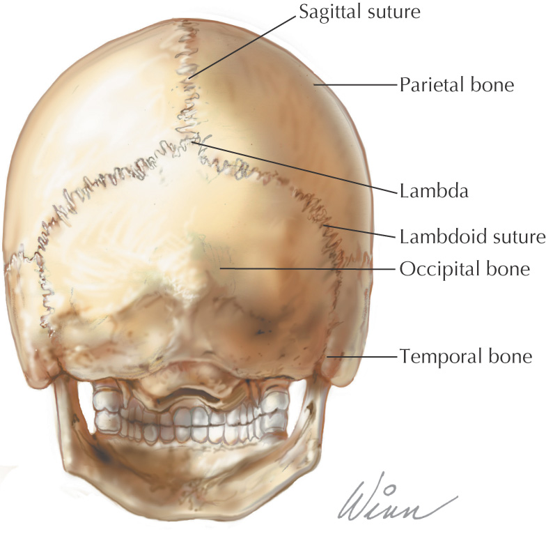
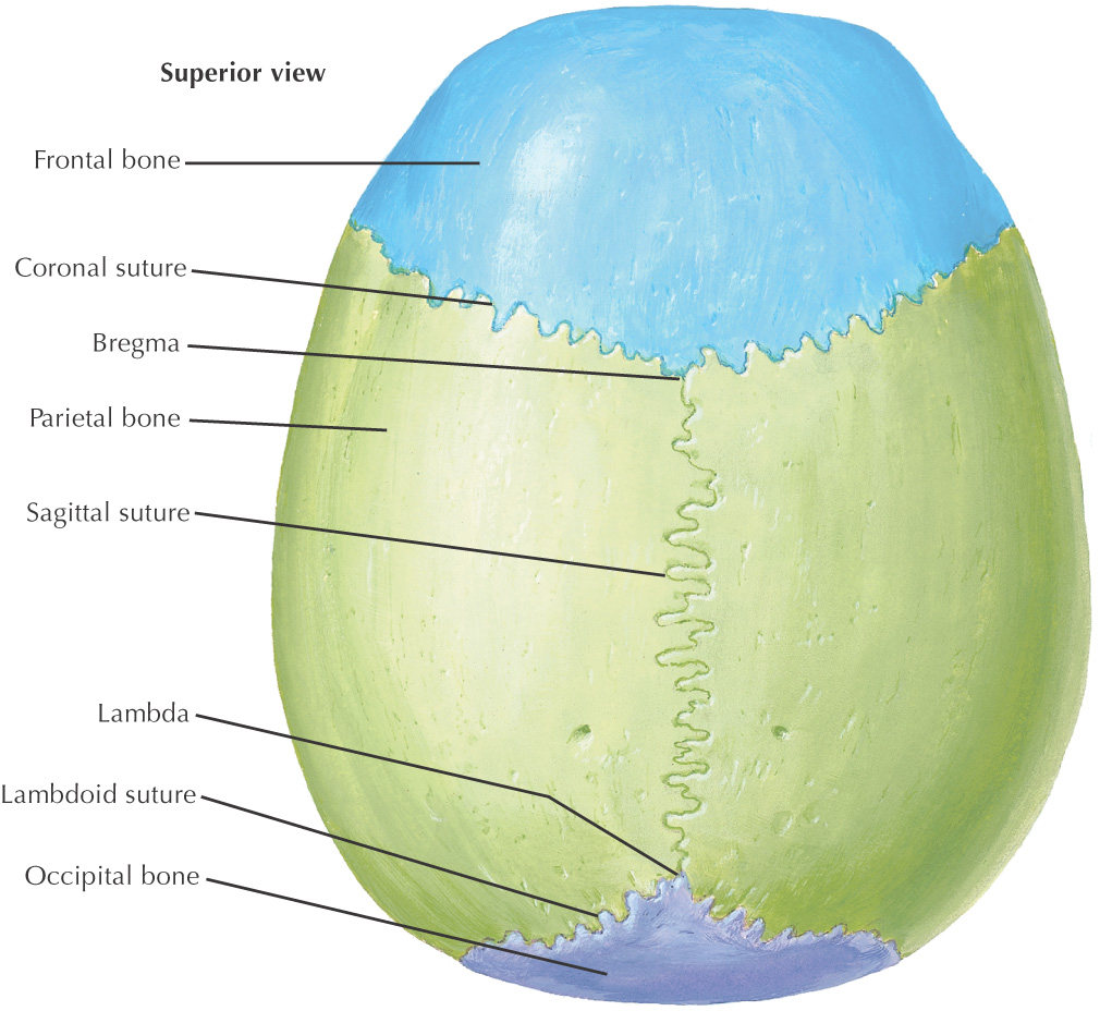
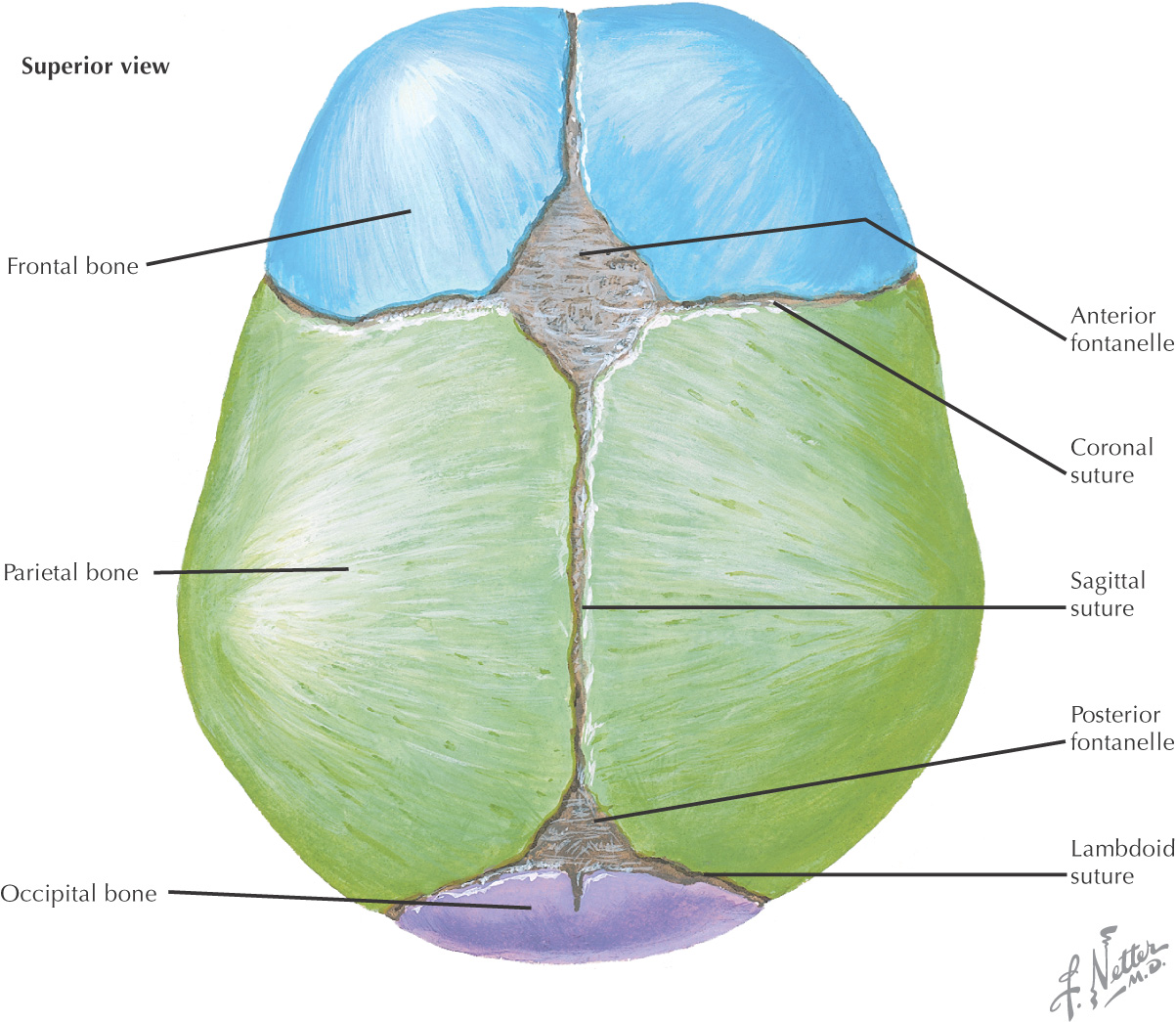
Skull is depicted by observing it from 5 views:
• Norma frontalis–the anterior view
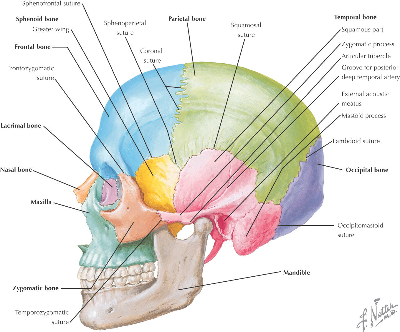
• Norma lateralis–the lateral view
• Norma occipitalis–the posterior view
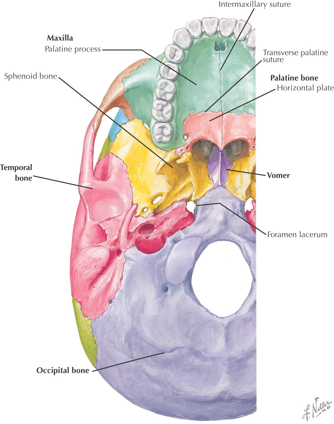
• Norma basalis–the inferior view
• Norma verticalis–the superior view
Bones |
|
Sutures |
Bones |
|
Sutures |
Bones |
|
Sutures |
Bones |
|
Sutures |
Bones |
|
Sutures |
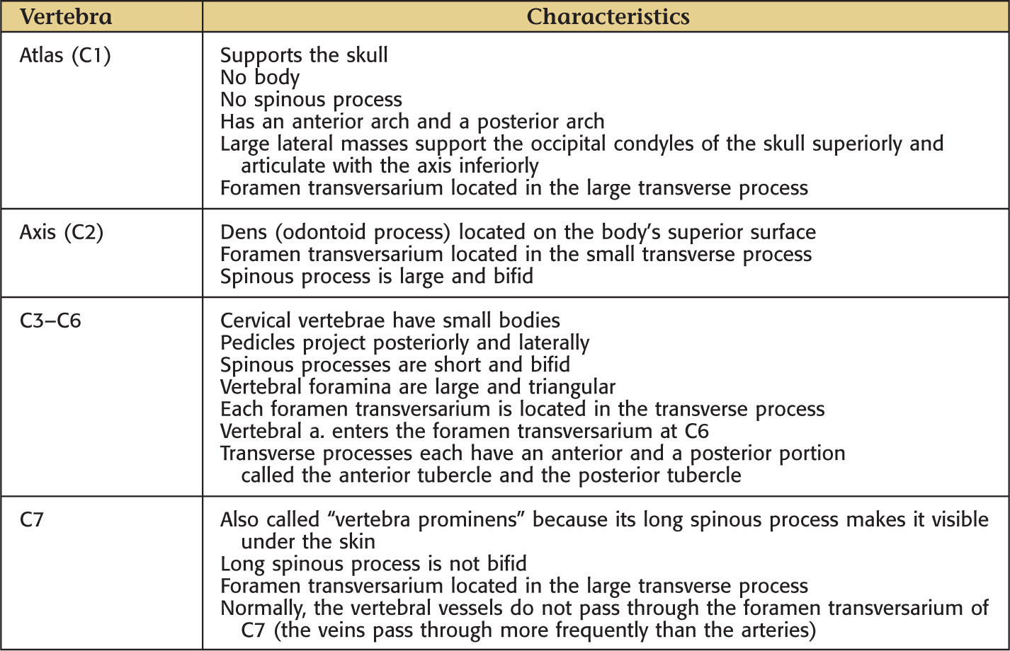
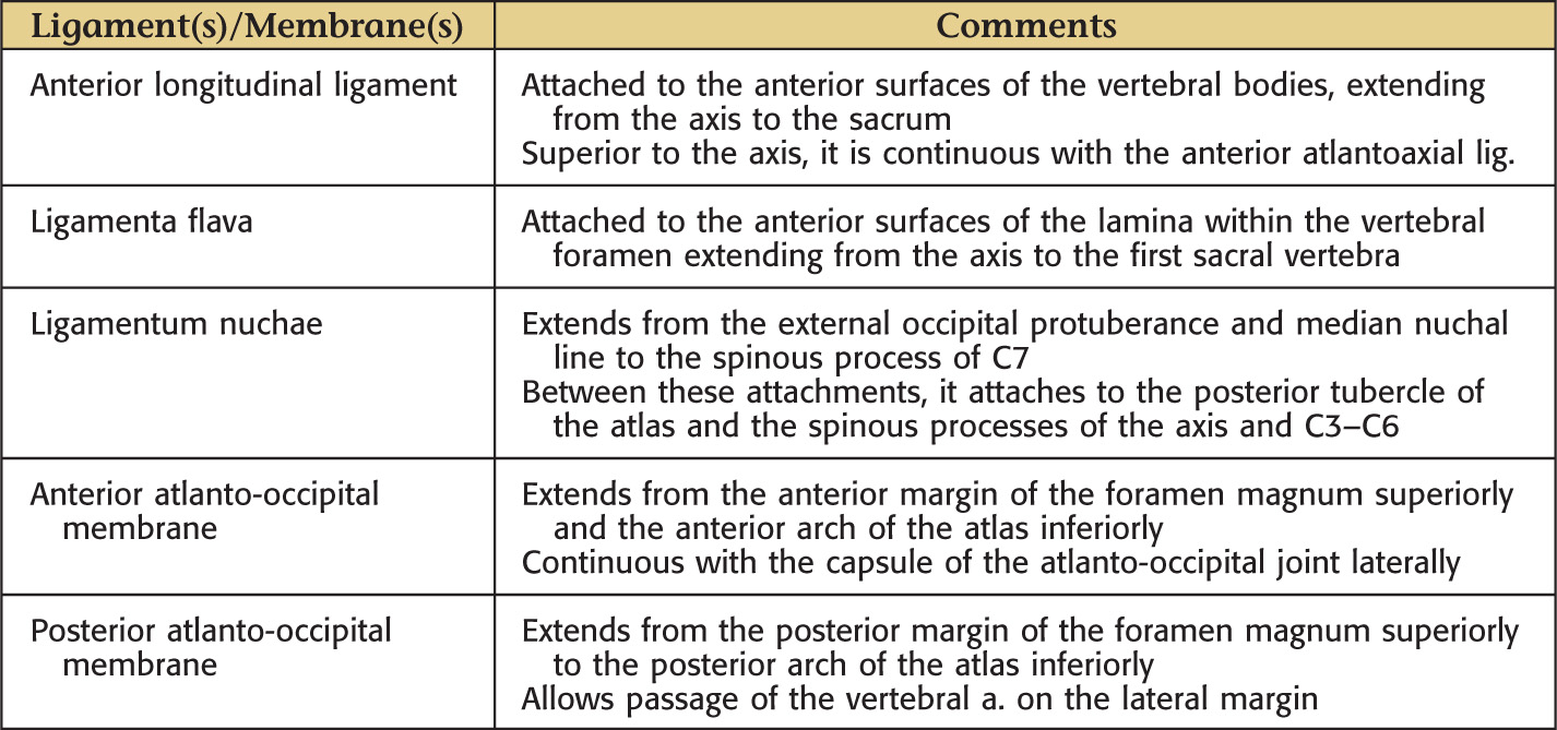
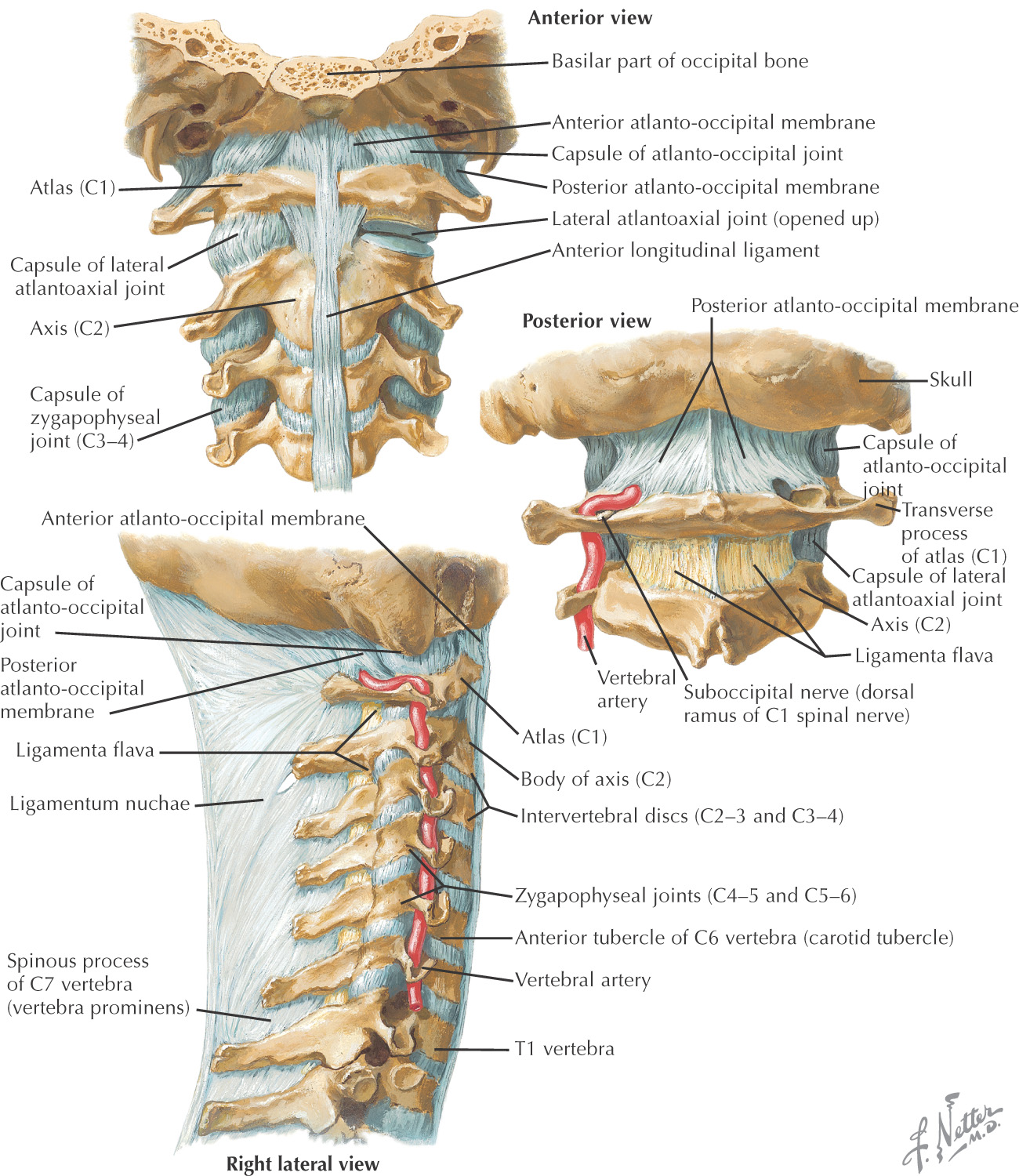
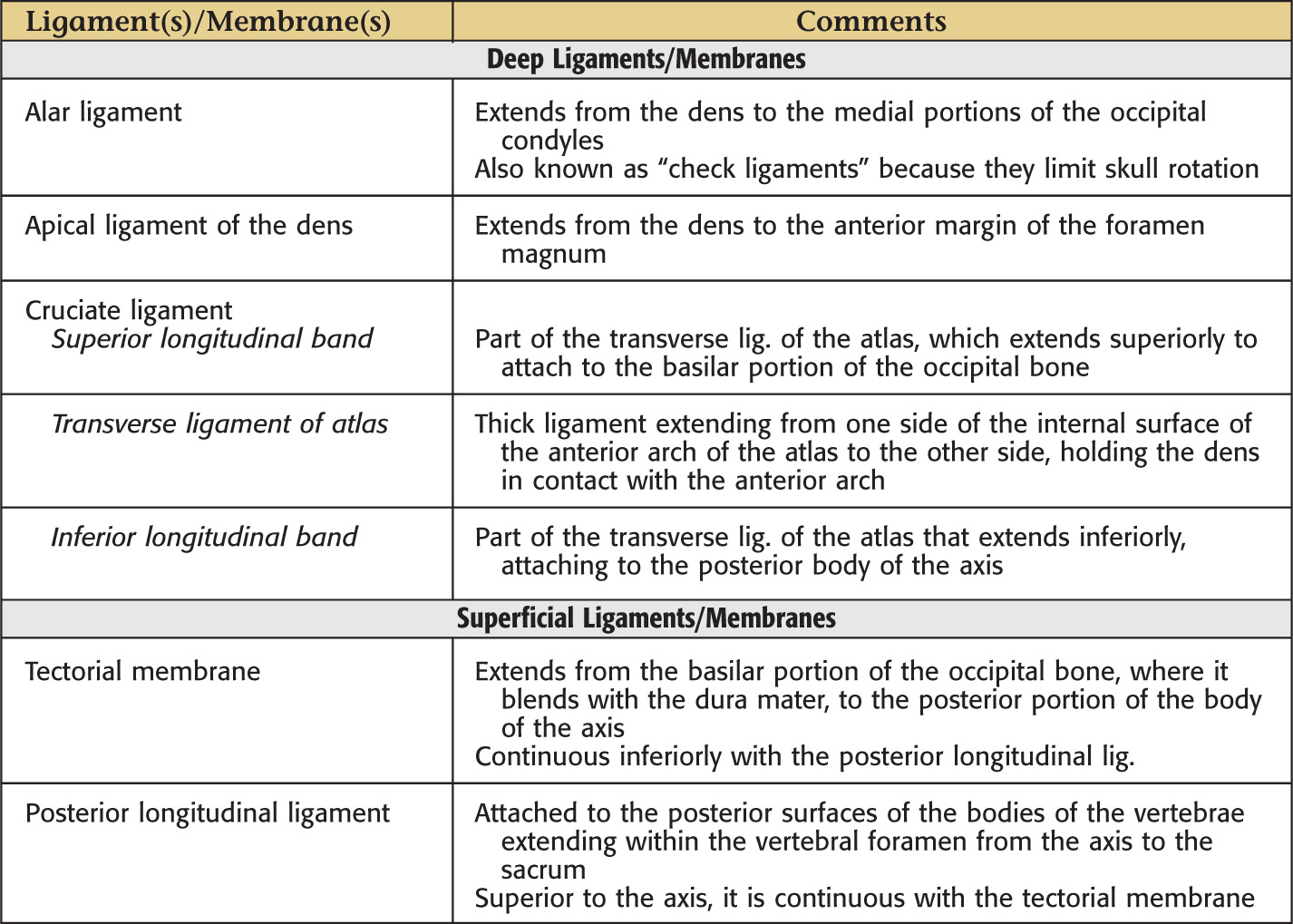
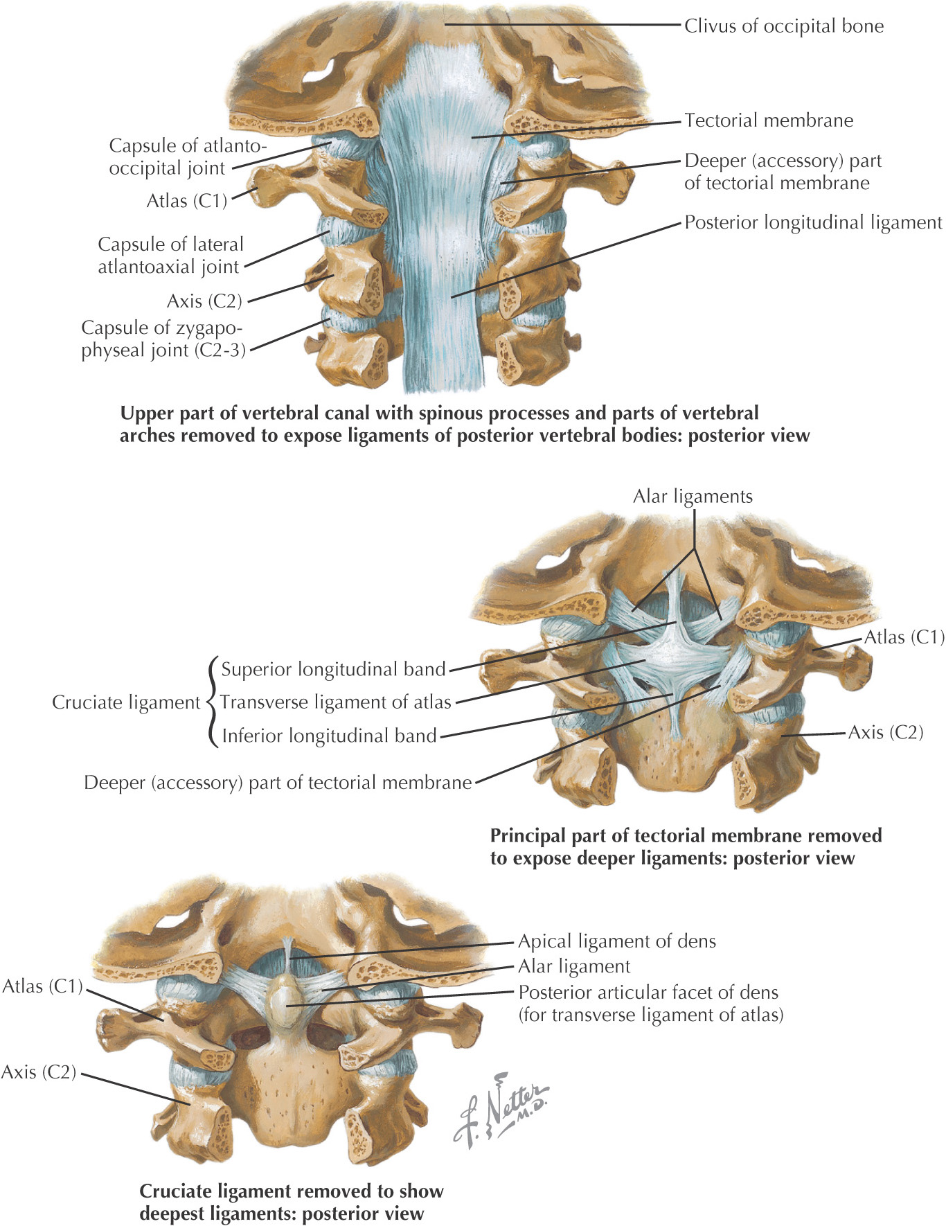
7 cervical vertebrae (C1 to C7)
The smallest vertebrae in the body
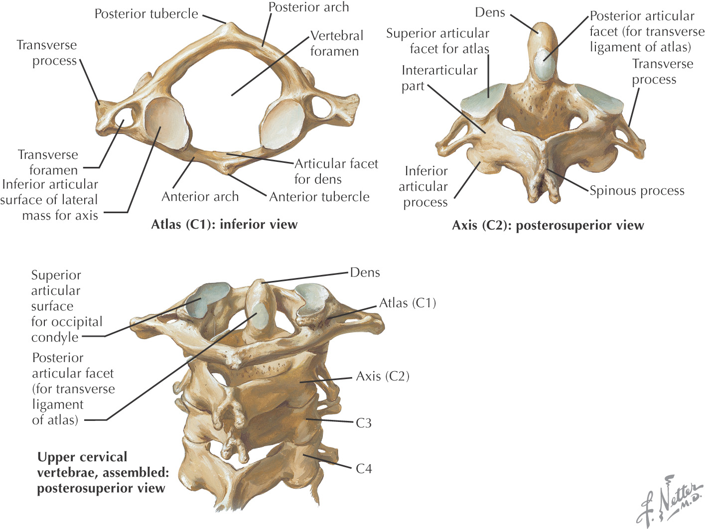
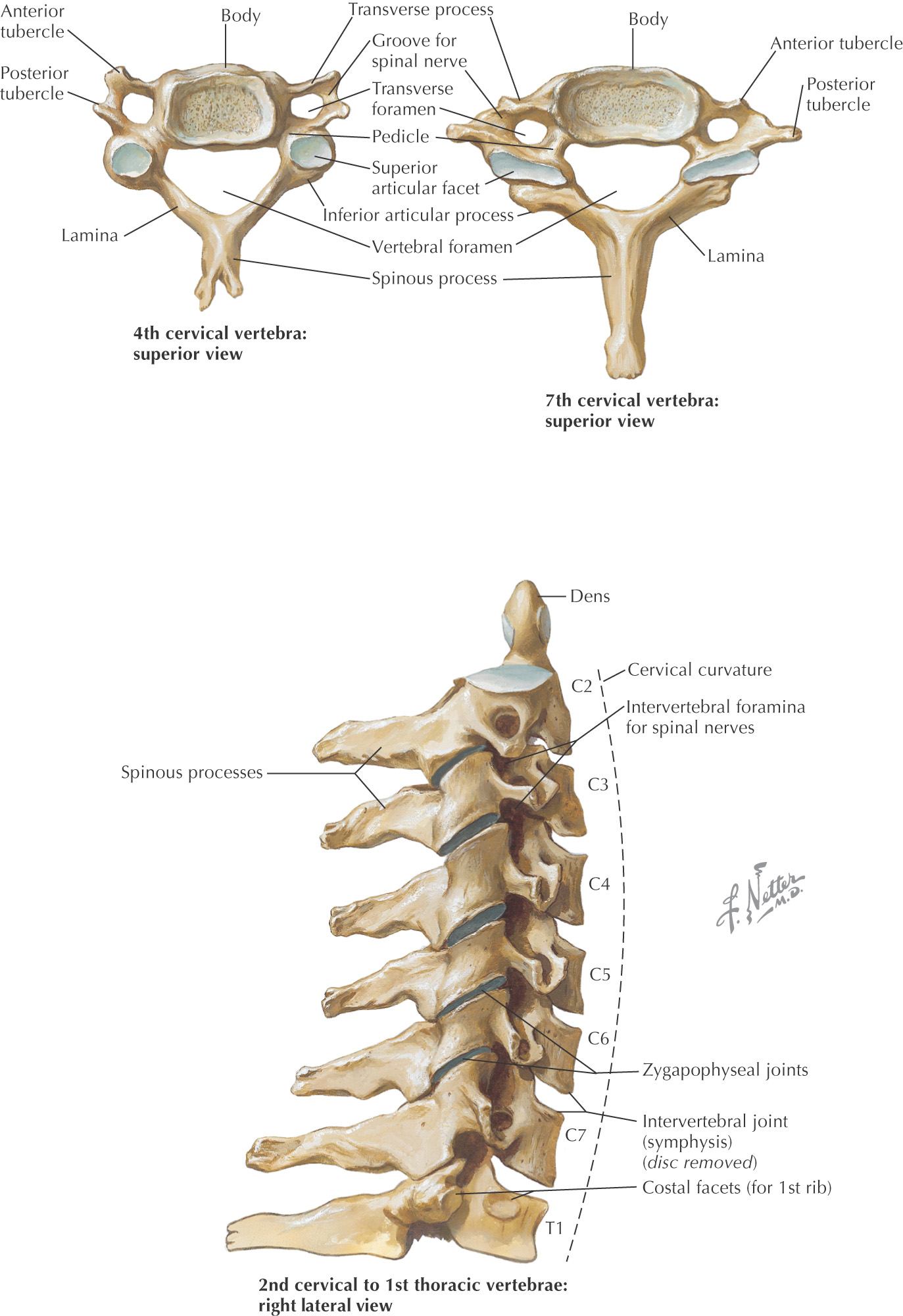
The 1st, 2nd, and 7th cervical vertebrae are unique in their shape; the 3rd to the 6th are similarly shaped
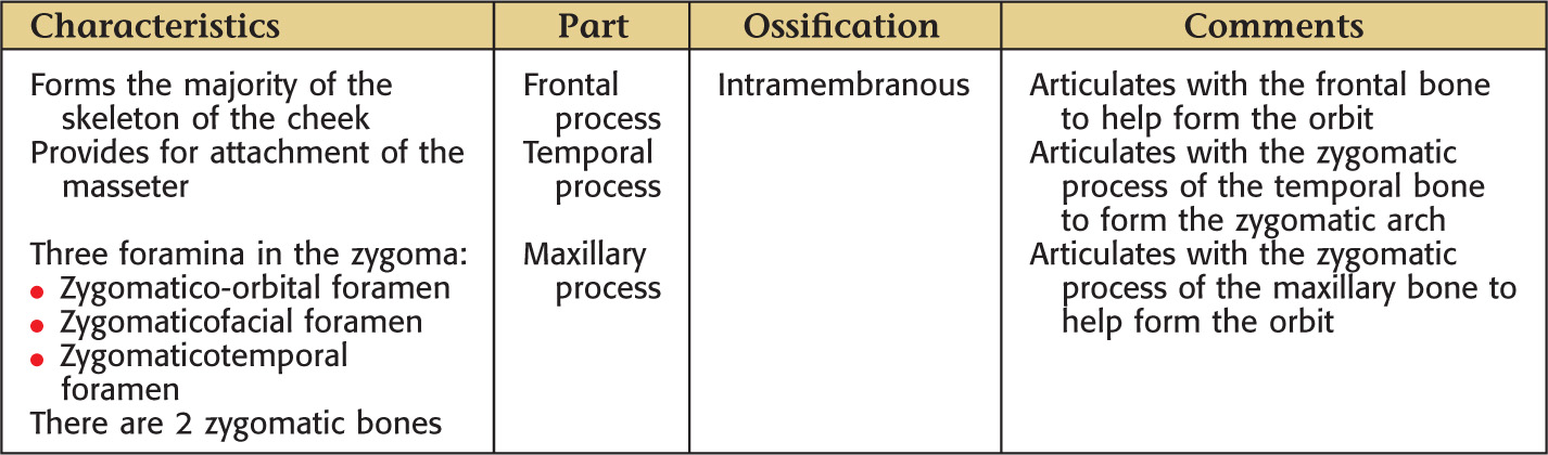
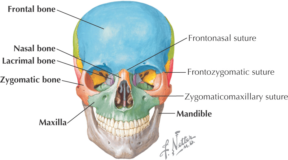
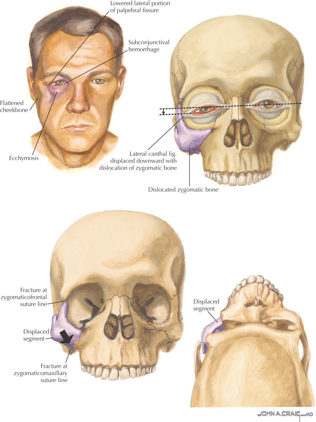
Zygoma is the second most commonly fractured bone of the face after the nasal bone
Susceptible to fracture, usually due to a facial blow from a fist or trauma related to a car accident
In fractures due to blows from a fist, the left zygomatic bone is more frequently fractured than the right
Most fractures are unilateral
May displace the zygomatic bone along the sutures, or more severe displacement in a posterior, medial, and inferior direction may occur
Common clinical manifestations include:
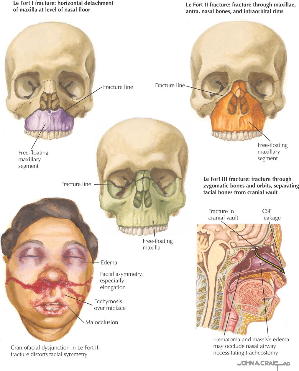
Trauma to the midface usually follows 1 of 3 patterns of fracture:
Horizontal, extending from the lateral margin of the piriform aperture to the pterygoid plates just superior to the apices of the teeth
Gives rise to a detached upper jaw relative to the rest of the maxillofacial skeleton
Pyramidal in outline, extending from the bridge of the nose at or inferior to the nasofrontal suture or maxilla, then inferiorly and laterally through the inferior orbital floor near the infraorbital foramen, through the anterior wall of the maxillary sinus, to the pterygoid plates
Transverse, extending from the nasofrontal suture and frontomaxillary suture and passing posteriorly along the medial wall of the orbit through the nasolacrimal groove and ethmoid, then following the inferior orbital fissure to the lateral wall of the orbit, and extending through the frontozygomatic suture
Within the nose, the fracture extends along the perpendicular plate, vomer, and pterygoid plates
In a Le Fort III fracture, the facial skeleton is detached from the base of the skull
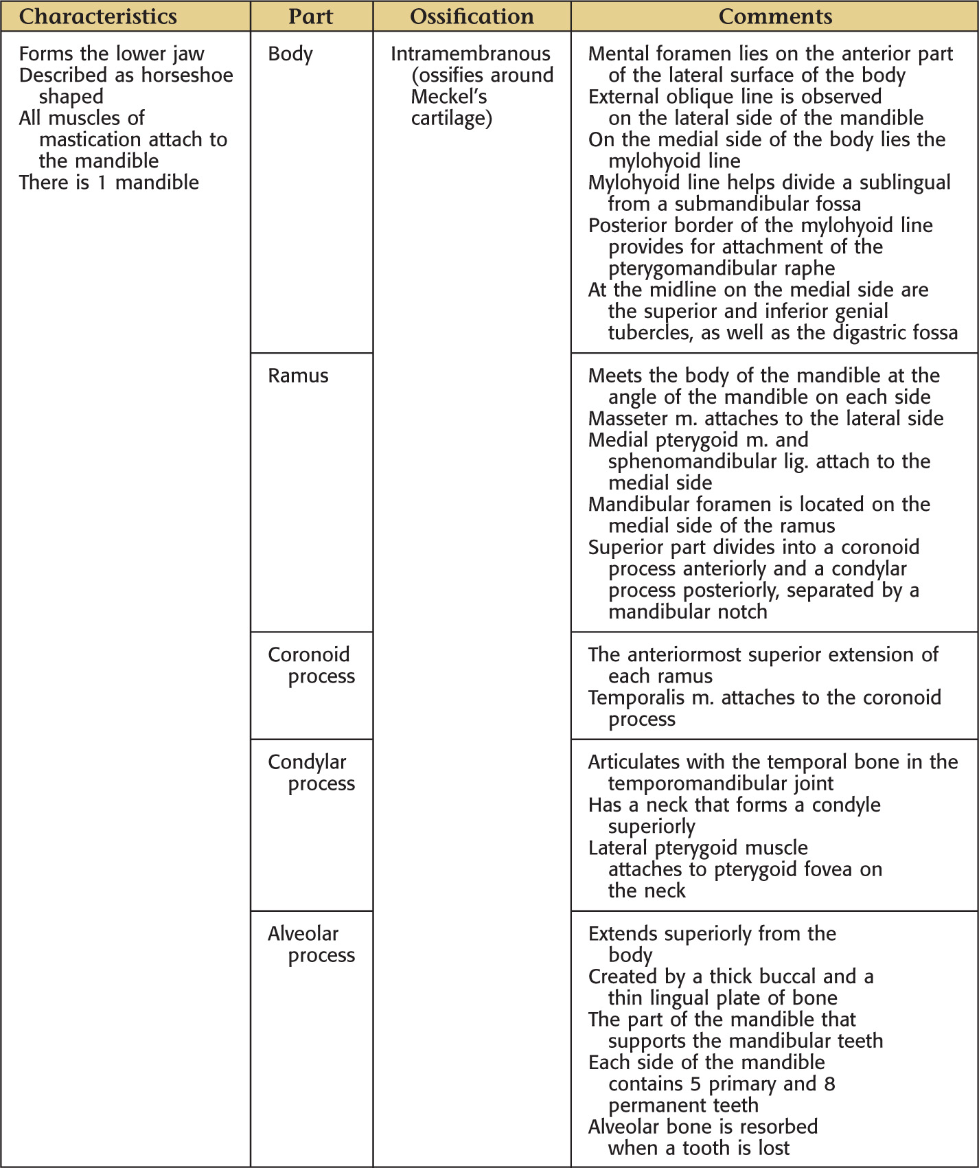
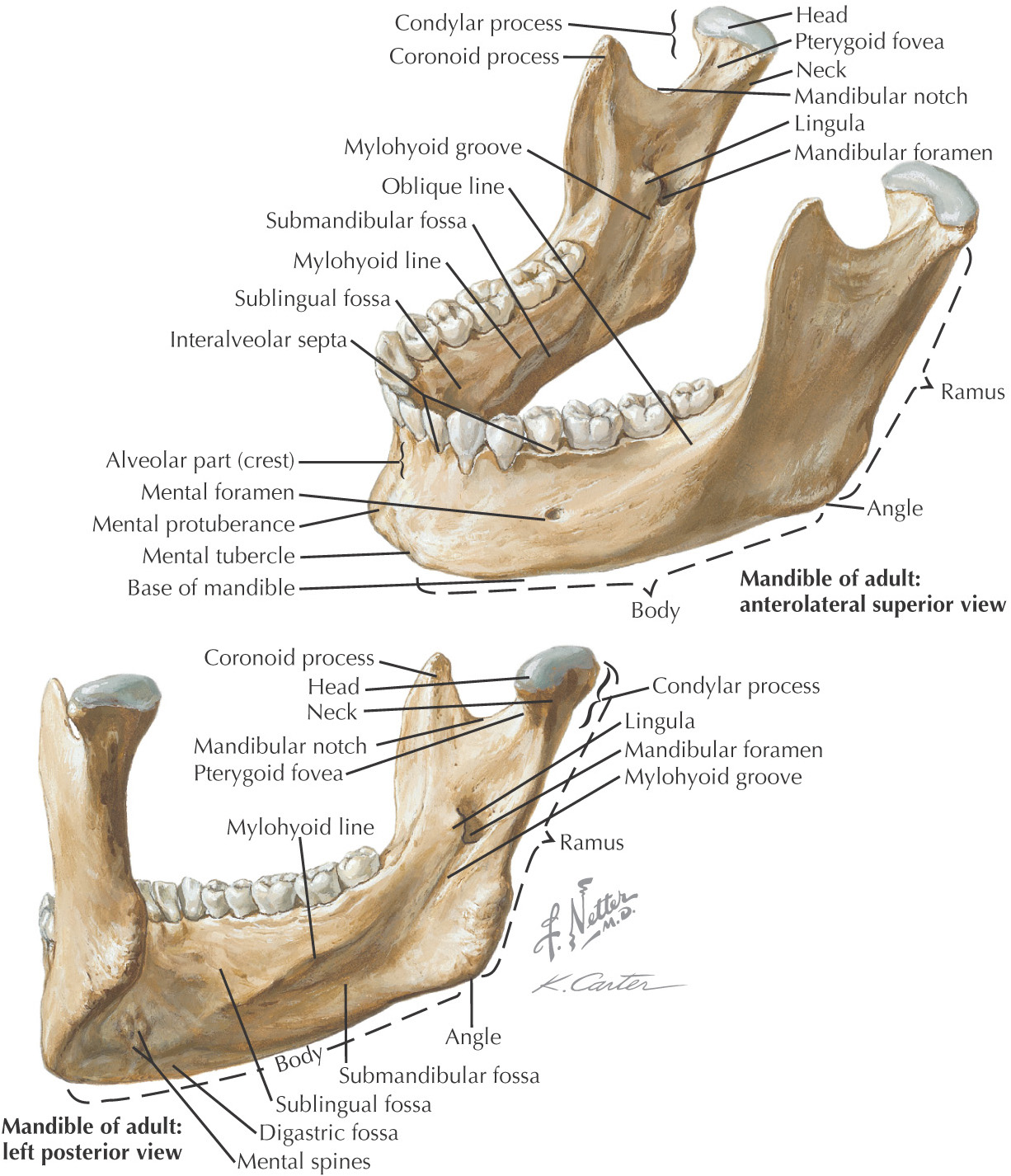
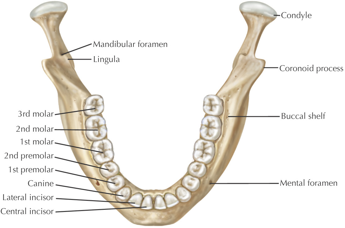
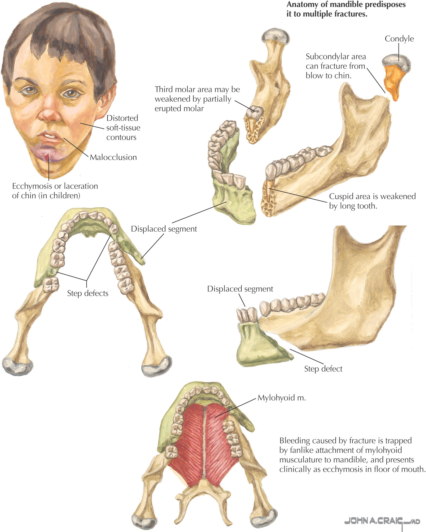
Mandible is a frequently fractured bone
Fractures result from blow from a fist or trauma incurred in motor vehicle accidents
Common sites (in decreasing order of frequency):
With double mandibular fractures, the second usually is contralateral
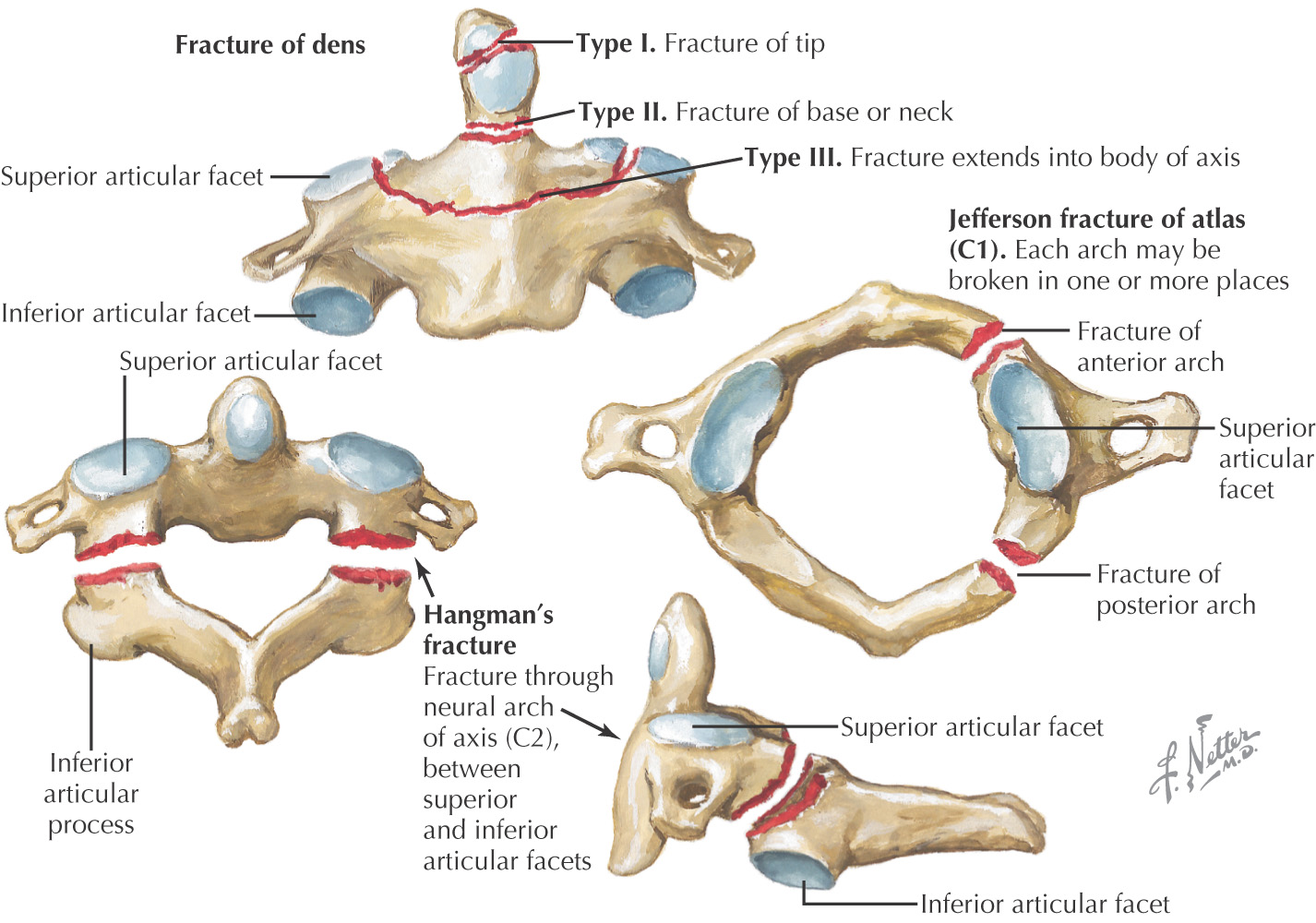
Two common types of cervical fractures:
Involves the atlas
Results from skull compression due to axial loading, causing the atlas to burst
Most patients are neurologically intact but have severe neck pain
Vertebral artery can be compromised
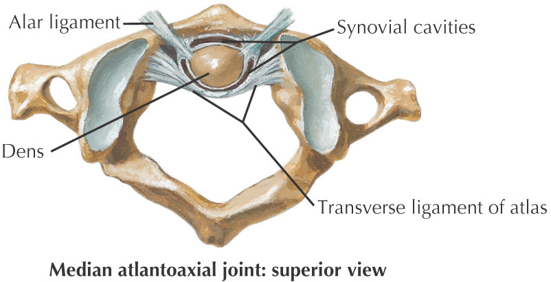
Classified as stable or unstable according to whether the transverse ligament of the atlas is intact:
• Stable fractures can be treated with an orthosis such as a soft collar
• Unstable fractures are more problematic; may require cranial traction applied with use of a halo, as well as cervical fusion
Occurs through the vertebral arch of the axis between the superior and the inferior articulating facets
A traumatic spondylolisthesis often is caused by extension of the neck with axial compression, common in car accidents
The historical hangman’s fracture is caused by extension and distraction of the neck
Involves the axis
Classification into 3 types:
• Type 1—fracture at the tip of the odontoid process
• Type 2—fracture along the base or the neck of the odontoid
• Type 3—fracture that passes through the body of the axis