Nervous tissue is divided into 2 major cell types:
• Neuroglial cells (the neuroglia)
The structural and functional cells in the nervous system
Respond to a nervous stimulus and conduct the stimulus along the length of the cell
A neuron’s cell body is called the perikaryon, or soma
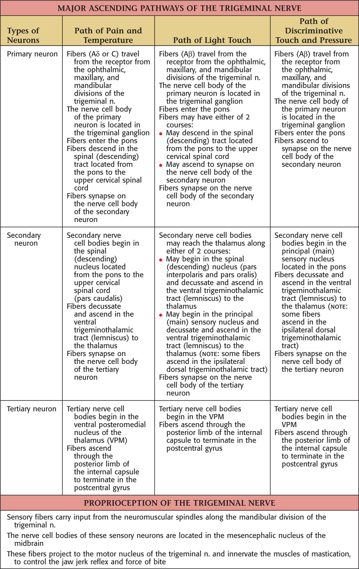
Cell bodies are classified by their location:
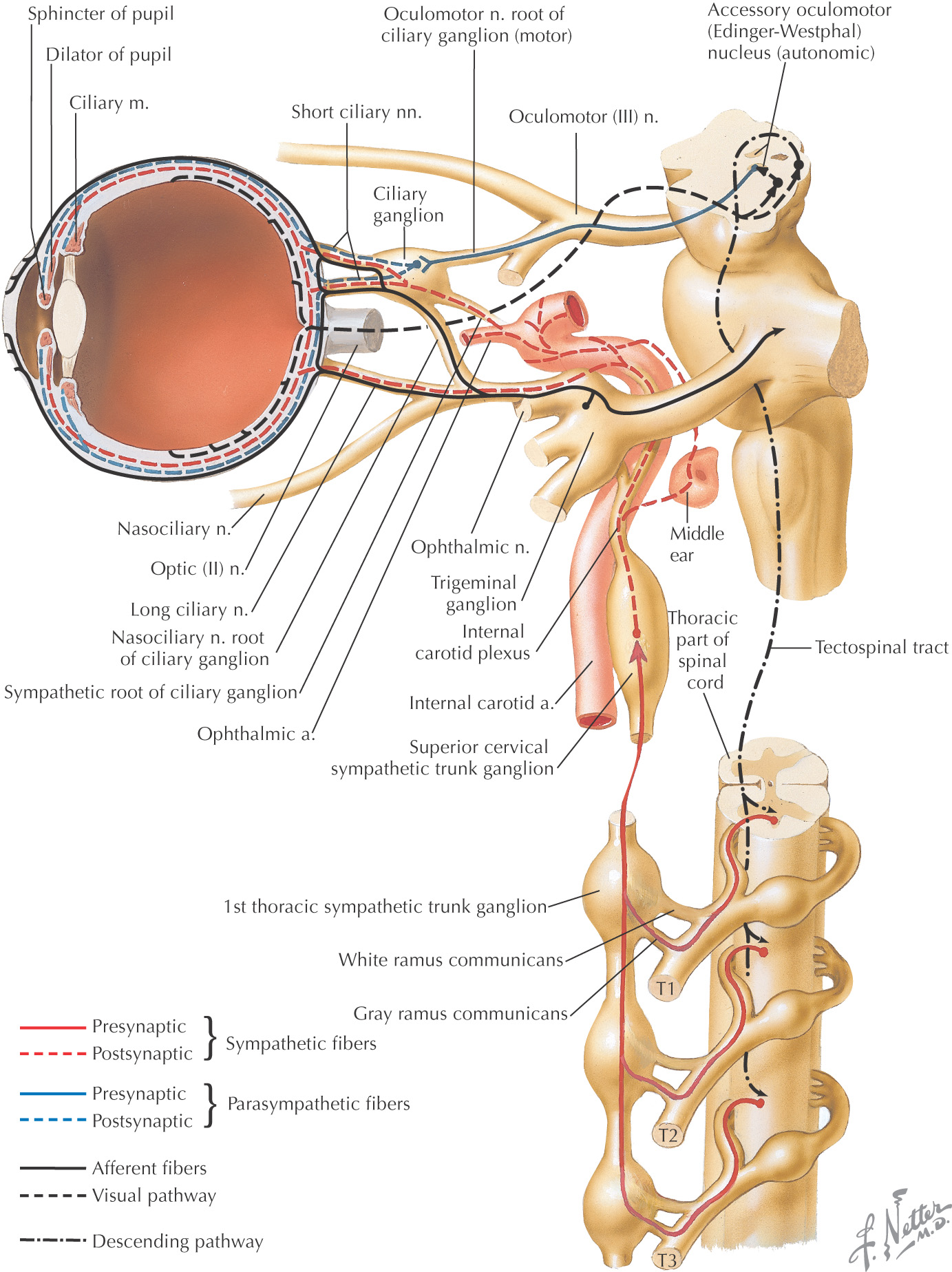
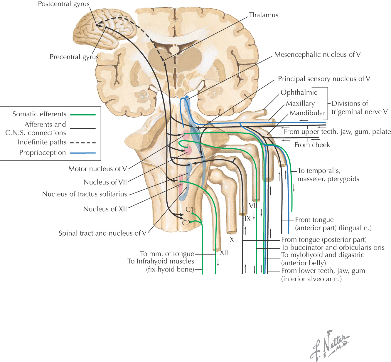
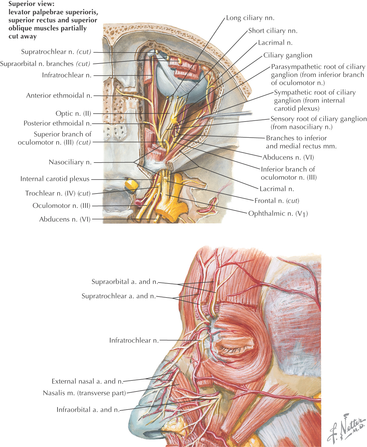
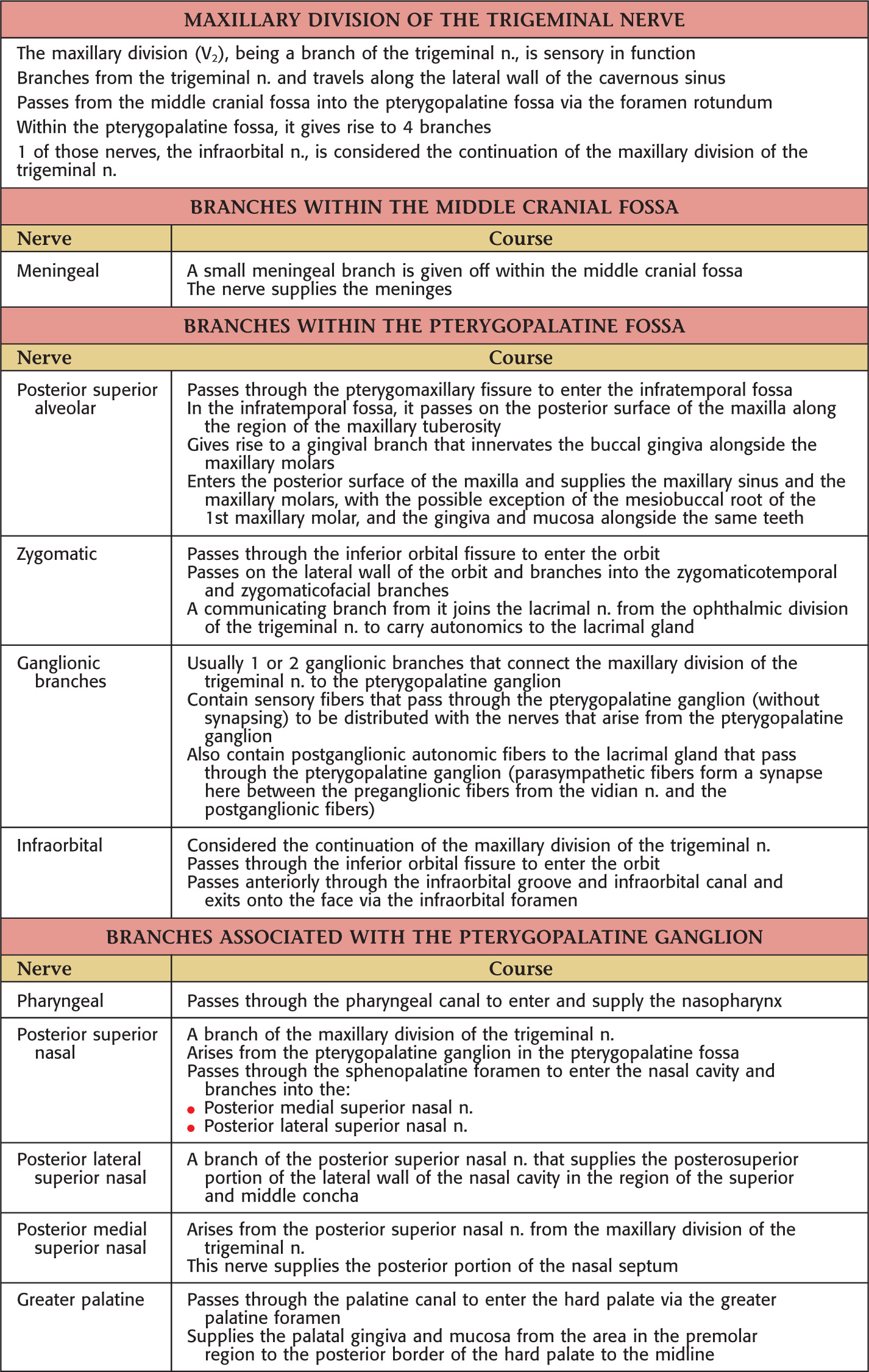
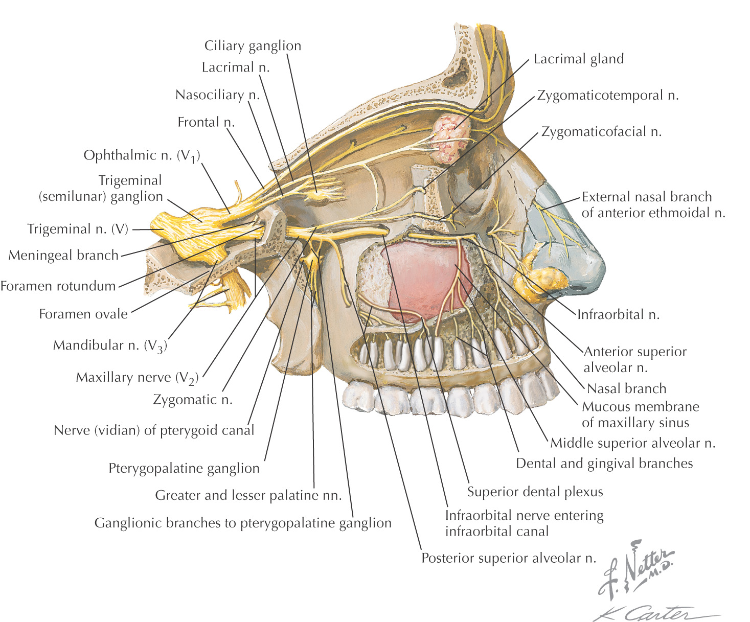
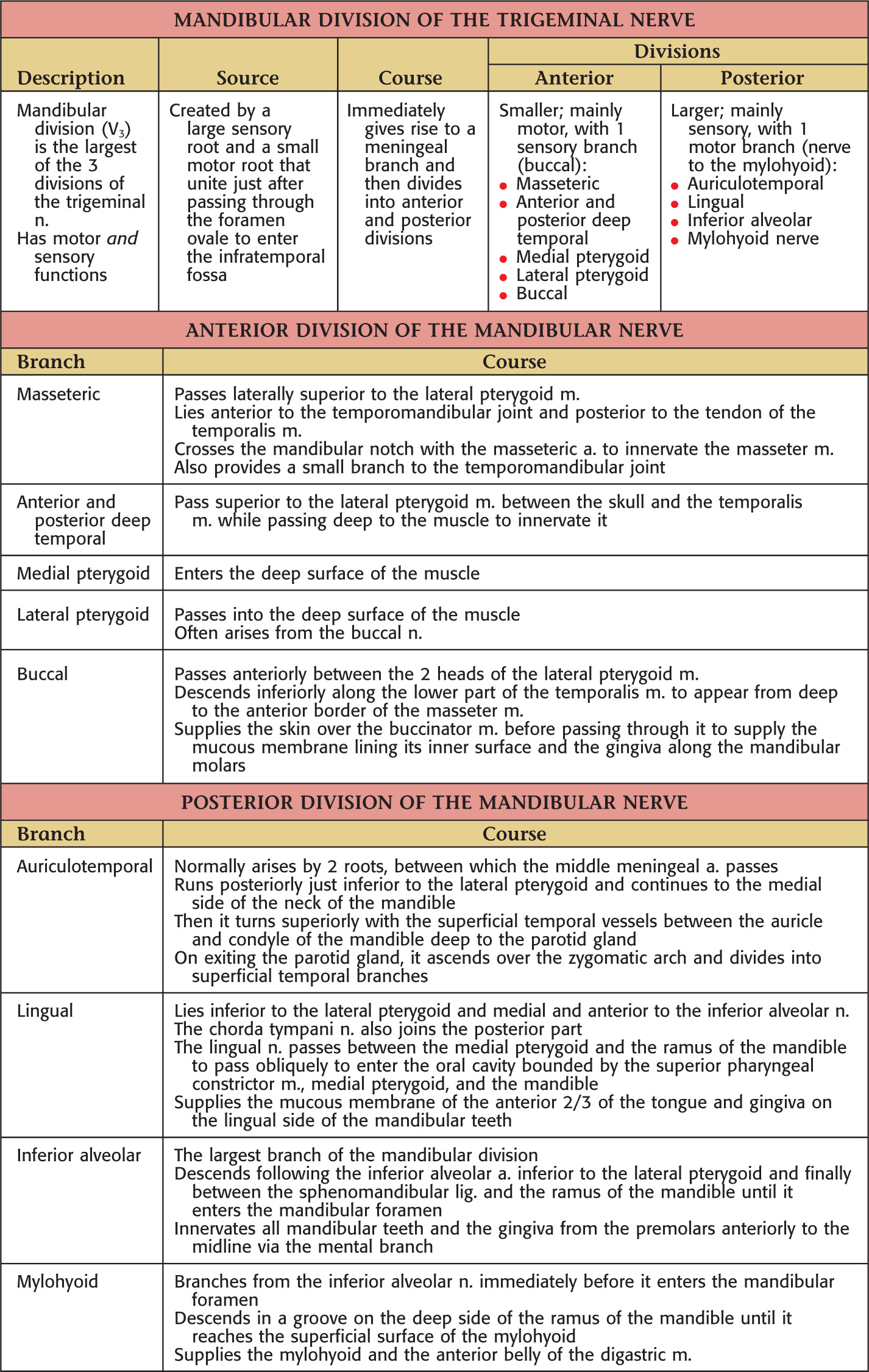
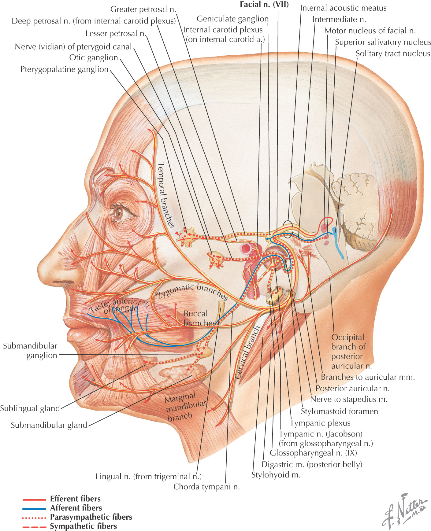
• Ganglion—a collection of nerve cell bodies located in the peripheral nervous system (e.g., dorsal root ganglion, trigeminal ganglion, ciliary ganglion)
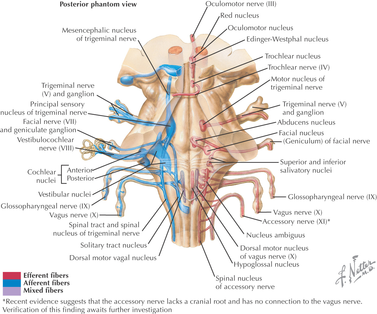
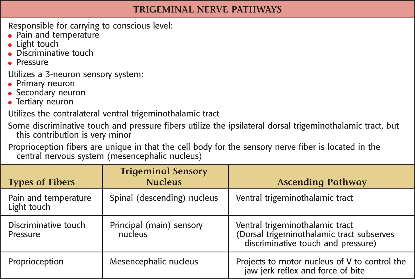
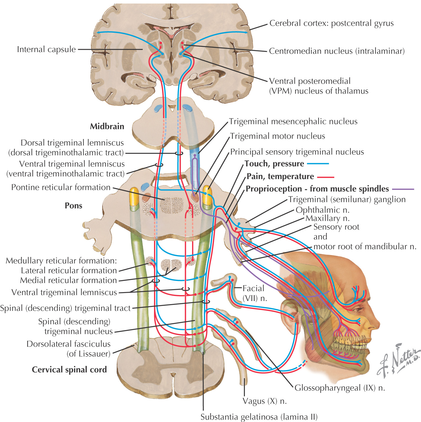
• Nucleus—a collection of nerve cell bodies located in the central nervous system (e.g., Edinger-Westphal nucleus, chief sensory nucleus of cranial nerve V, motor nucleus of cranial nerve VII)
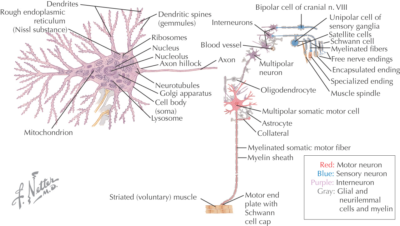
Neuron’s cell bodies contain typical cellular organelles within their cytoplasm:
• Rough endoplasmic reticulum (Nissl substance)
Neurons have 2 types of processes that extend from the nerve cell body:
• Dendrite—process that carries nerve impulses toward the nerve cell body; neurons may have multiple dendrites
• Axon—process that carries nerve impulses away from the nerve cell body; neurons can have only 1 axon
3 major types of neurons:
• Unipolar—has only 1 process from the cell body (sensory neurons)
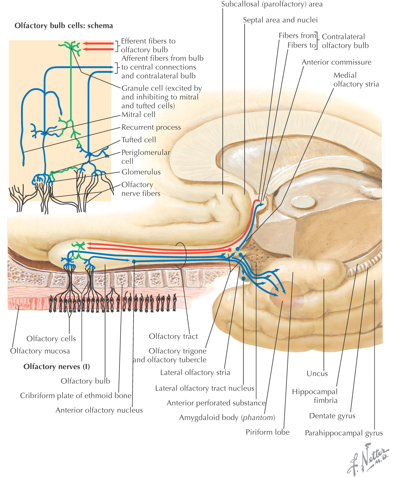
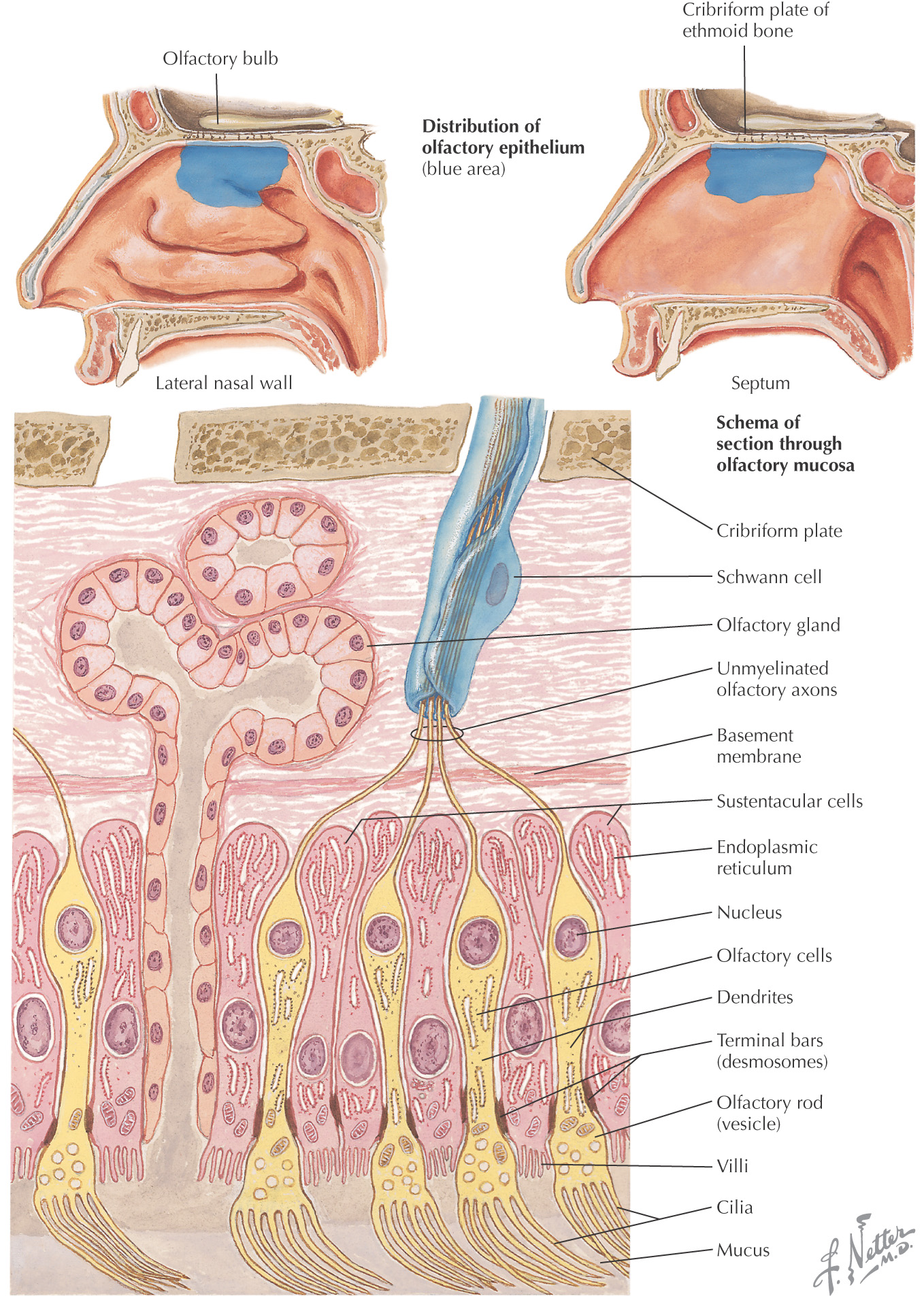
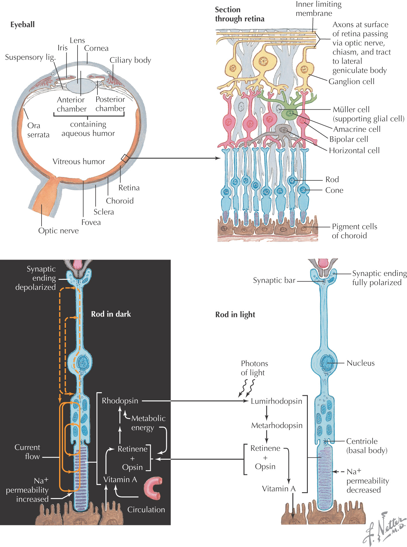
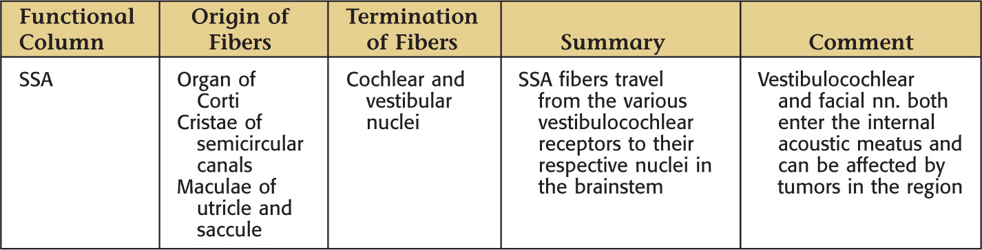
• Bipolar—has 2 processes from the cell body: 1 dendrite and 1 axon (sensory neurons; located only in the retina, olfactory epithelium, and the vestibular and cochlear ganglia)
• Multipolar—has 3 or more processes from the cell body: 2 or more dendrites and 1 axon (motor neurons and interneurons)
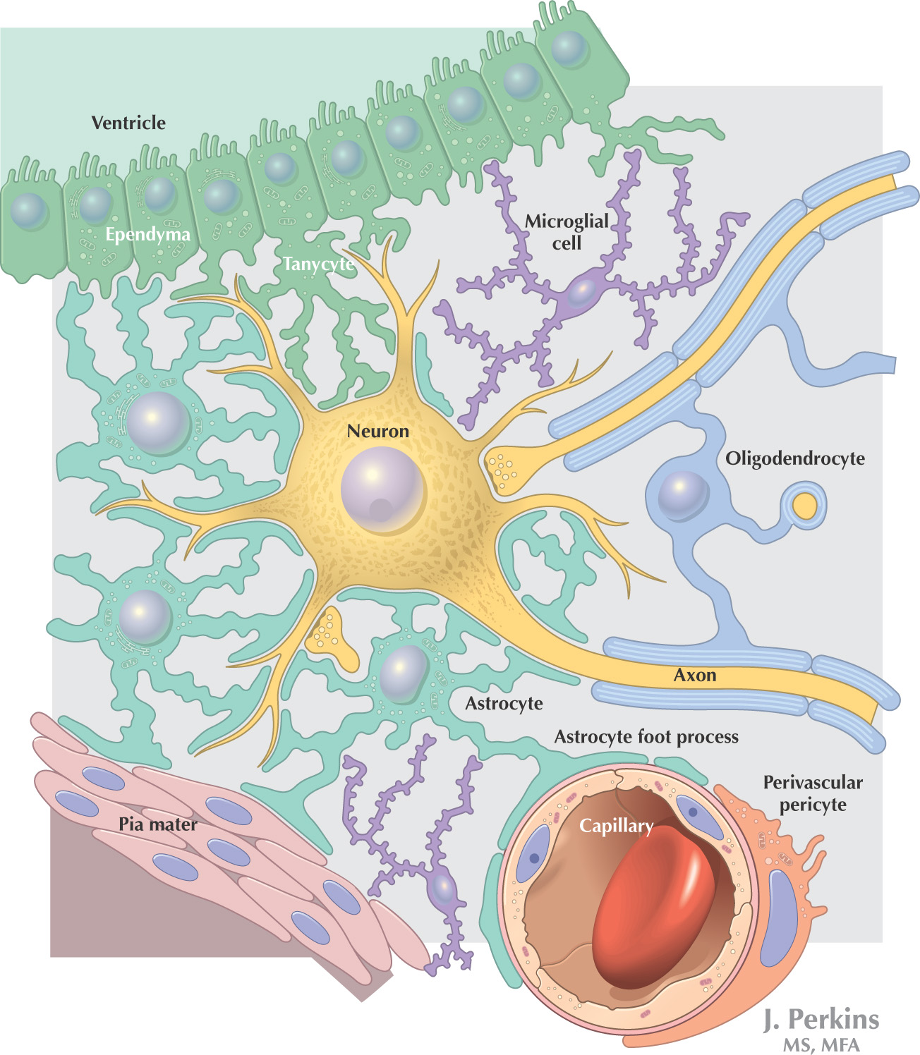
Neuroglia is the supporting nervous tissue for neurons, although neuroglial cells also have assistive roles in neuron function
Neuroglial cells have only 1 type of process
Classification:
• Astrocytes—located in the central nervous system; help keep neurons in place, provide nutritional support, regulate the extracellular matrix, form part of the blood-brain barrier
• Oligodendrocytes—located in the central nervous system; responsible for axon myelination in the central nervous system; 1 oligodendrocyte can myelinate 1 segment of multiple axons
• Microglia—located in the central nervous system; responsible for phagocytosis to remove waste
• Schwann cells—located in the peripheral nervous system; responsible for axon myelination in the peripheral nervous system; 1 schwann cell can myelinate 1 segment of 1 axon
• Satellite cells—located in the peripheral nervous system; surround the nerve cell bodies of ganglia
The central nervous system is composed of the:
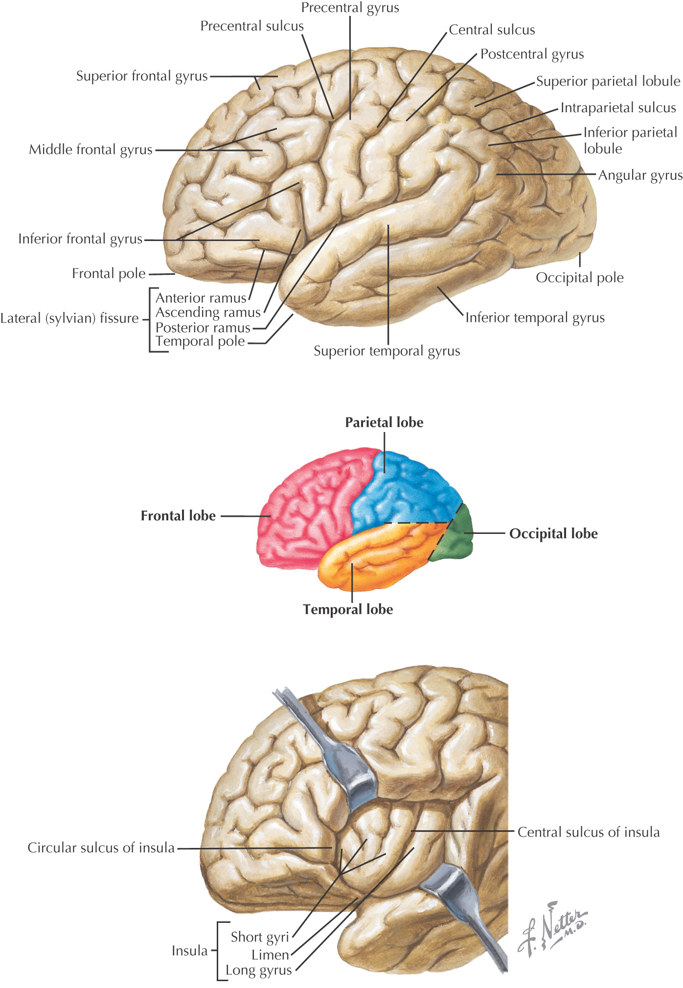
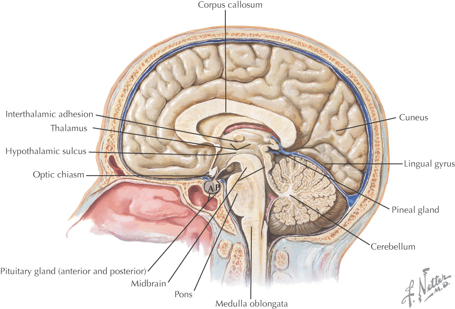
The surface of the cerebral cortex of the brain is divided by:
• Gyri (singular gyrus)—the elevations of brain tissue on the surface
• Sulci (singular sulcus)—the grooves or fissures located between the gyri
There are 3 large sulci that help divide the cerebral hemispheres into 4 of its lobes:
• Central sulcus (of Rolando)—divides frontal lobe from parietal lobe
• Lateral sulcus (of Sylvius)—divides the frontal and parietal lobes from the temporal lobe
• Parieto-occipital sulcus—divides the parietal lobe from the occipital lobe
The brain is divided into 5 lobes:
• Frontal—motor movement, motor aspect of speech (Broca’s area), reasoning, emotions, personality, and problem solving
• Parietal—sensory perceptions related to pain, temperature, touch and pressure, spatial orientation and perception, sensory aspect of language (Wernicke’s area)
• Temporal—auditory perceptions, learning, and memory
• Insula—associated with visceral functions including taste
Composed of 4 parts:
• Thalamus—major relay center of the somatosensory system and parts of the motor system
• Hypothalamus—controls the autonomic nervous system and endocrine system
• Epithalamus—major structures include the pineal gland (which controls circadian rhythms) and the habenula
• Subthalamus—an extrapyramidal nucleus of the motor system; if lesioned, will result in a contralateral hemiballismus
Composed of 3 parts:
Part of the motor system
Receives sensory input of all forms that use the deep cerebellar nuclei
Associated with:
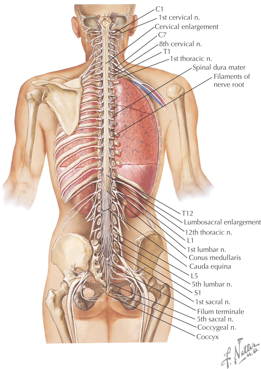
The caudal continuation of the central nervous system
Begins at the caudal end of the medulla and ends at vertebral level L1—2, tapering into the conus medullaris
Has 2 enlargements associated with the limbs:
• Cervical—associated with the upper limb and found between the spinal cord at levels C4 to T1
• Lumbosacral—associated with the lower limb and found between the spinal cord at levels L1 to S2
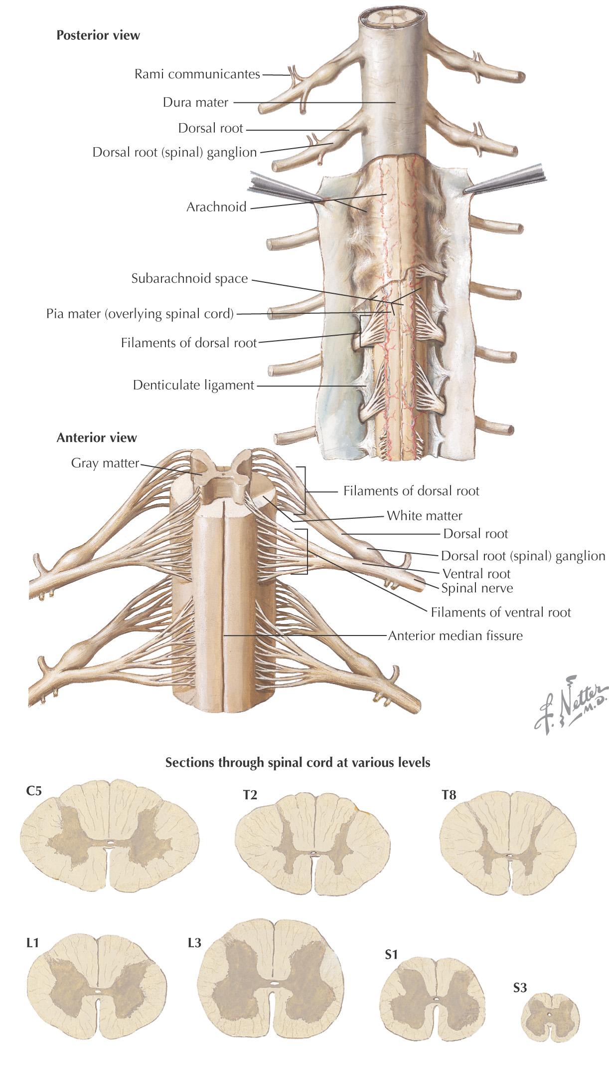
Composed of:
• Gray matter—location of nerve cell bodies and neuroglial cells
• White matter—location of the axons and neuroglial cells
Has 5 levels:
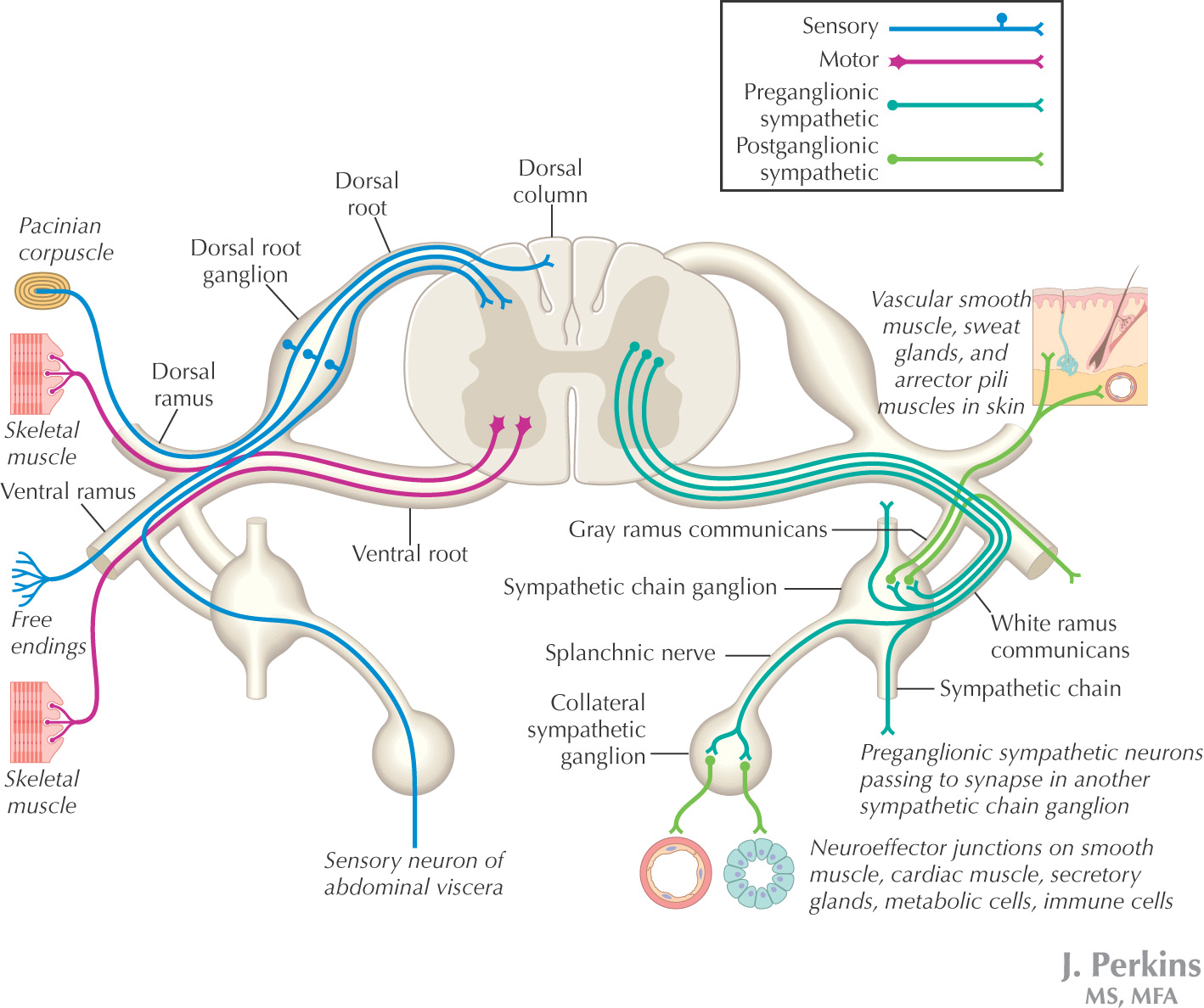
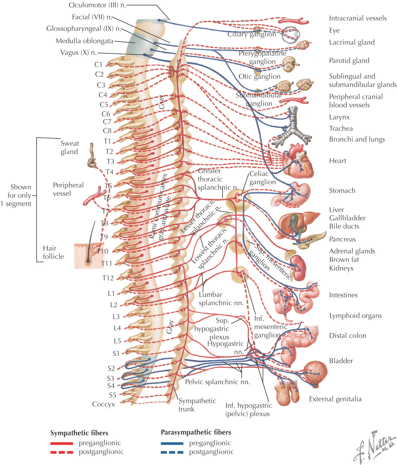
Peripheral nervous system is that portion of the nervous system located external to the central nervous system
Consists of:
Can be subdivided into:
• Somatic nervous system—voluntary system associated with afferent (sensory) and efferent (motor) fibers
• Autonomic nervous system—involuntary system associated with homeostasis of the body
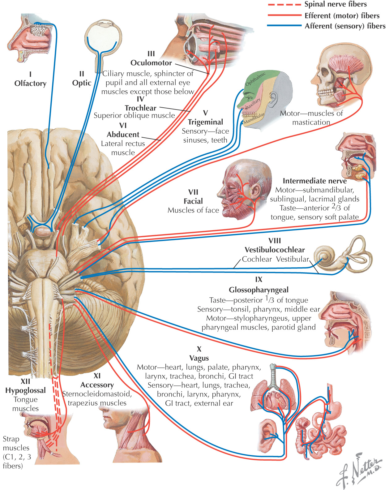
Cranial nerves or cerebral nerves are those peripheral nerves that leave the brain or brainstem
The cranial nerves customarily are subdivided into 12 pairs:
Because of the high degree of differentiation in the brain of humans, cranial nerves are more complex in structure and function than spinal nerves
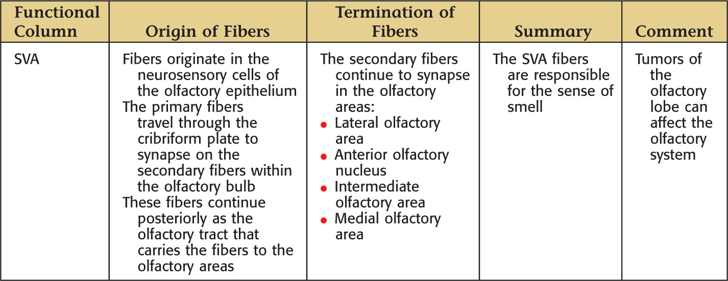
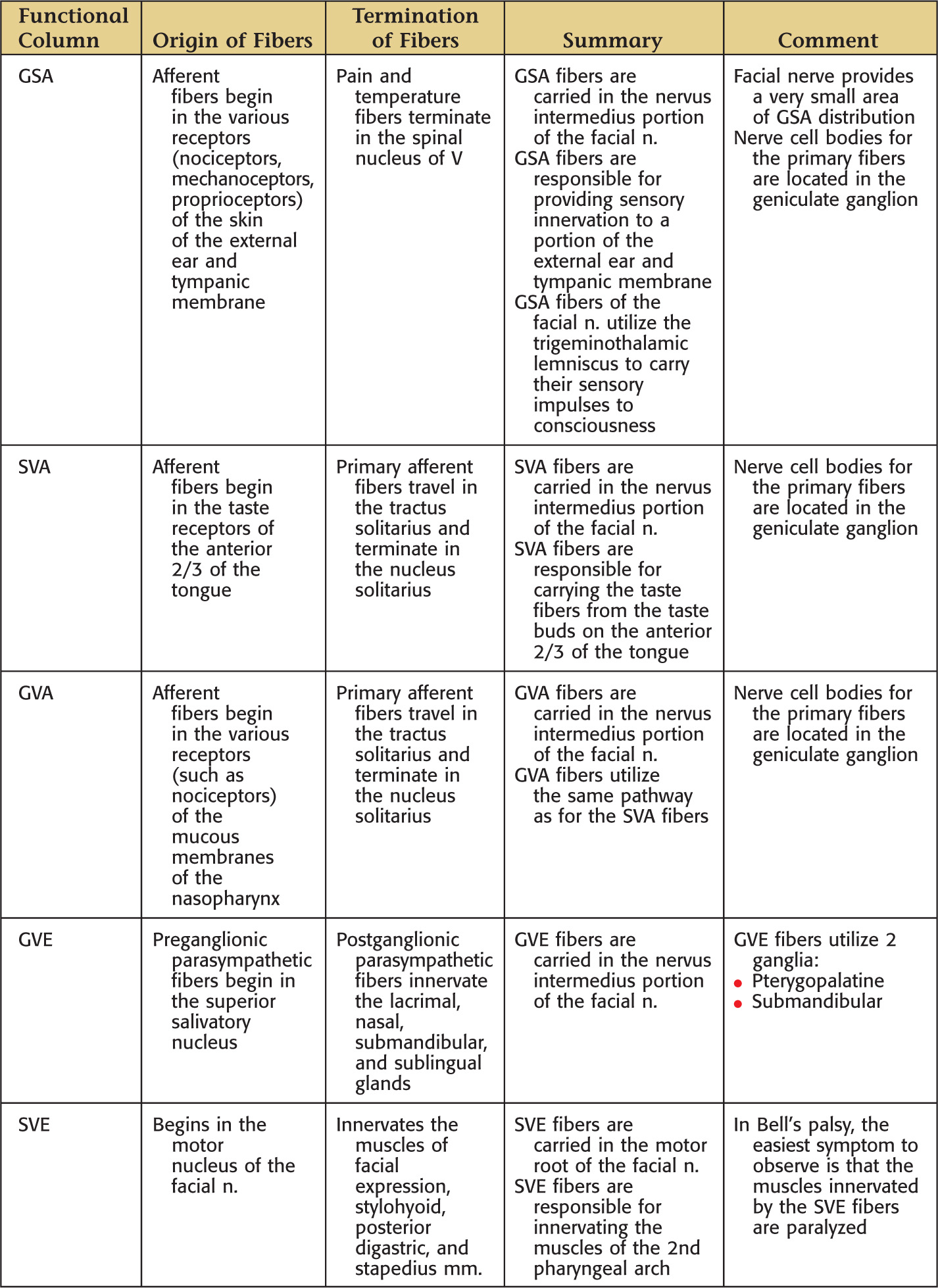
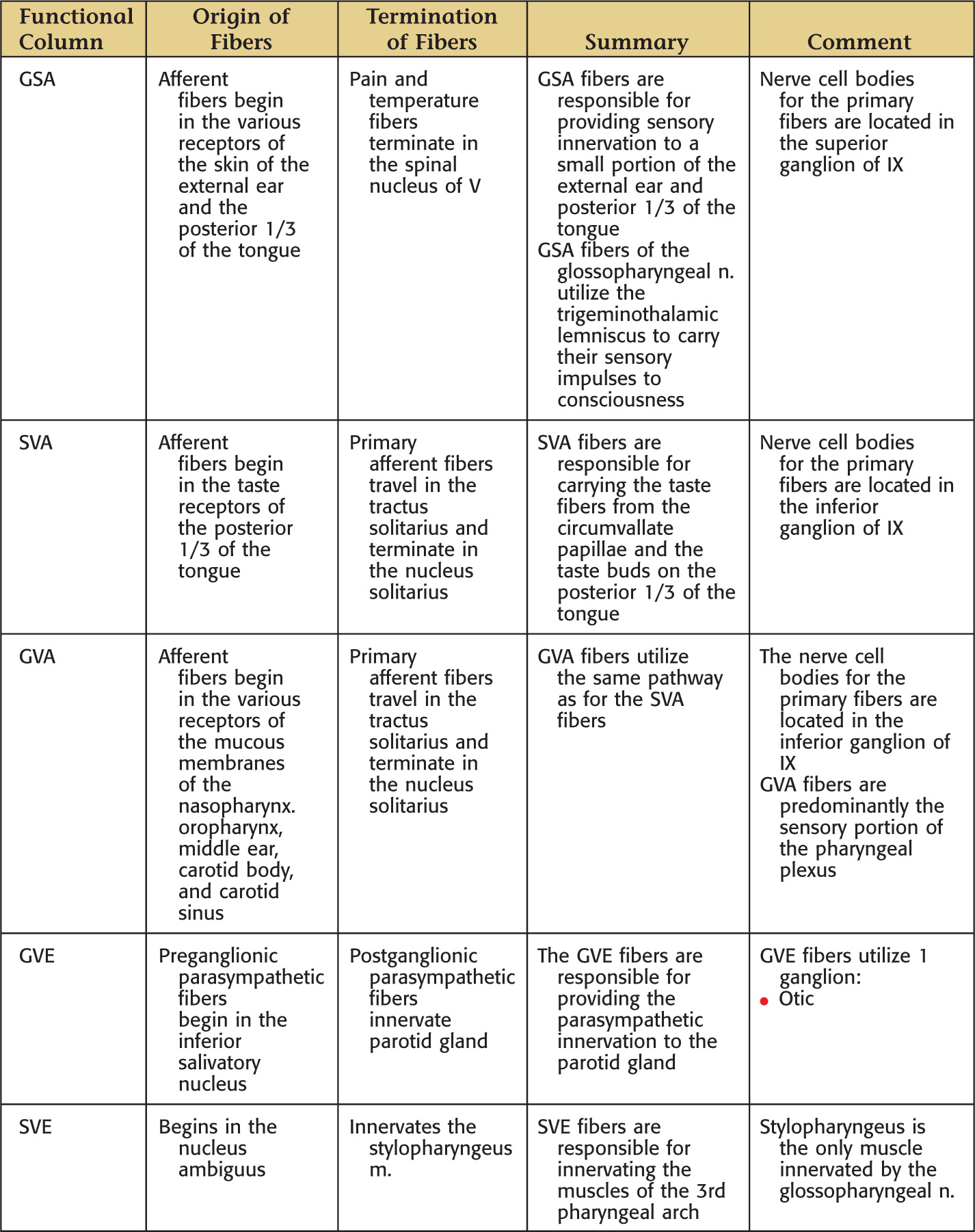
7 functional components (or functional columns) of the cranial nerves are recognized
• Concept of functional columns comes from studies of spinal nerves—functions associated with different neurologic pathways along spinal column are assigned corresponding „columns”
A given cranial nerve may have 1 to 5 functional columns
The functional columns are classified as general or special:
• General—these functional columns have the same functions as those for spinal nerves
• Special—these functional columns are specific only to cranial nerves
General and special functional columns each are subdivided into 2 additional categories:
• Afferent (sensory) and efferent (motor)
• Somatic (body-related) and visceral (organ-related)
*Within each designation: G or S, general or special; S or V, somatic or visceral; A or E, afferent or efferent.

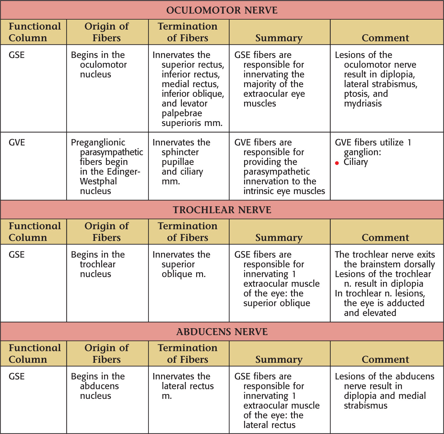
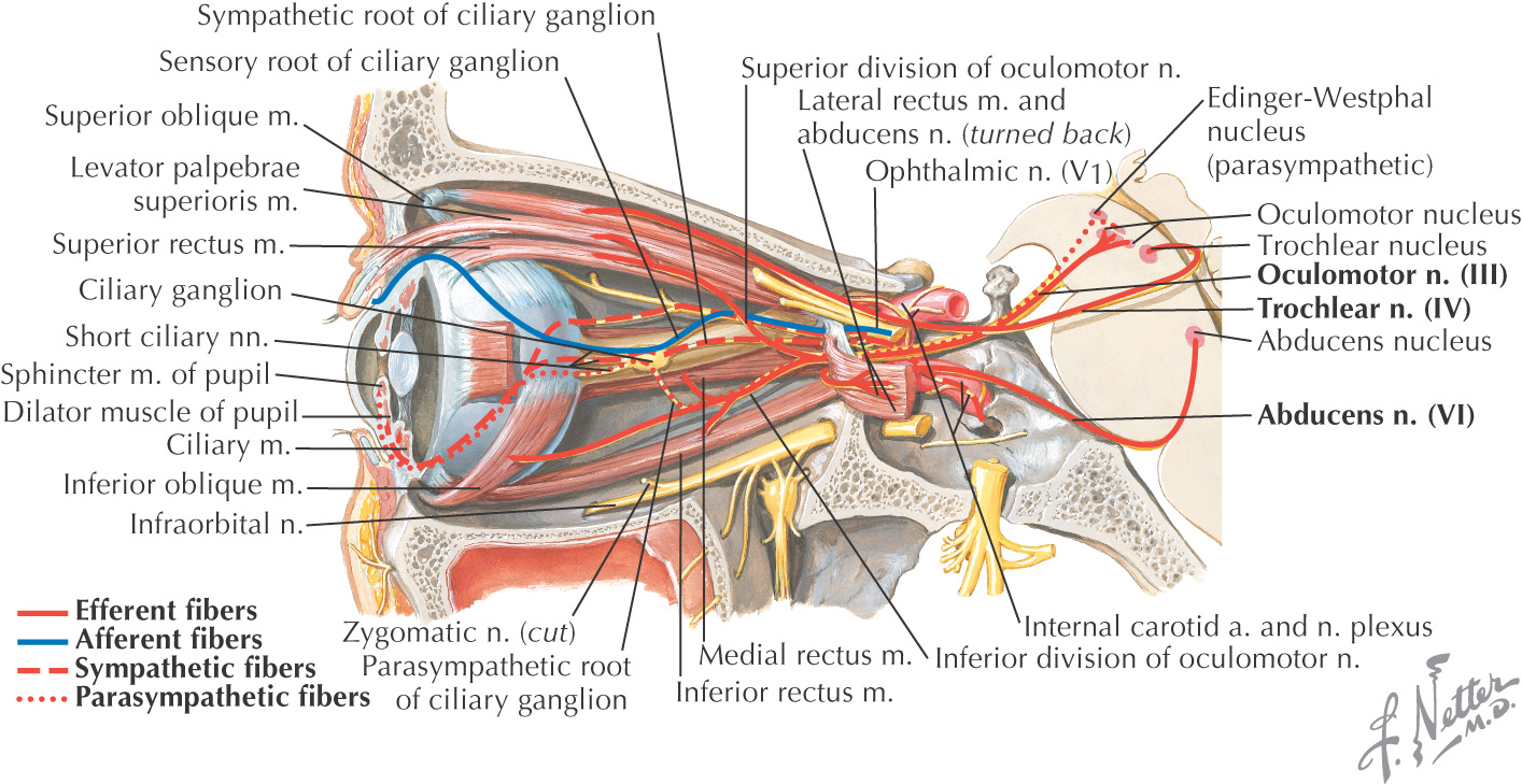
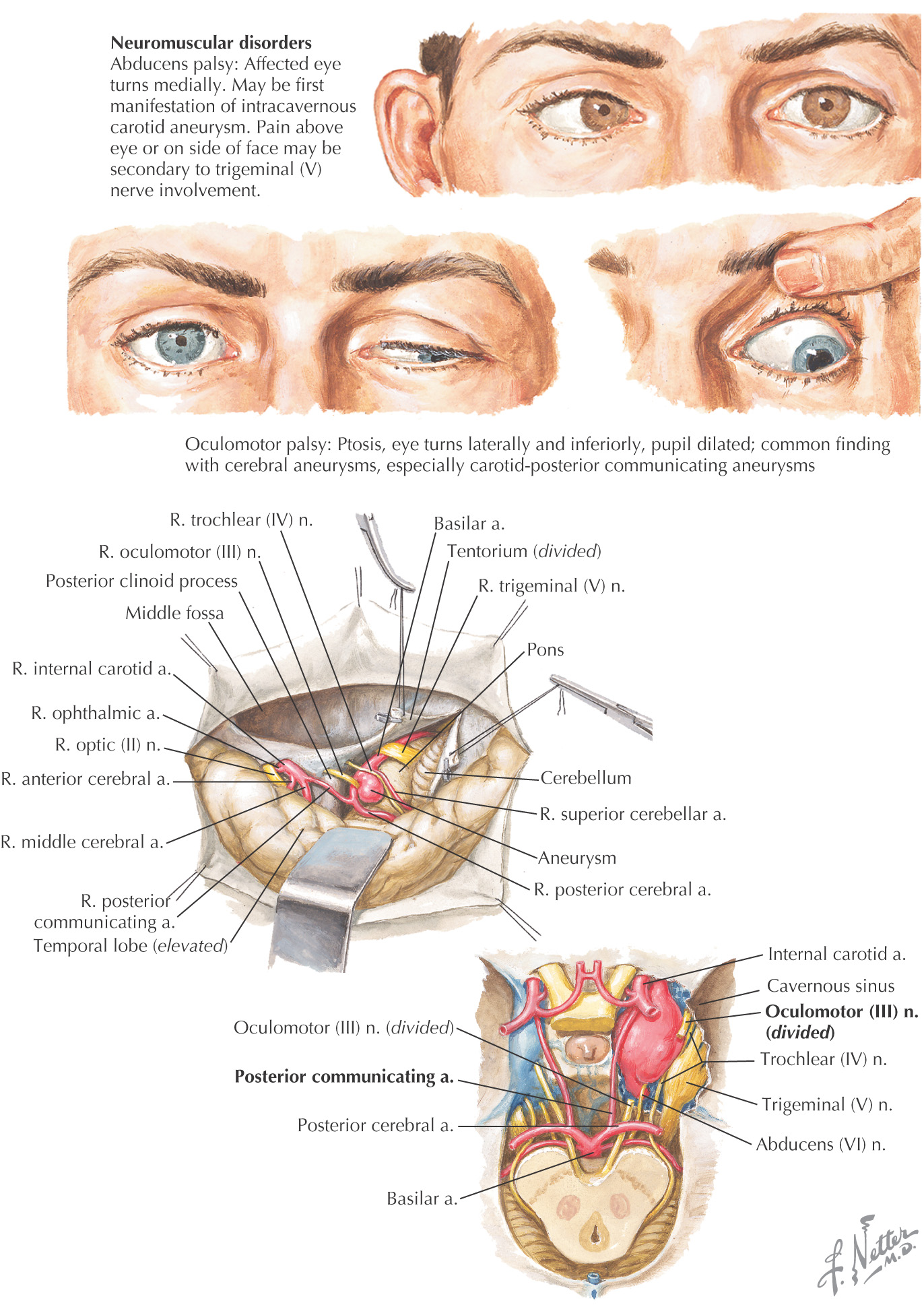
Because of the close proximity of the oculomotor, trochlear, and abducens nerves to blood vessels supplying the brain, aneurysms along these vessels may lead to a paralysis of the muscles that they innervate
Commonly affected vessels include the basilar, posterior cerebral, and posterior communicating arteries
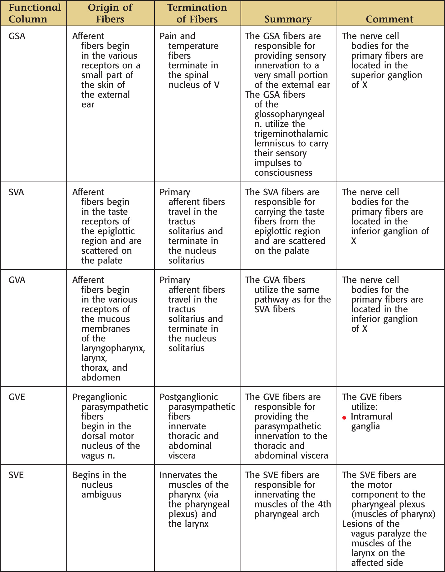
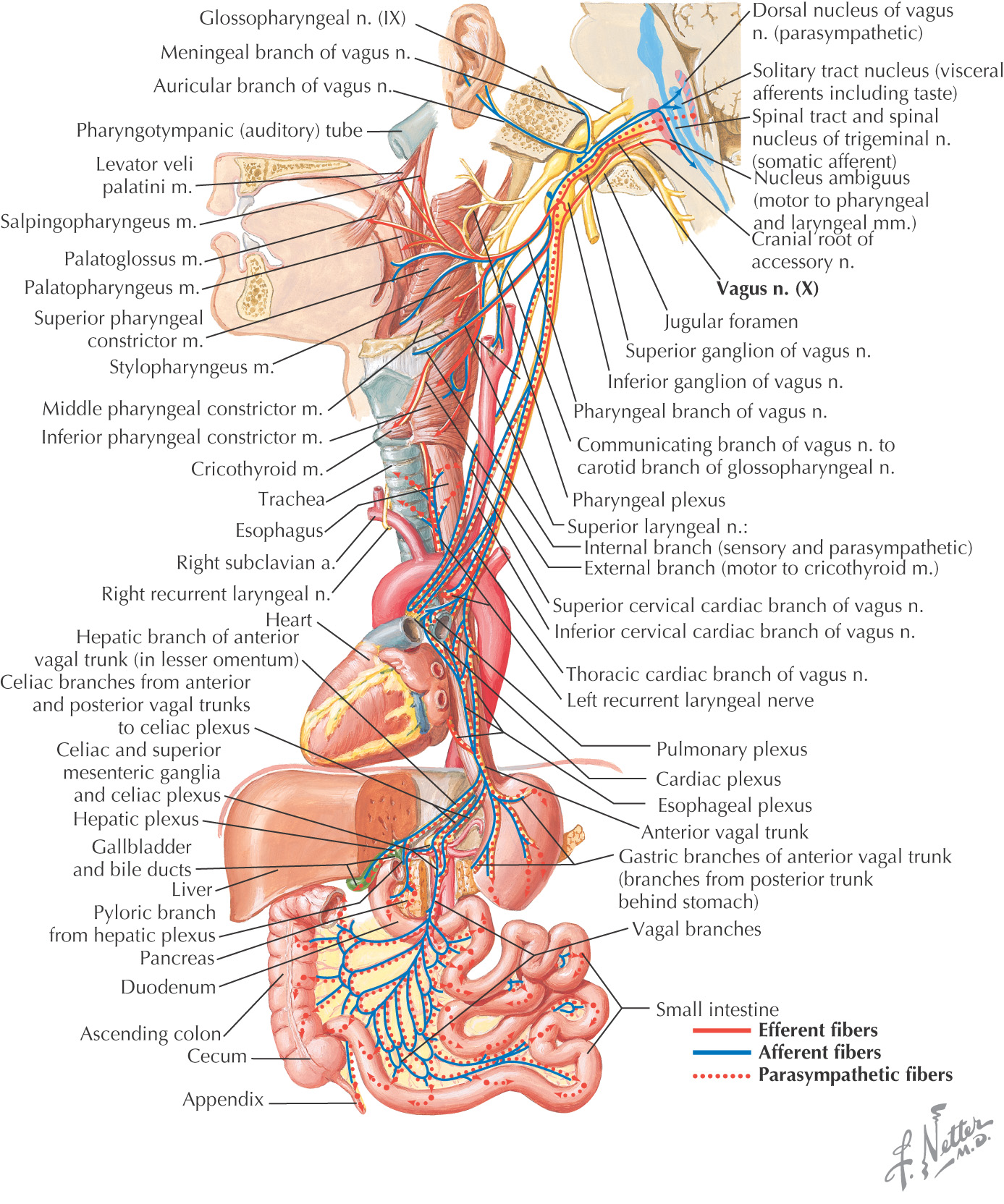
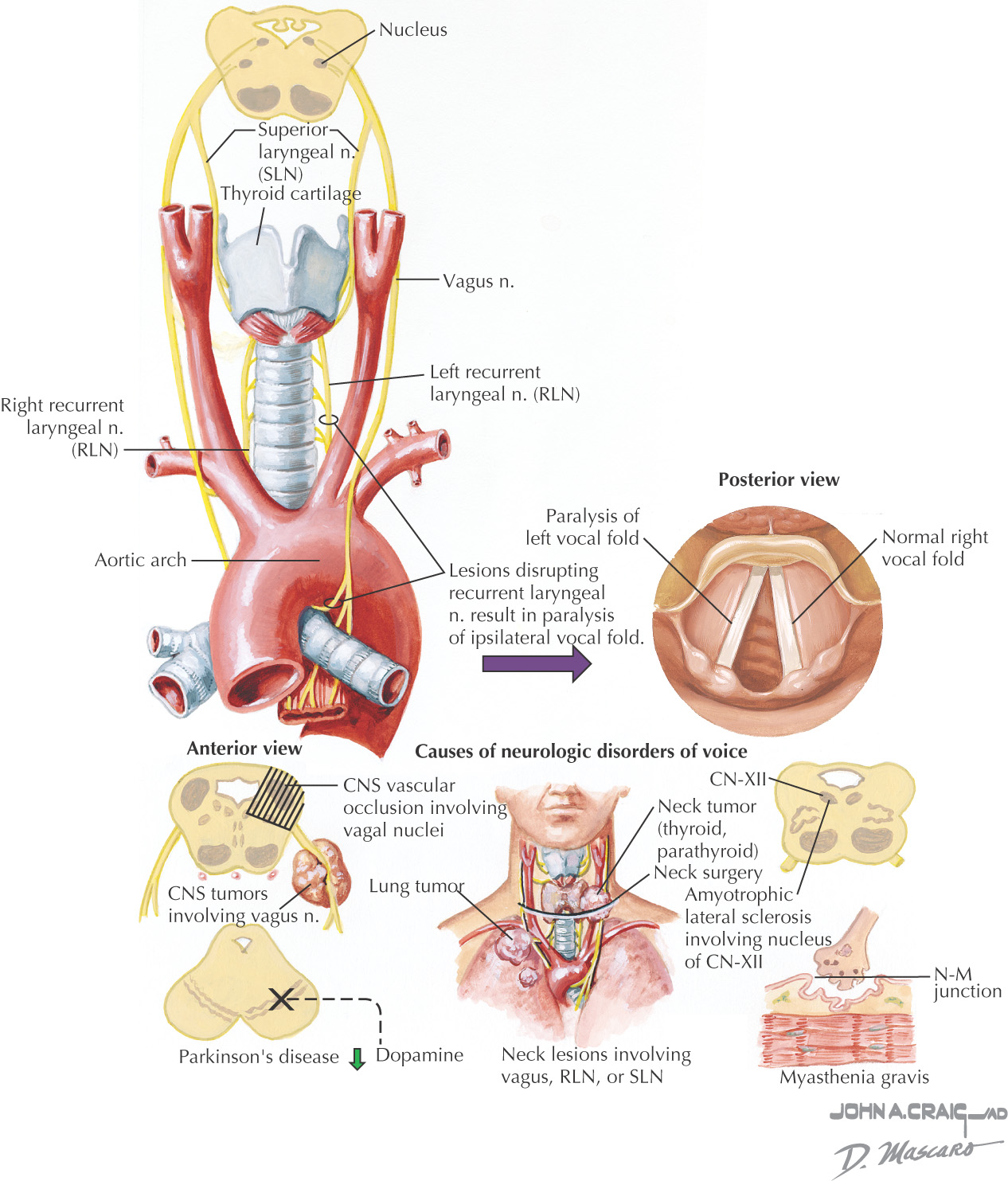
The vagus nerve provides all of the motor and sensory innervation to the larynx
The superior laryngeal nerve divides into the internal laryngeal (sensory) and external laryngeal (motor to the cricothyroid)
The recurrent laryngeal provides sensory and motor innervation to the remainder of the muscles of the larynx
Lesions of the recurrent laryngeal nerve result in a paralysis of the ipsilateral vocal fold
This problem usually manifests clinically as hoarseness with an ineffective cough
Common causes include:
The voice also may be affected in Parkinson’s disease and myasthenia gravis
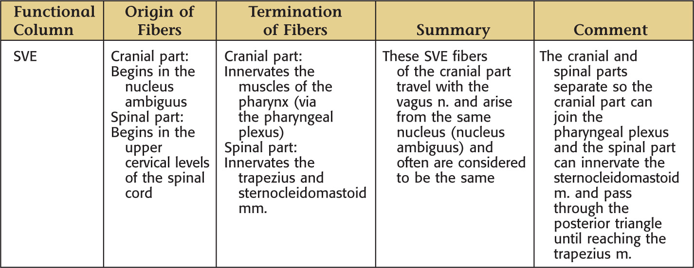
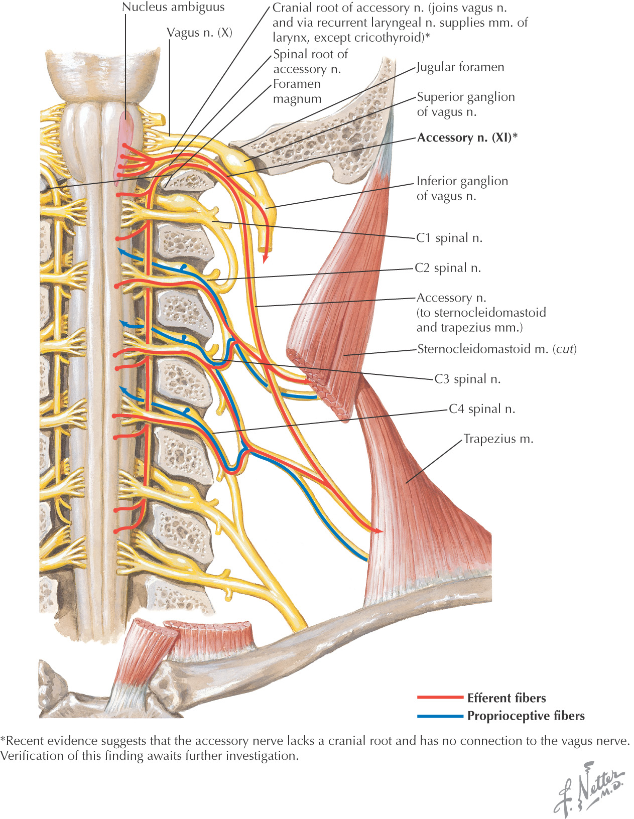
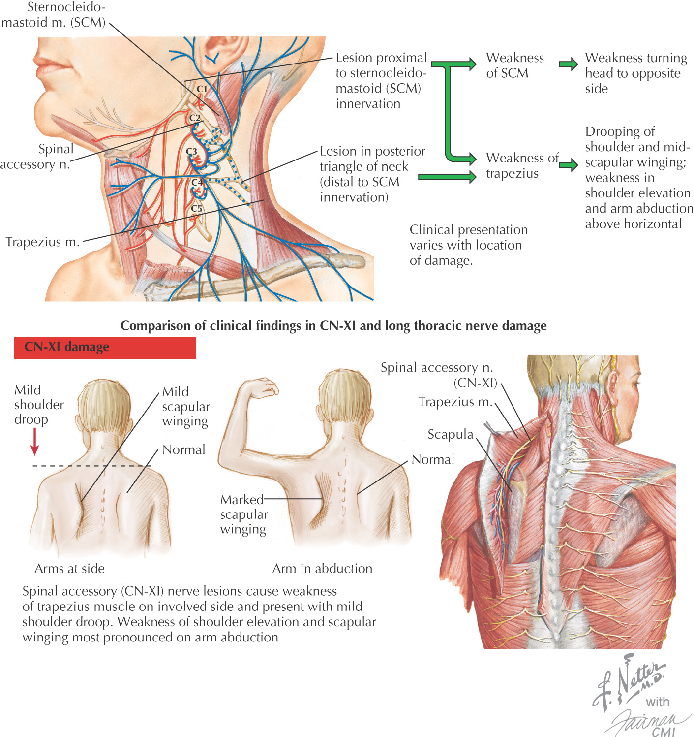
The spinal accessory nerve provides motor innervation to the sternocleidomastoid and trapezius muscles
The spinal accessory nerve courses close to the superficial cervical lymph nodes
• This course makes it vulnerable to damage during biopsy or radical neck dissection in the posterior triangle
• Damage to the spinal accessory nerve also may result from a carotid endarterectomy
In lesions located in the posterior triangle, the sternocleidomastoid muscle is unaffected, but the trapezius muscle is deinnervated
• The shoulder droops, with mild winging of the scapula
• Abduction of the arm also is affected when patient attempts to raise it above the horizontal plane
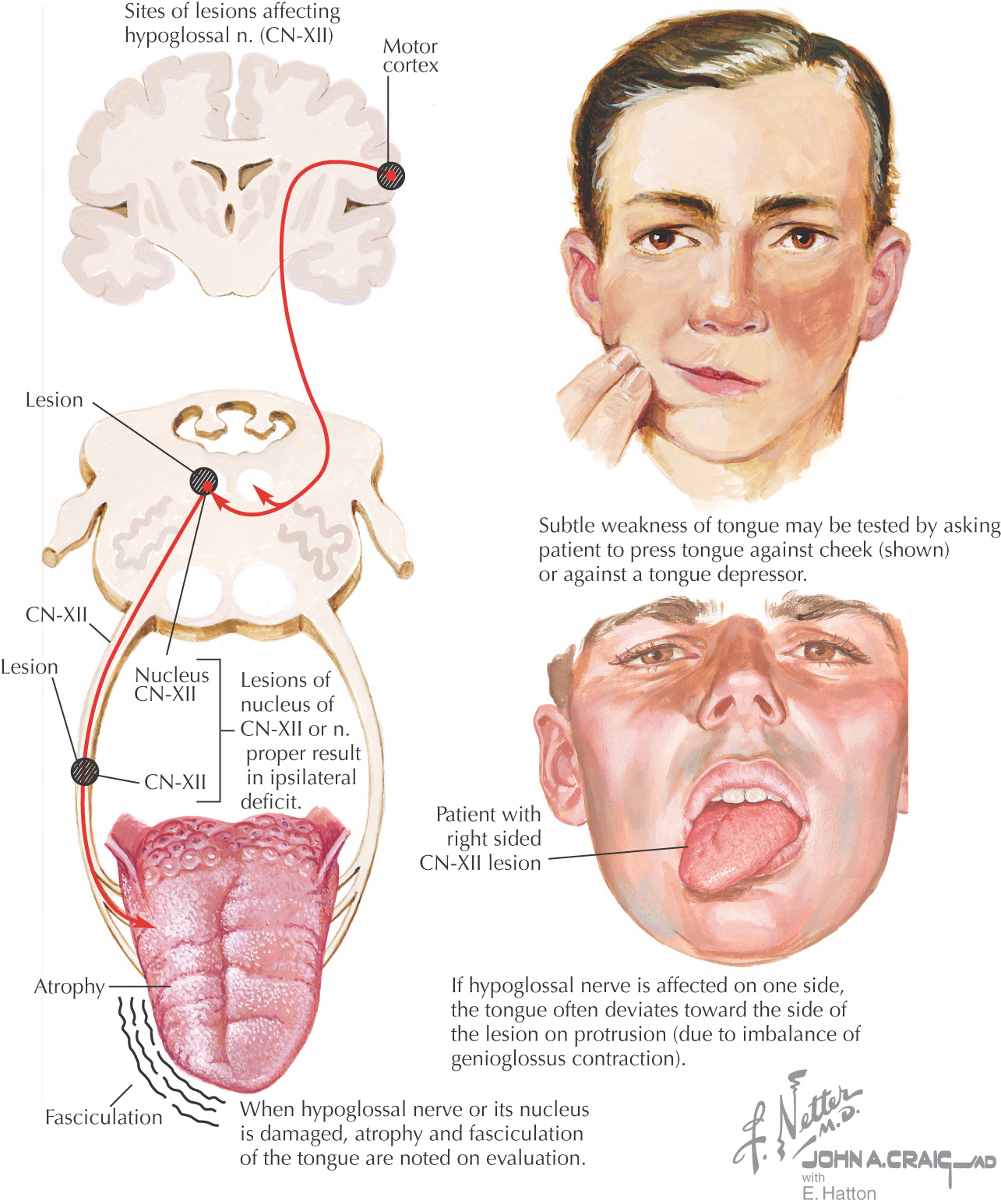
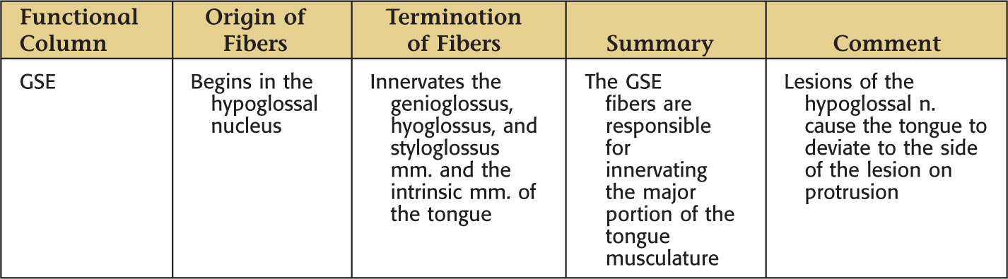
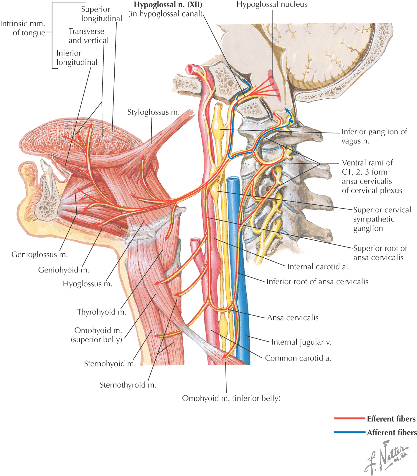
The hypoglossal nerve provides motor innervation to a majority of the muscles of the tongue, including:
Protrusion of the tongue is accomplished by the bilateral actions of the genioglossus muscles
Paralysis of a genioglossus muscle causes the protruded tongue to deviate to the paralyzed side
Paralysis of the hypoglossal nerve can be caused by:
A similar paralysis can be caused by a stroke affecting the upper motor neurons on the side contralateral to the paralyzed muscles, owing to the crossing fibers of the upper motor neurons