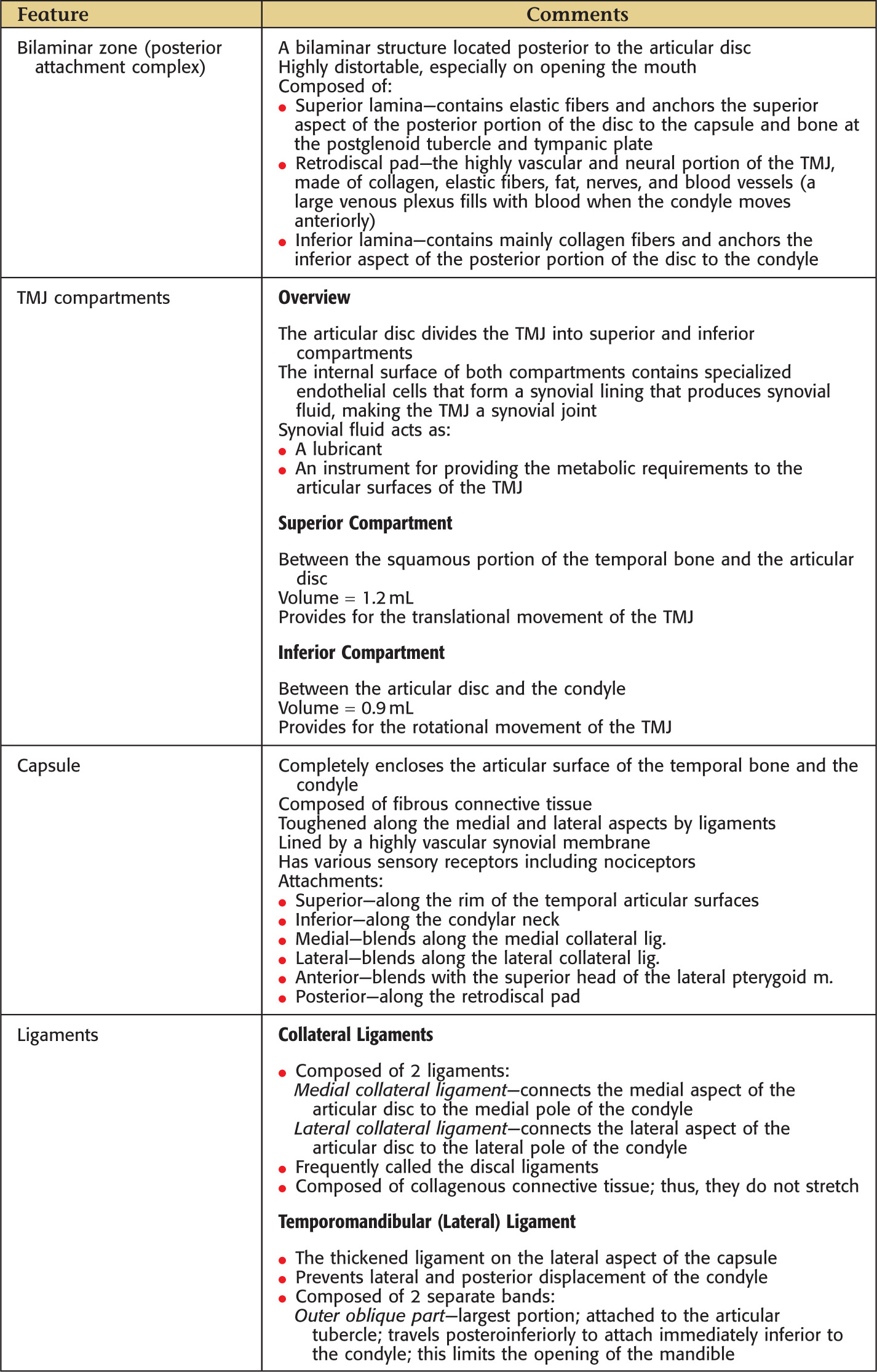
Overview and Topographic Anatomy
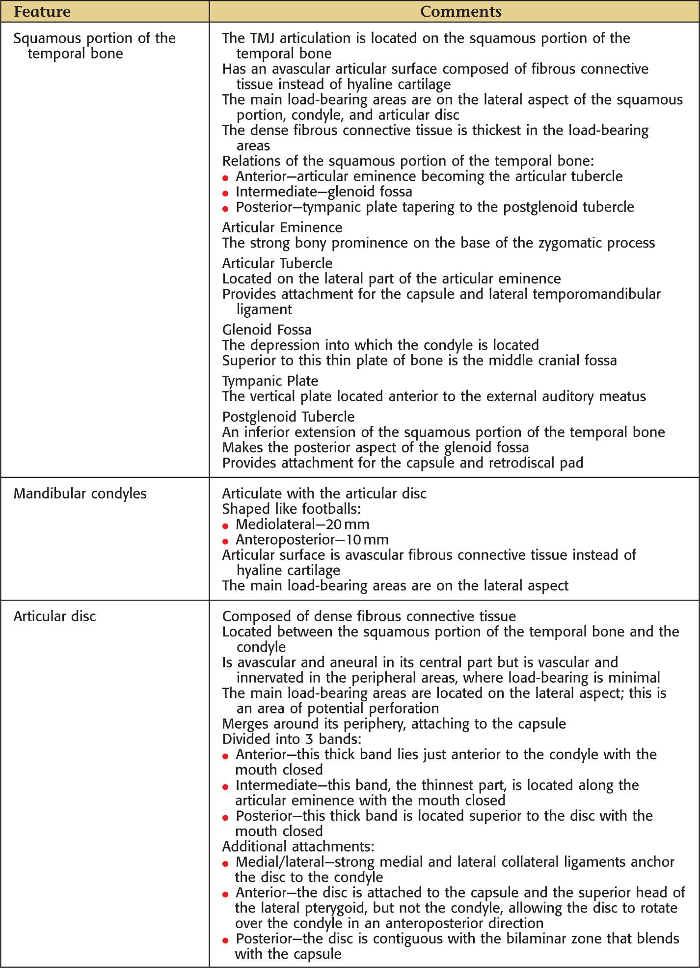
The temporomandibular joint (TMJ) is the articulation between the squamous portion of the temporal bone and the condyle of the mandible
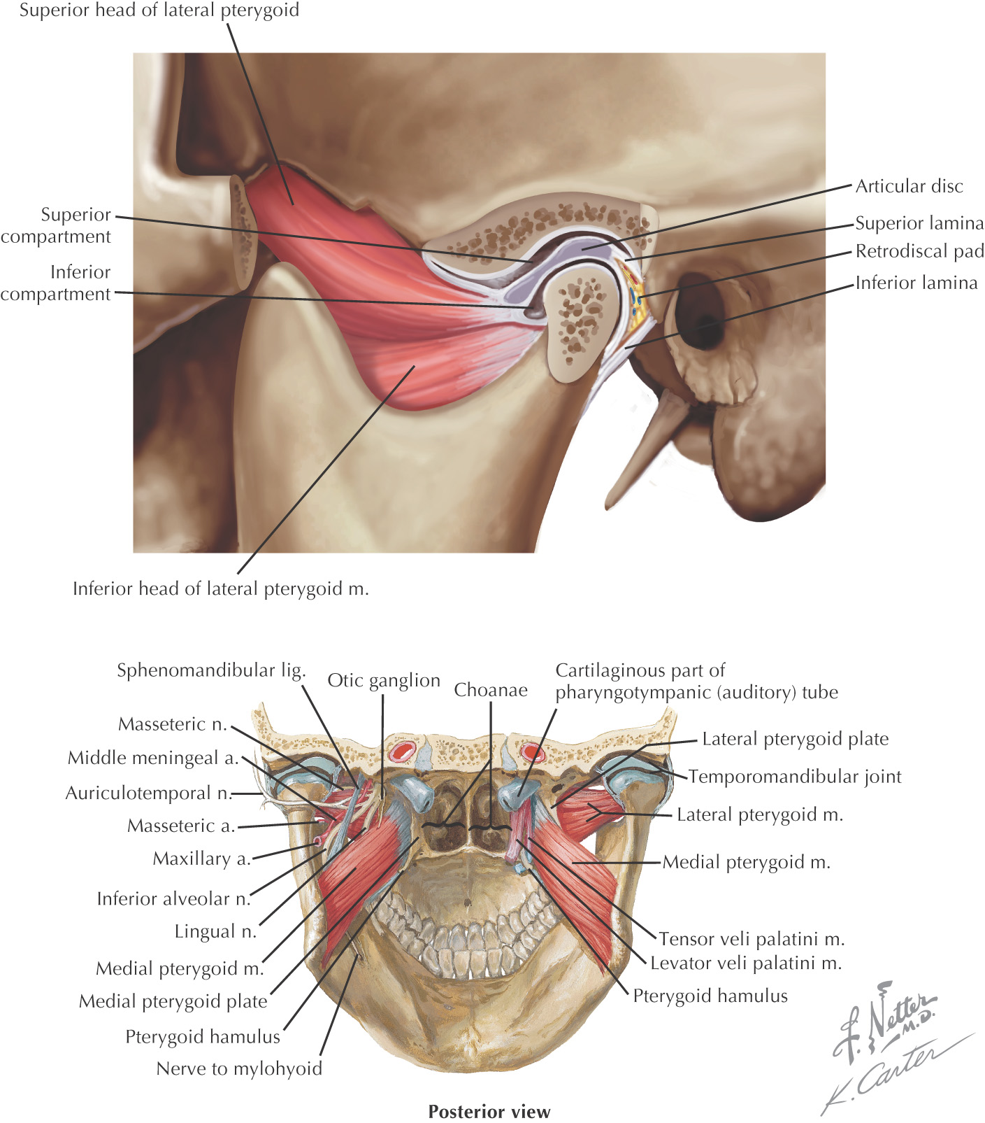
The TMJ comprises 2 types of synovial joints—hinge and sliding—and consists of the following:
• Squamous portion of the temporal bone
• Articular disc (contained within the TMJ)
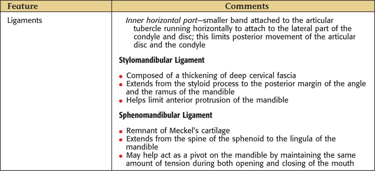
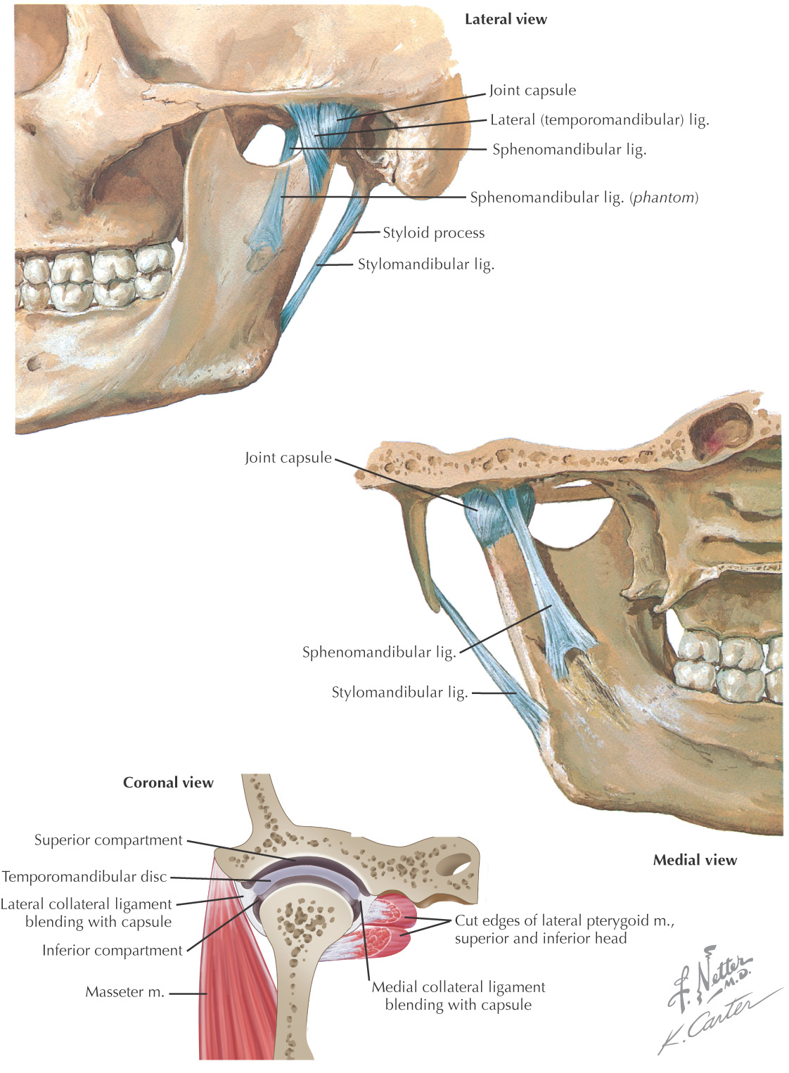
• Ligaments (serve as boundaries)
Affects approximately 25% of the population and may be severe in a small subgroup
Causes include arthritis, trauma, infection, bruxism, and disc displacement
More common in females
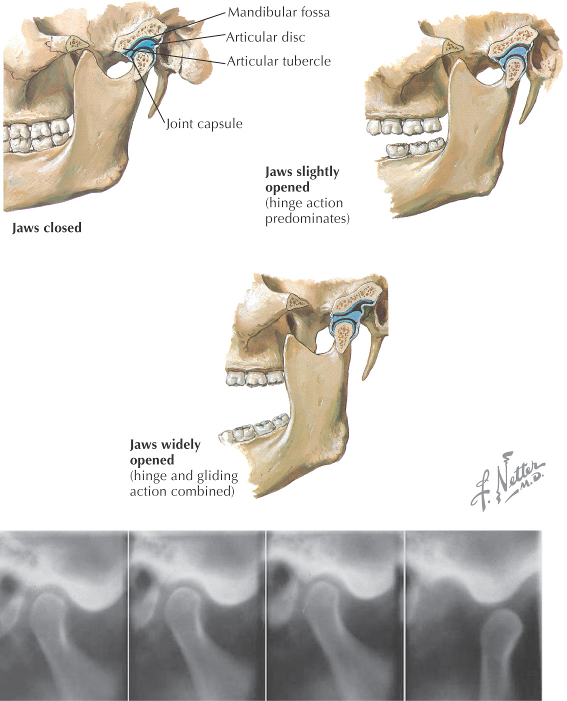
Opening the mandible involves a complex series of movements
Initial movement is rotational, which occurs in the lower TMJ compartment:
• Lateral pterygoid (inferior head) initiates the opening of the jaw (the superior head of the lateral pterygoid is described as being active during elevation of the mandible in a “power stroke”)
• As the mandible is depressed, the medial and collateral ligaments tightly attach the condyle to the articular disc, thereby allowing only for rotational movement
• Once the TMJ becomes taut, no further rotation of the condyle can occur
• Normally, rotational movement continues until the upper and the lower teeth are about 20 mm away from each other
For additional movement of the mandible, translational movement must occur:
• A translational movement occurs in the upper TMJ compartment and provides for most of the mandible’s ability to open
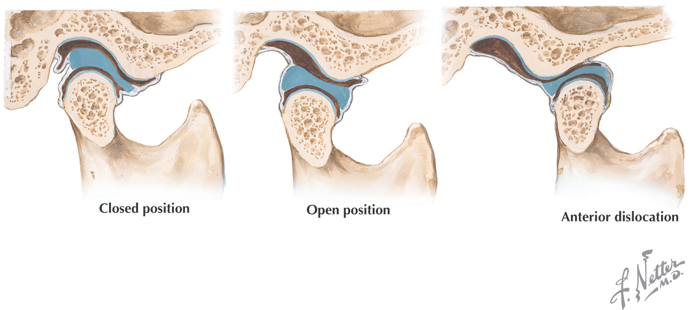
• In this movement, the articular disc and the condyle complex slide inferiorly on the articular eminences, allowing for maximum depression of the mandible
Mandibular dislocation (or subluxation of the TMJ) occurs when the condyle moves anterior to the articular eminence
• With dislocation, the mouth appears “wide open”
• Because the condyle is displaced anterior to the articular eminence, a depression can be palpated posterior to the condyle
Spontaneous dislocations can occur from a variety of actions ranging from an extended dental treatment to a simple yawn
Because the mandible is dislocated, the patient has a great deal of difficulty verbalizing his or her predicament
Relocation involves repositioning the condyle posterior to the articular eminence
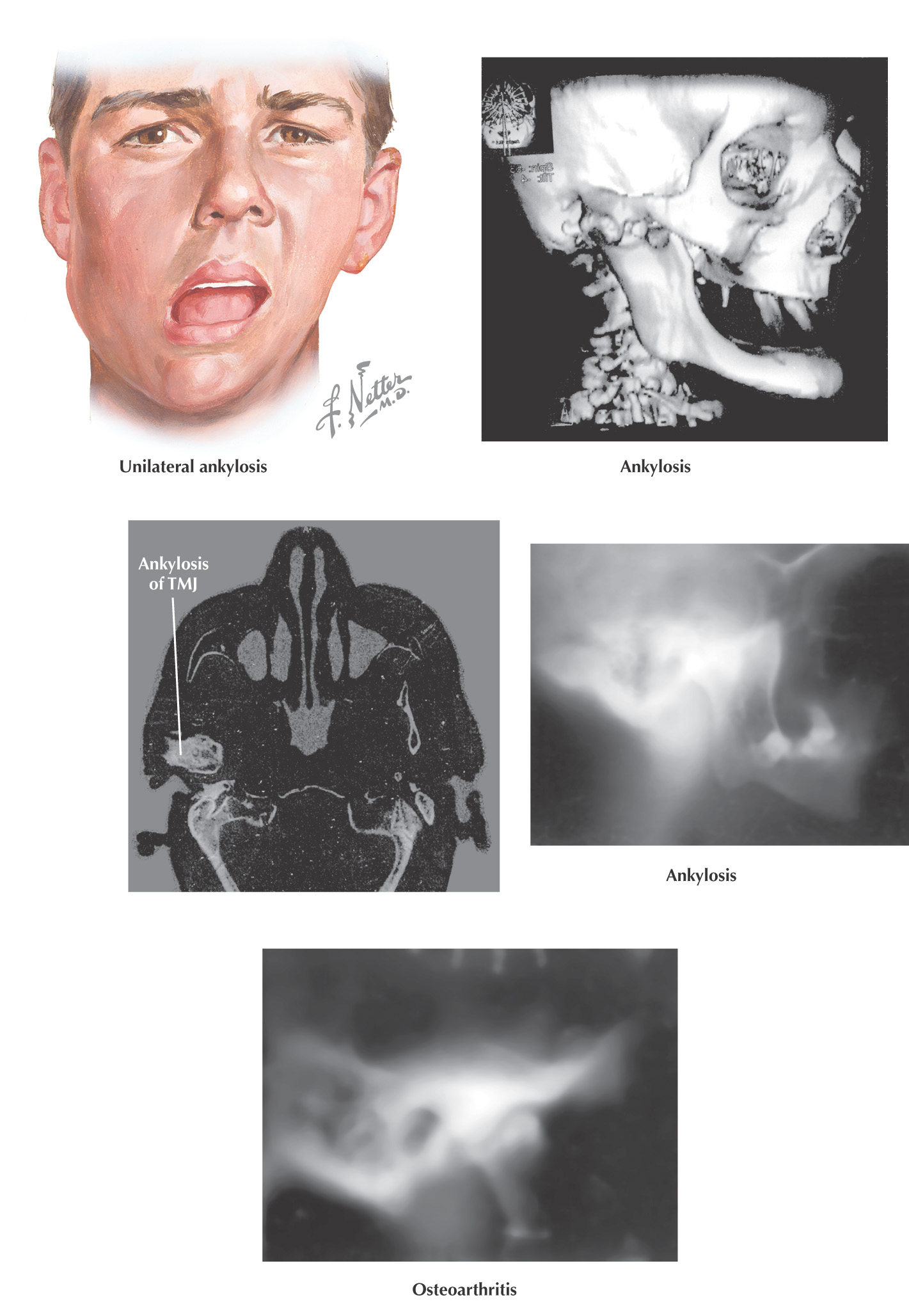
Arthritis is the most common cause of pathologic changes in the TMJ
When rheumatoid arthritis occurs, usually both TMJs are affected, and other joints tend to be affected before the TMJ
Radiologic images in the initial disease stages show decreased joint space without osseous changes
Radiologic images in the late disease stages show decreased joint space with osseous changes, possibly including ankylosis
In osteoarthritis, causes include normal wear, trauma, and bruxism, and clinical manifestations may range from mild to severe
Ankylosis is an obliteration of the TMJ space with abnormal osseous morphologic features, which often occurs as a result of trauma or infection
Classified as either true (intracapsular) or false ankylosis (extracapsular condition usually associated with an abnormally large coronoid process or zygomatic arch)
Treatment varies in accordance with the cause but may include a prosthetic replacement or condylectomy