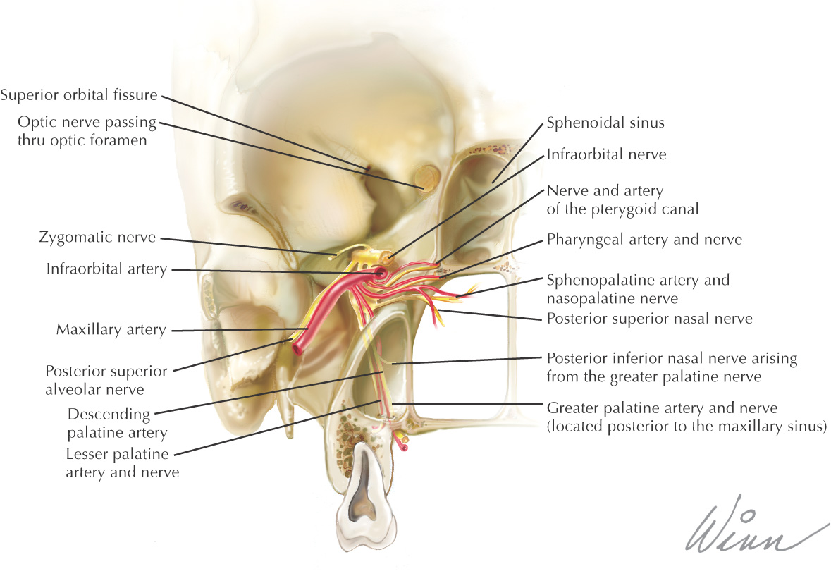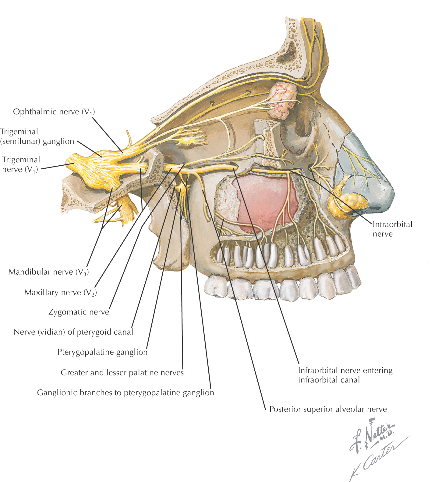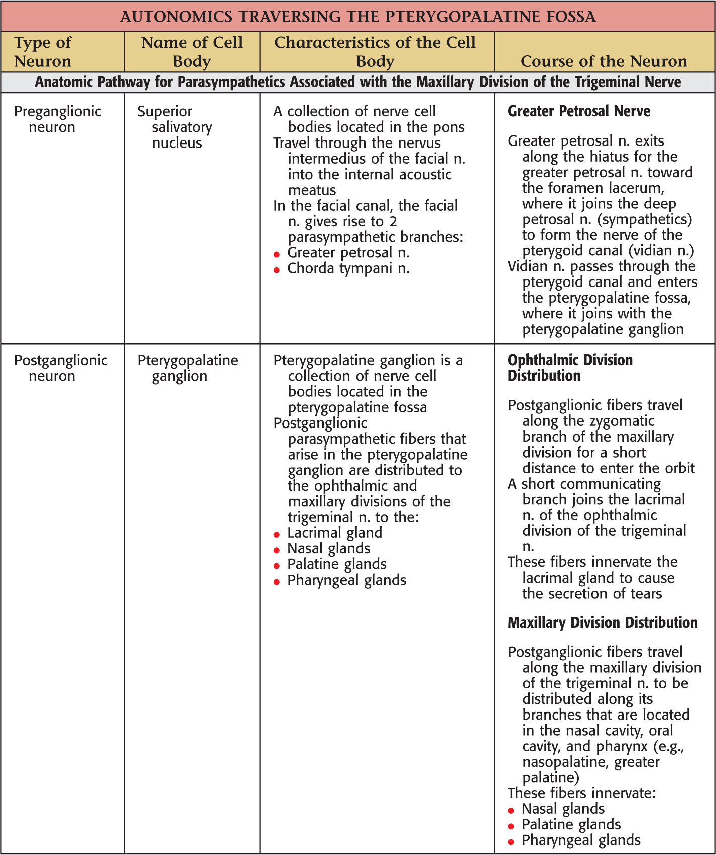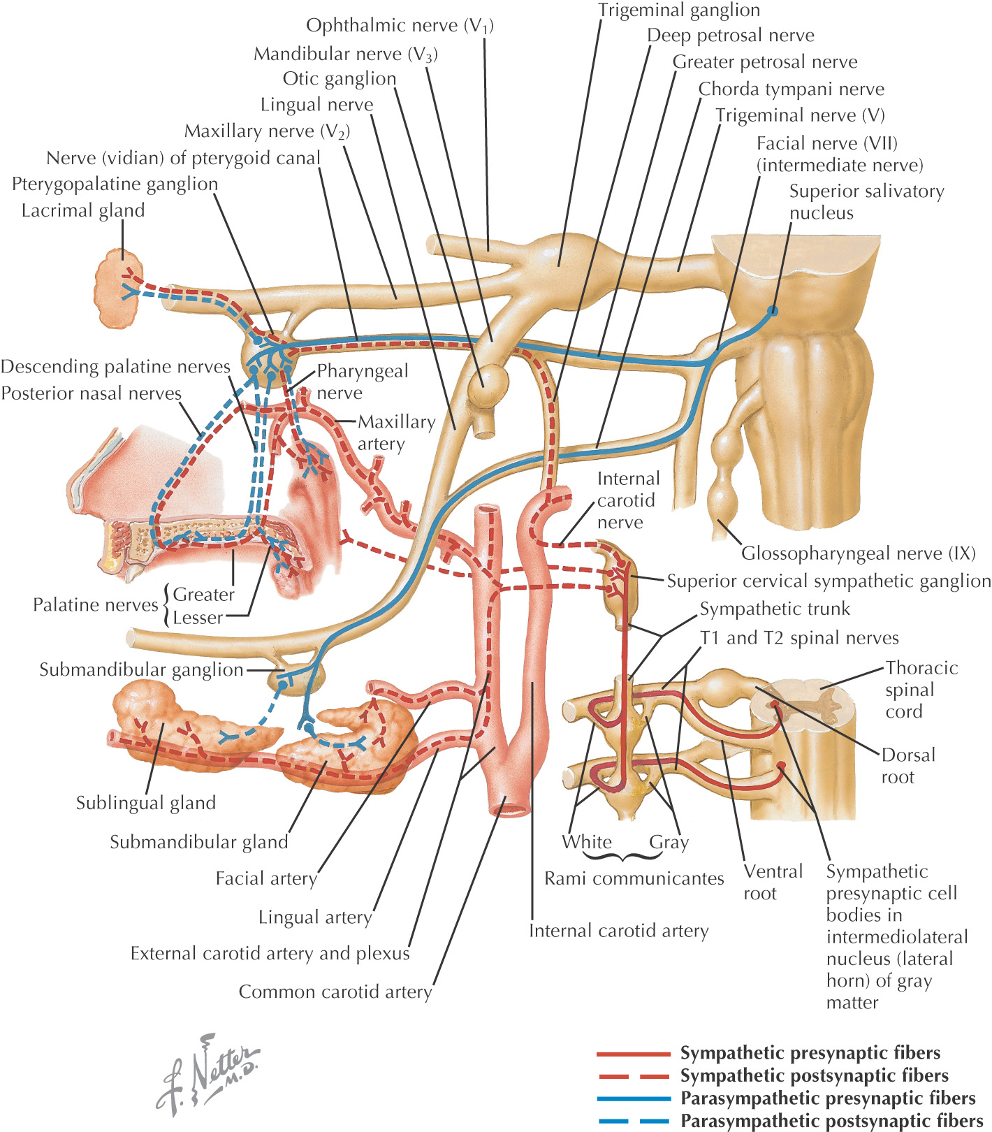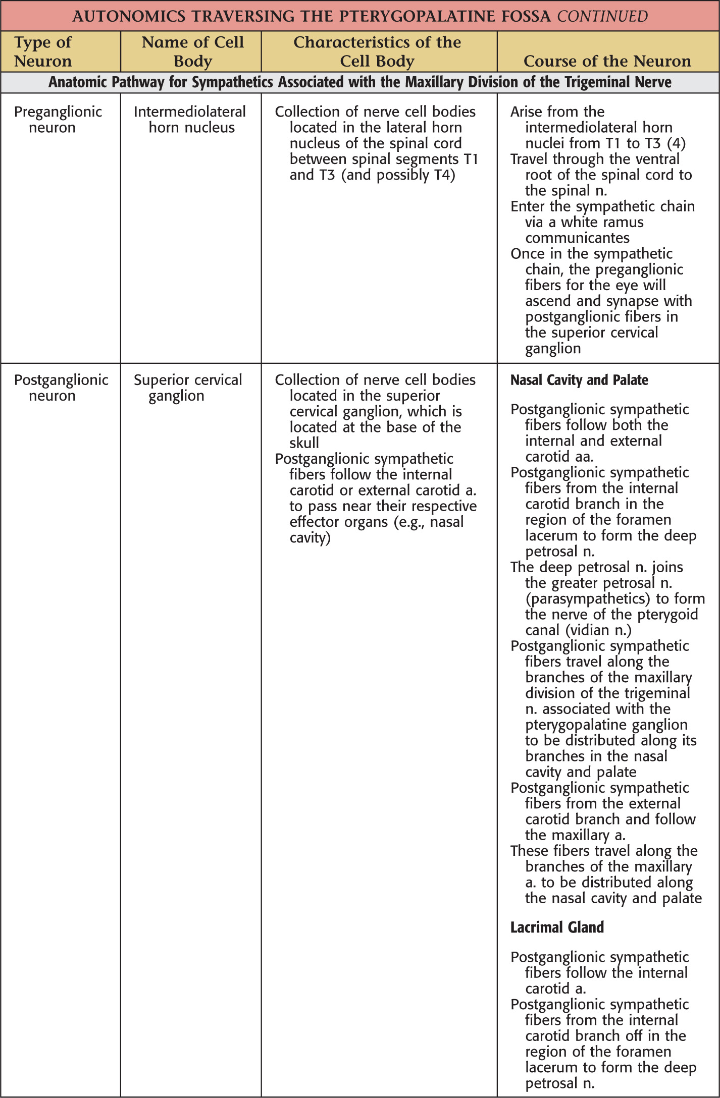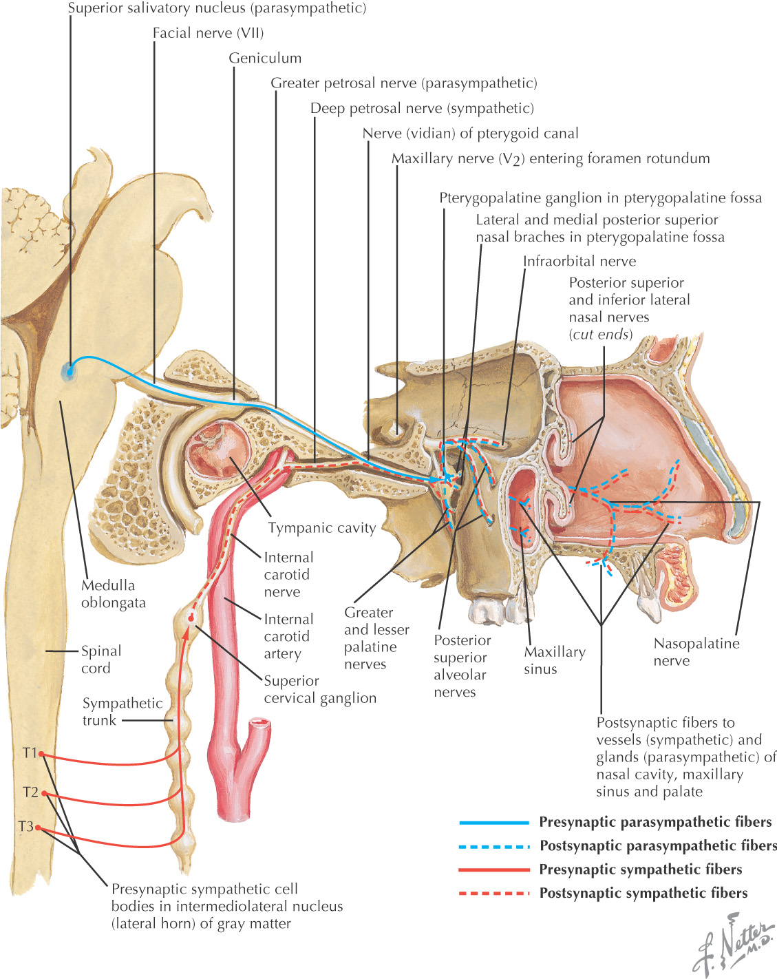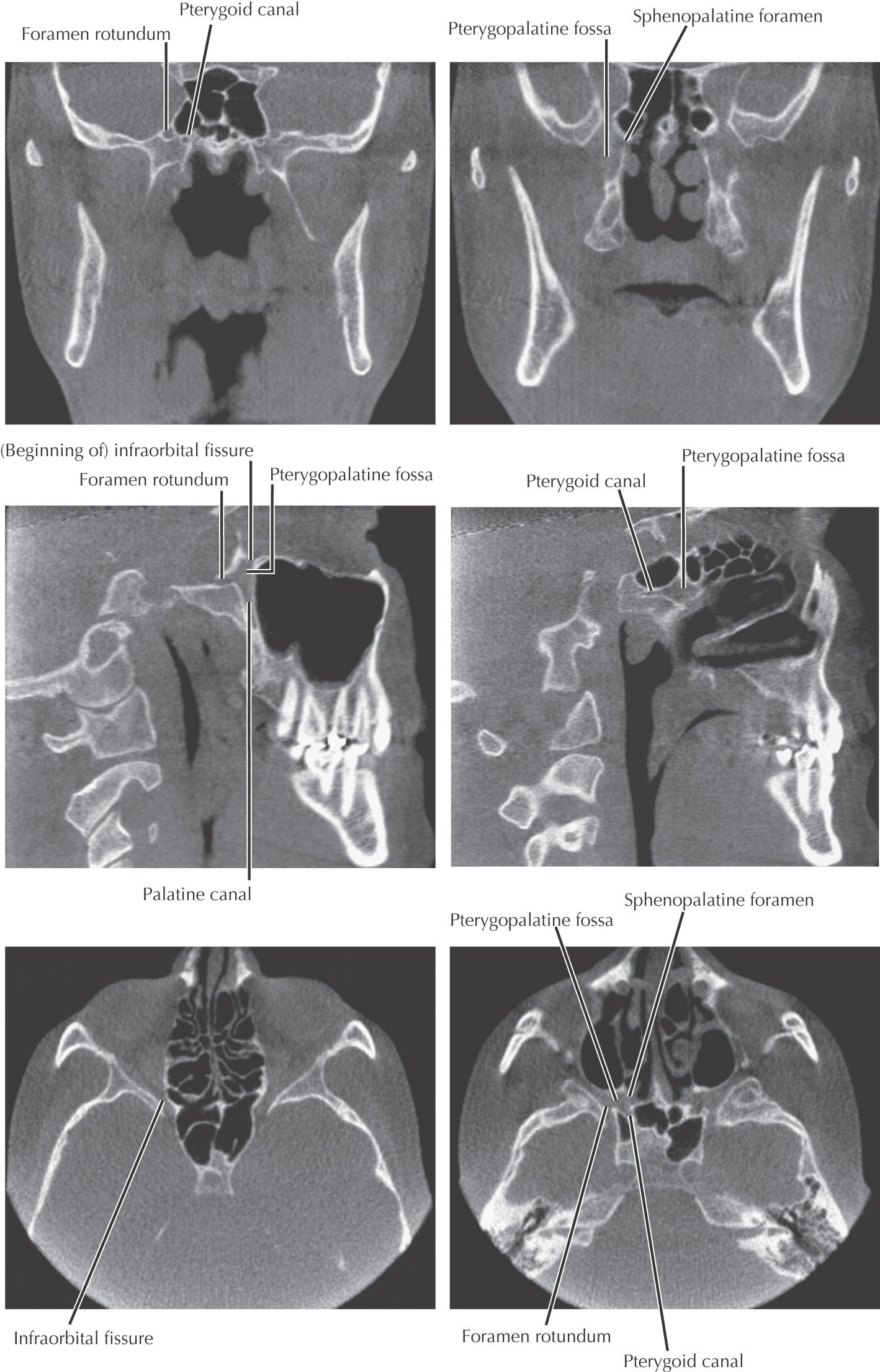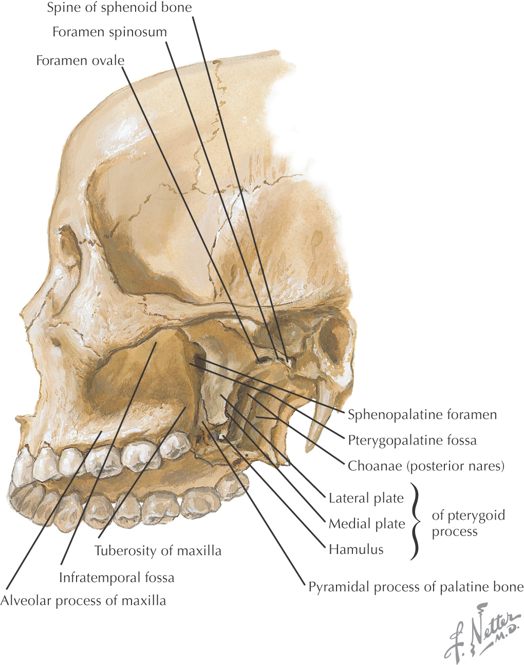
Overview and Topographic Anatomy
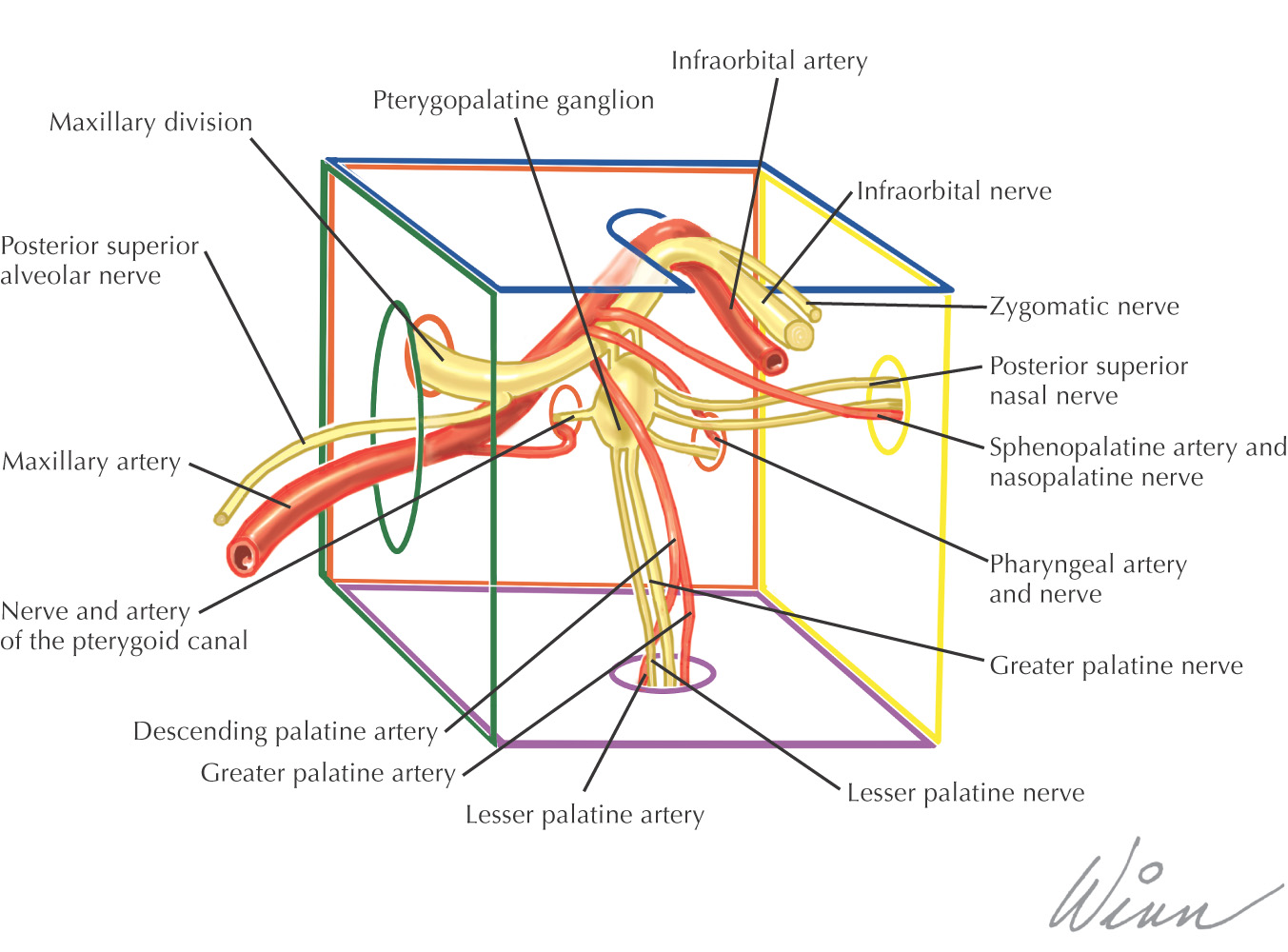
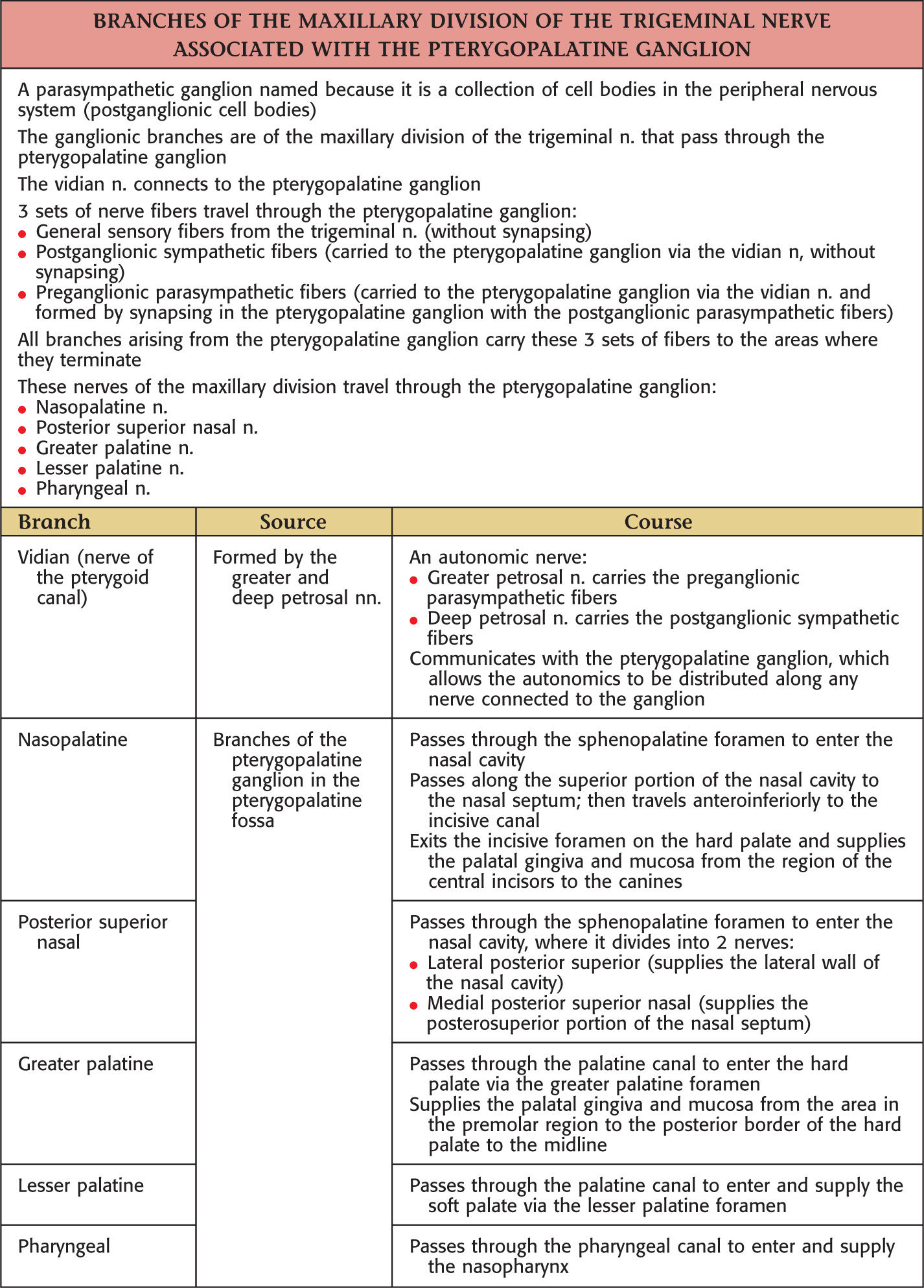
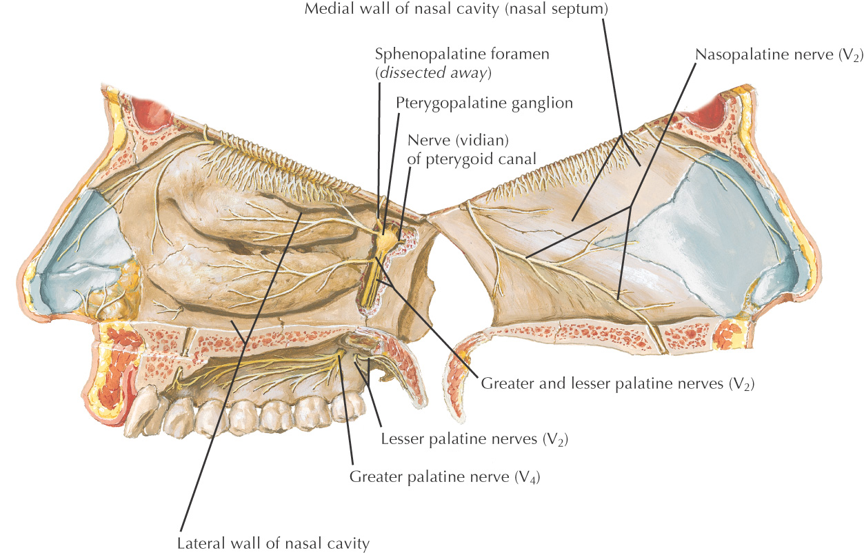
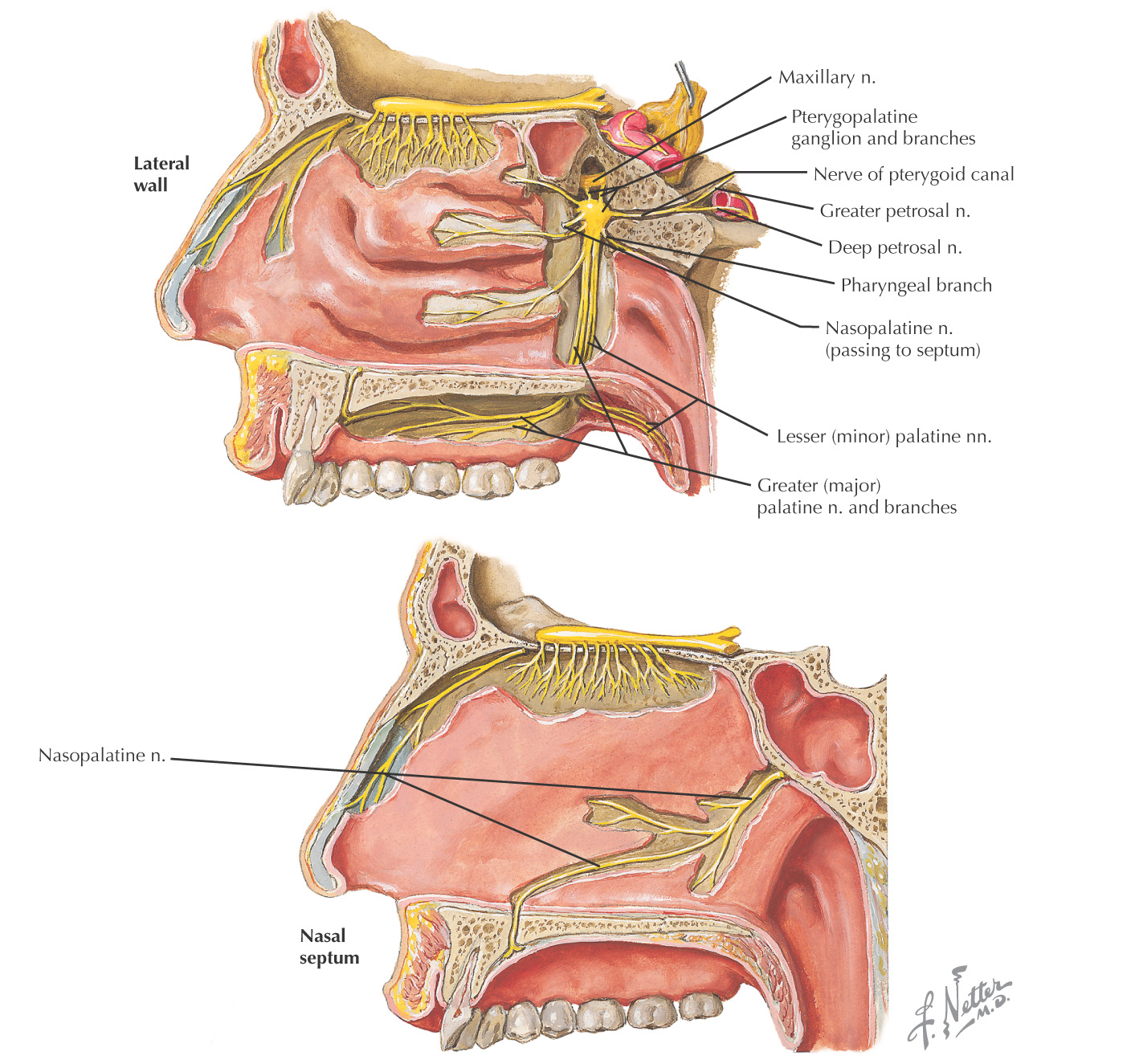
Contents of the Pterygopalatine Fossa
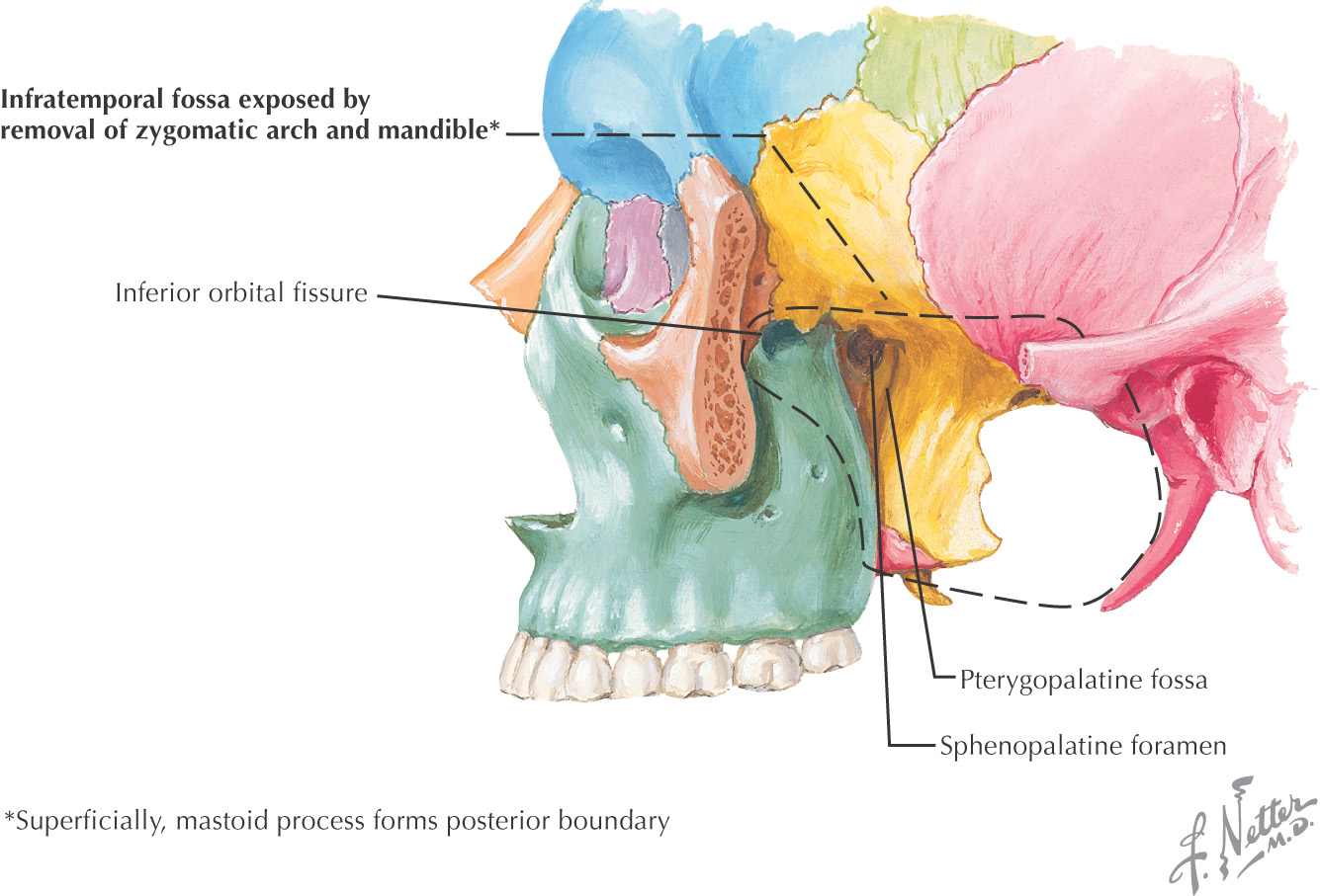
Pyramid-shaped fossa on the lateral aspect of the skull between the maxilla’s infratemporal surface and the pterygoid process of the sphenoid
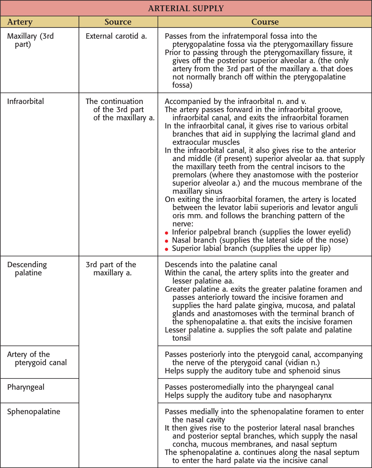
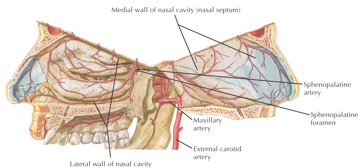
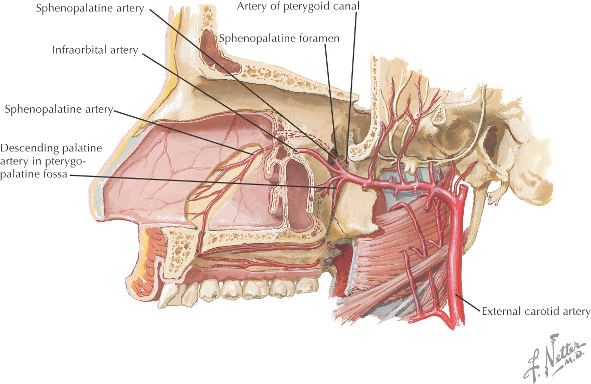
Contains major nerves and blood vessels that supply the nasal cavity, upper jaw, hard palate, and soft palate: the maxillary division of the trigeminal nerve, pterygopalatine (sphenopalatine, Meckel’s) ganglion, and 3rd portion of the maxillary artery
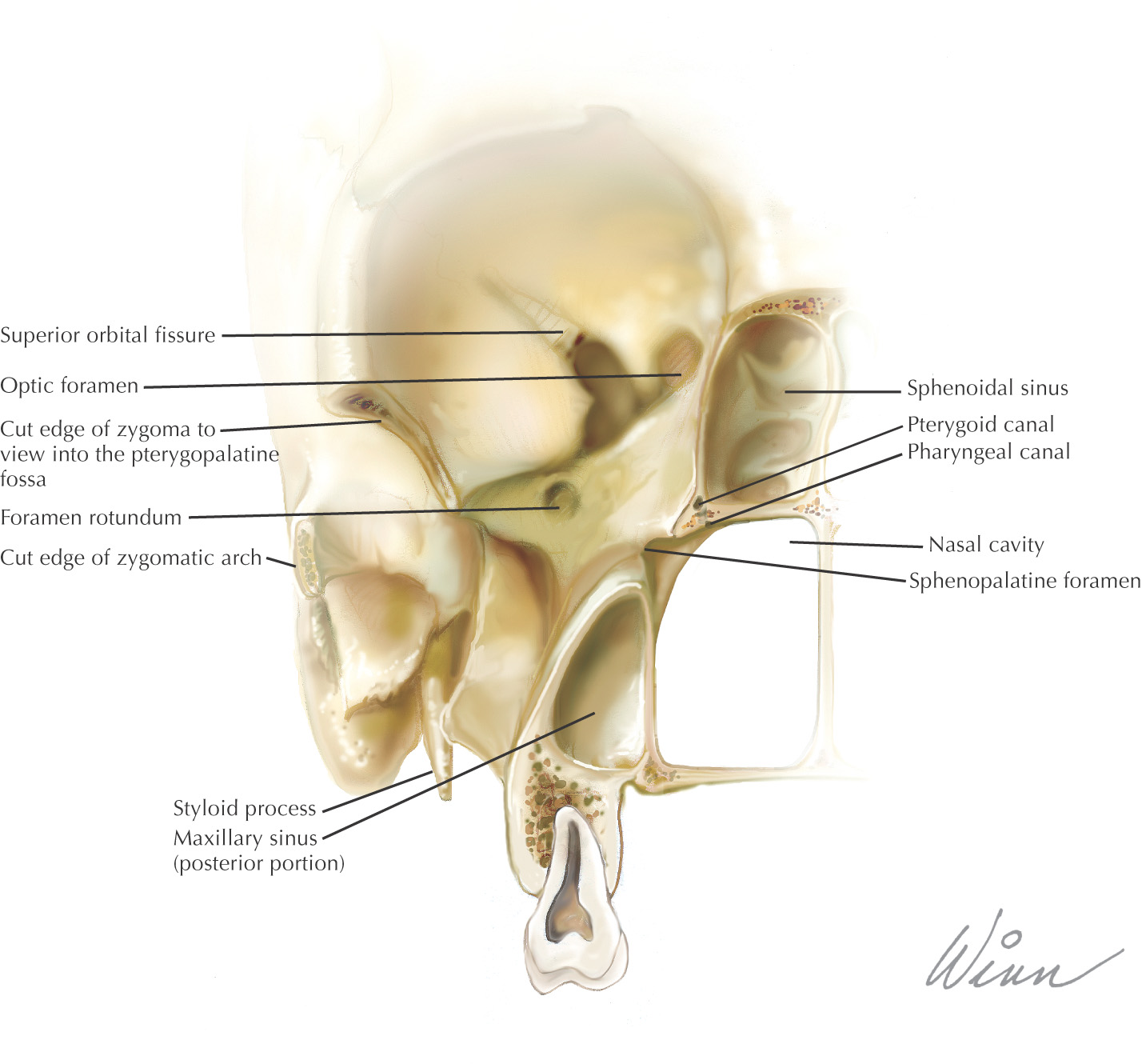
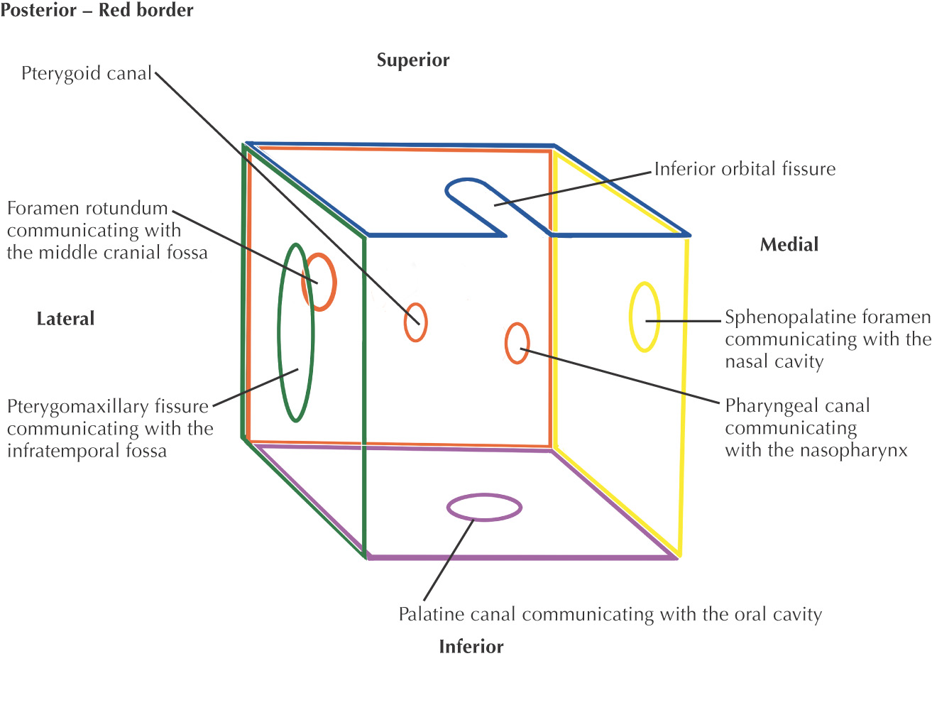
Allows the infratemporal fossa, middle cranial fossa, foramen lacerum, nasopharynx, nasal cavity, orbital cavity, and oral cavity to communicate
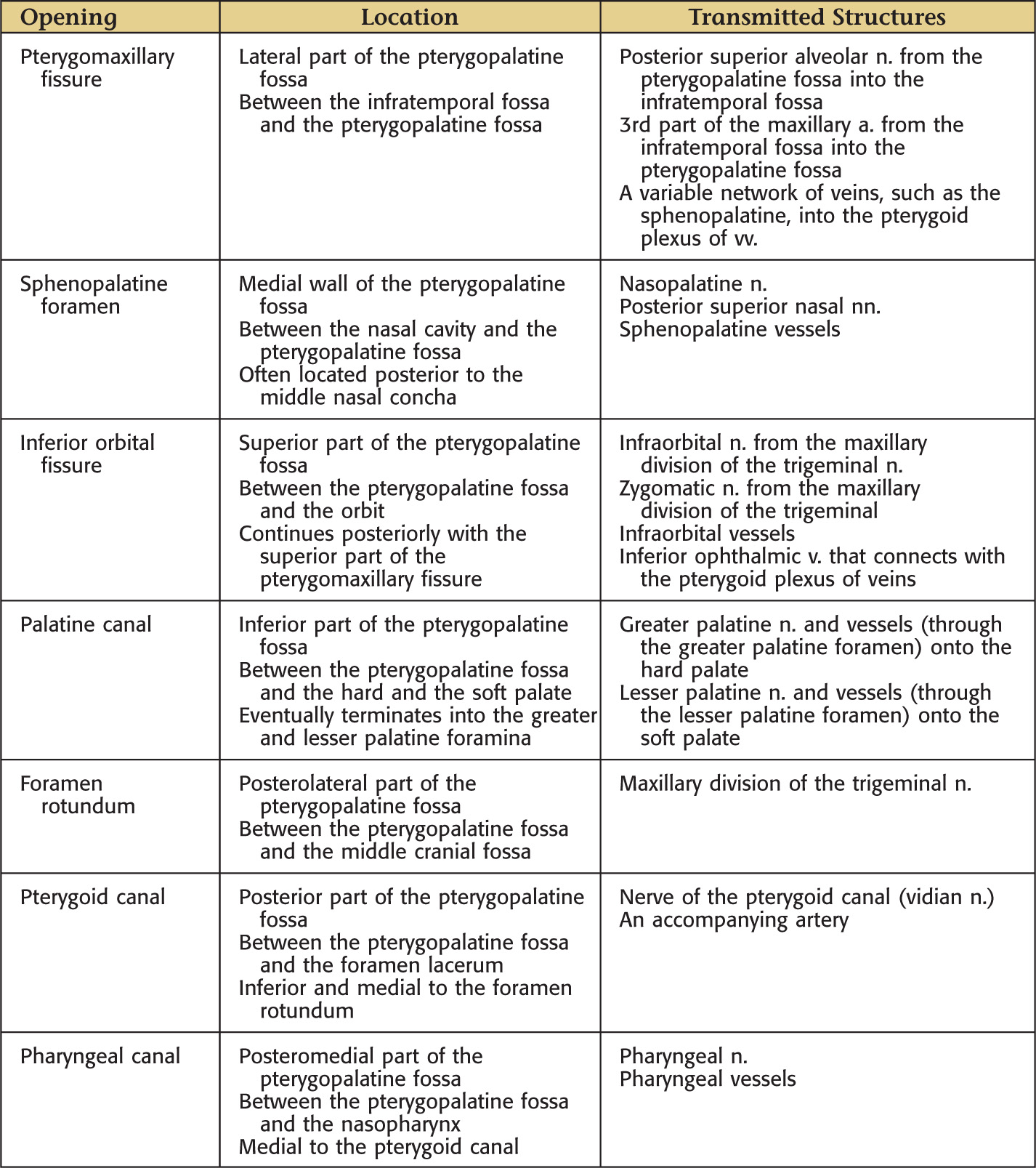
7 foramina/fissures allow passage of nerves and vessels
Border |
Structures |
Anterior wall |
Infratemporal surface of the maxilla |
Posterior wall |
Pterygoid process of the sphenoid |
Medial wall |
Perpendicular plate of the palatine |
Lateral wall |
None (open to the pterygomaxillary fissure) |
Superior wall |
Inferior surface of the sphenoid and the orbital plate of the palatine bone |
Inferior wall |
Pyramidal process of the palatine |