Overview and Topographic Anatomy
The prominent anatomic structure located inferior and medial to the eyes
Helps in breathing and olfaction
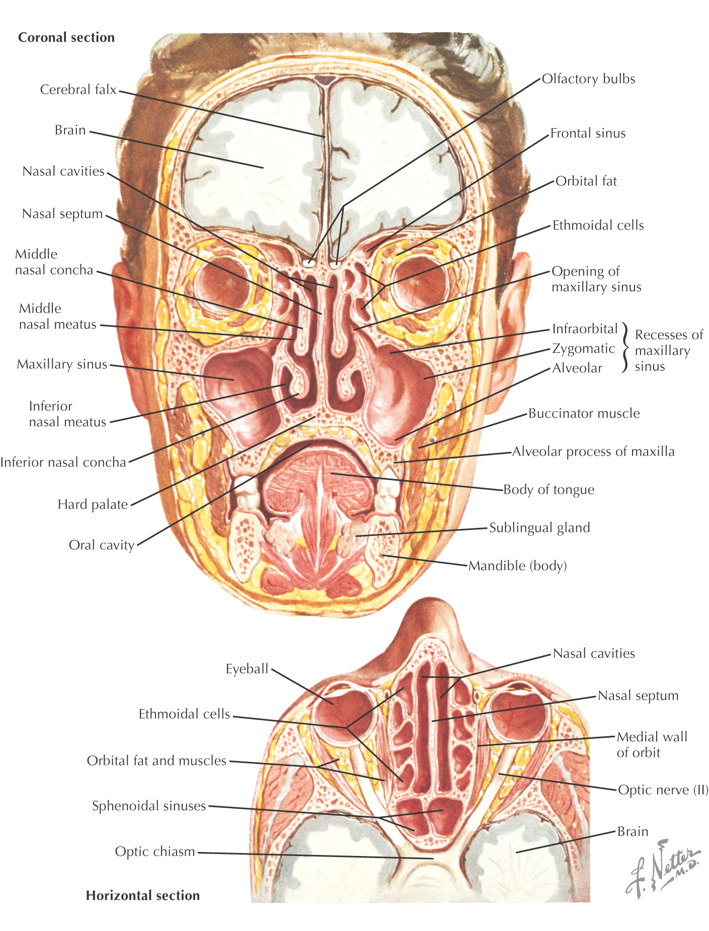
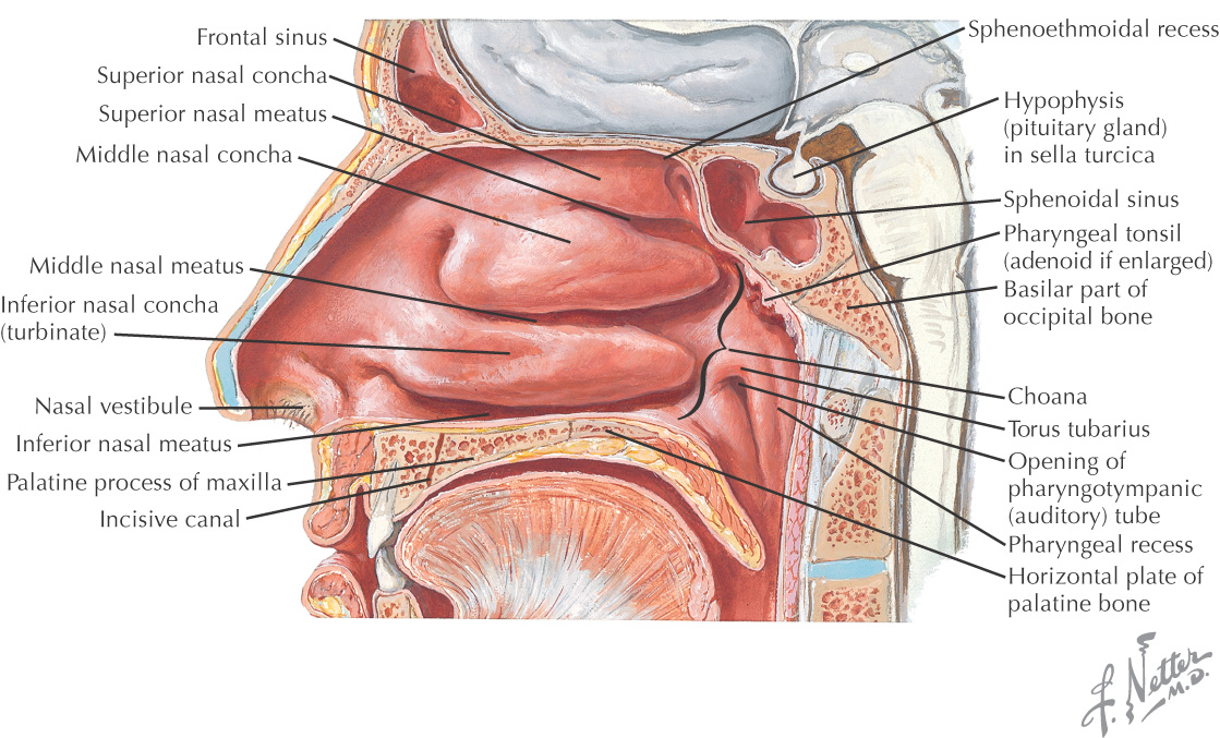
The complex chamber located posterior to the vestibule and atrium of the nose
Highly vascular and easily congested
When this tissue is irritated, its blood vessels reflexively dilate and the glands secrete, normally leading to sneezing
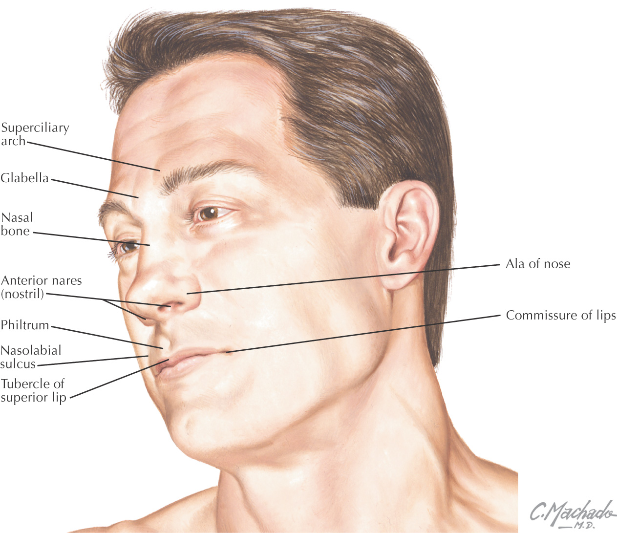
The nose is pyramidal in form
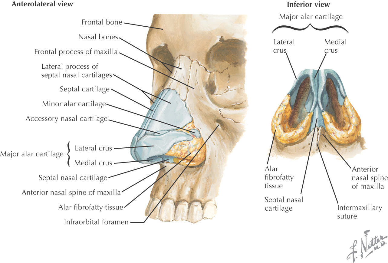
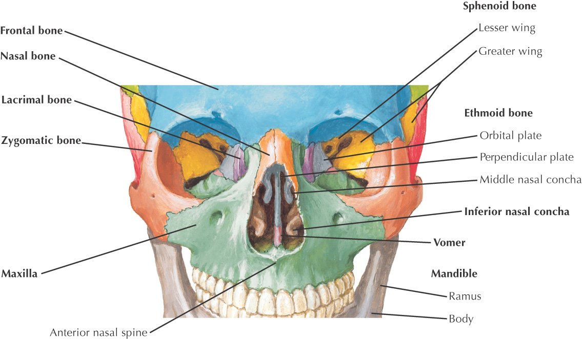
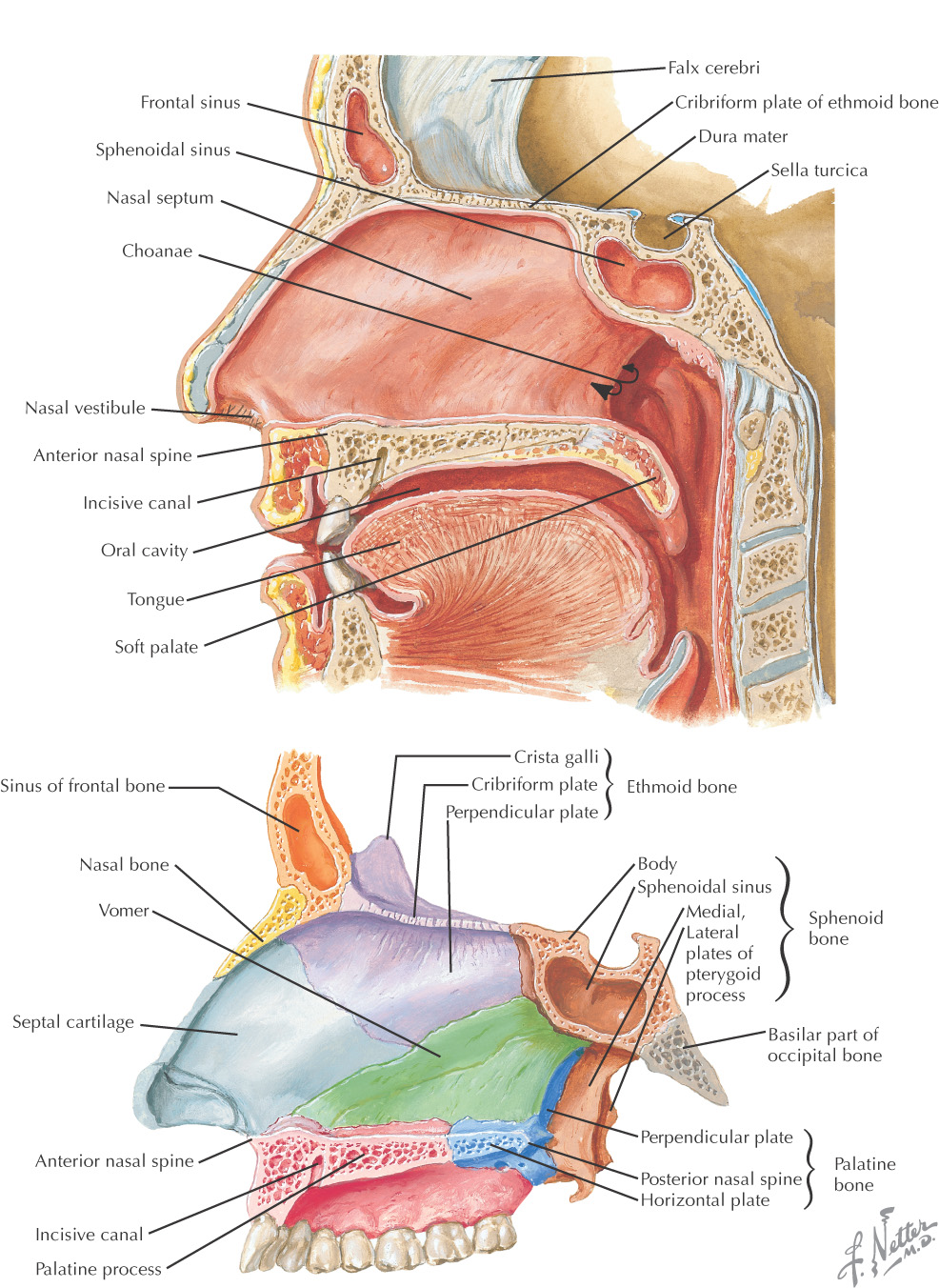
3 pairs of bones form the root of the nose:
Because the root of the nose is made of bone, it is fixed
3 different cartilages form the dorsum and apex of the nose:
Because the dorsum and apex are cartilaginous, the nose is quite mobile
The cavity of the nose opposite the alar cartilage is called the vestibule and is lined by many coarse hairs called vibrissae
The cavity superior to the vestibule is the atrium
At the apex are found the 2 nostrils, or anterior nares, which are separated by the septum connecting the apex to the philtrum of the upper lip
Fibrous tissue helps connect the cartilages together and posteriorly to the maxilla
The primary lymphatic drainage of the nose is into the submandibular lymph nodes
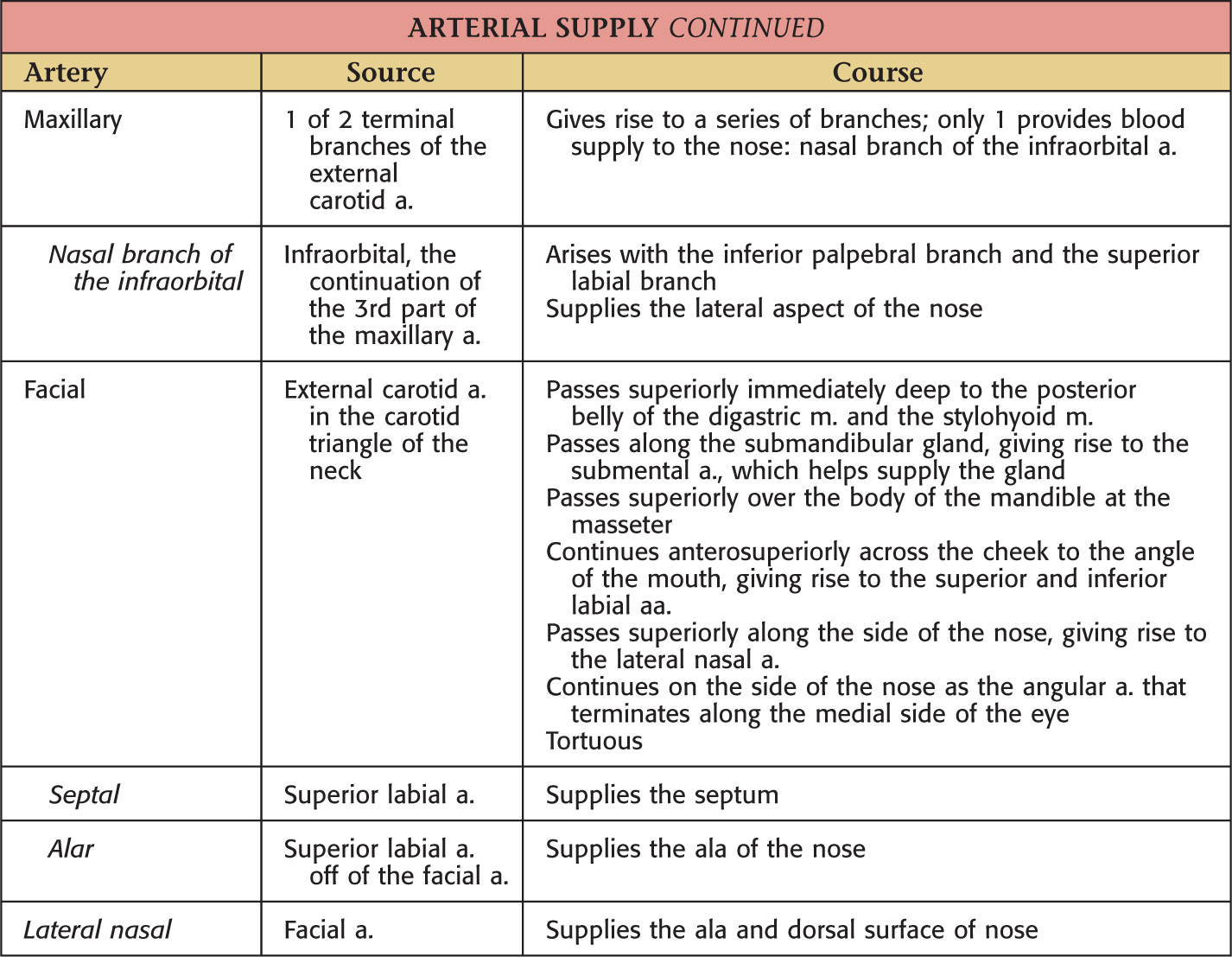
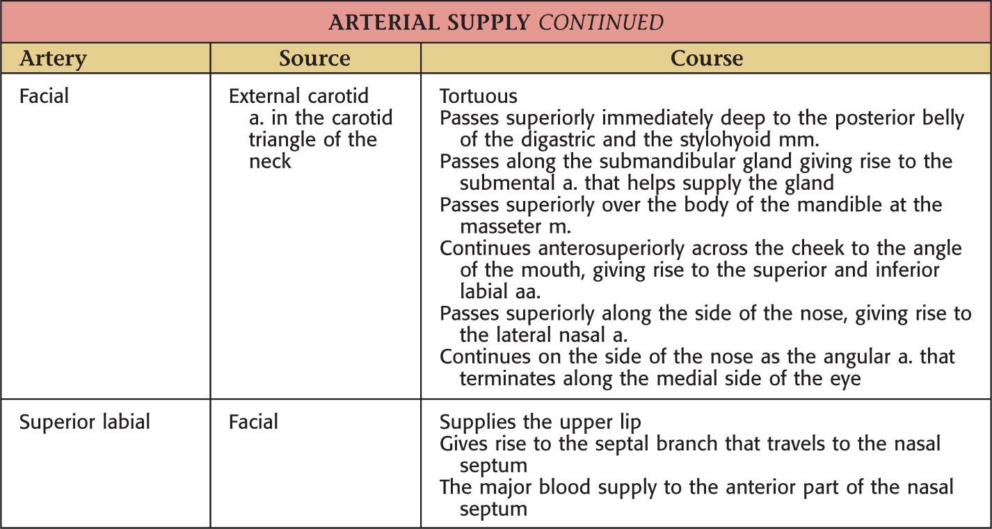
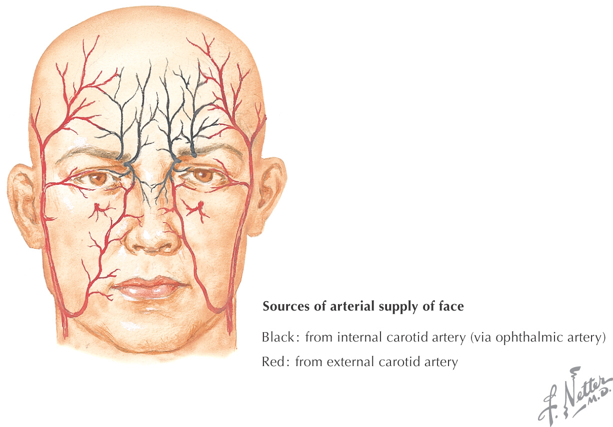
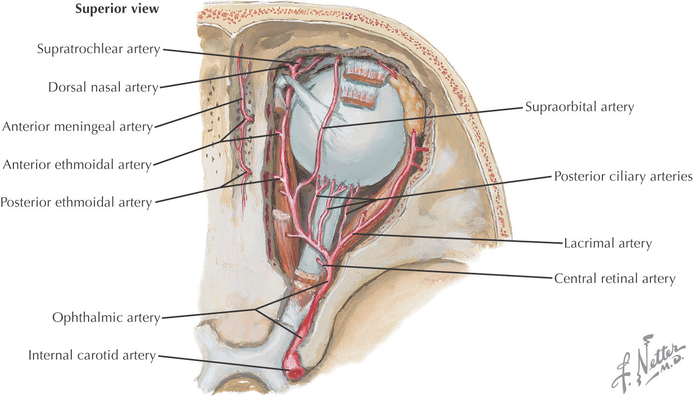
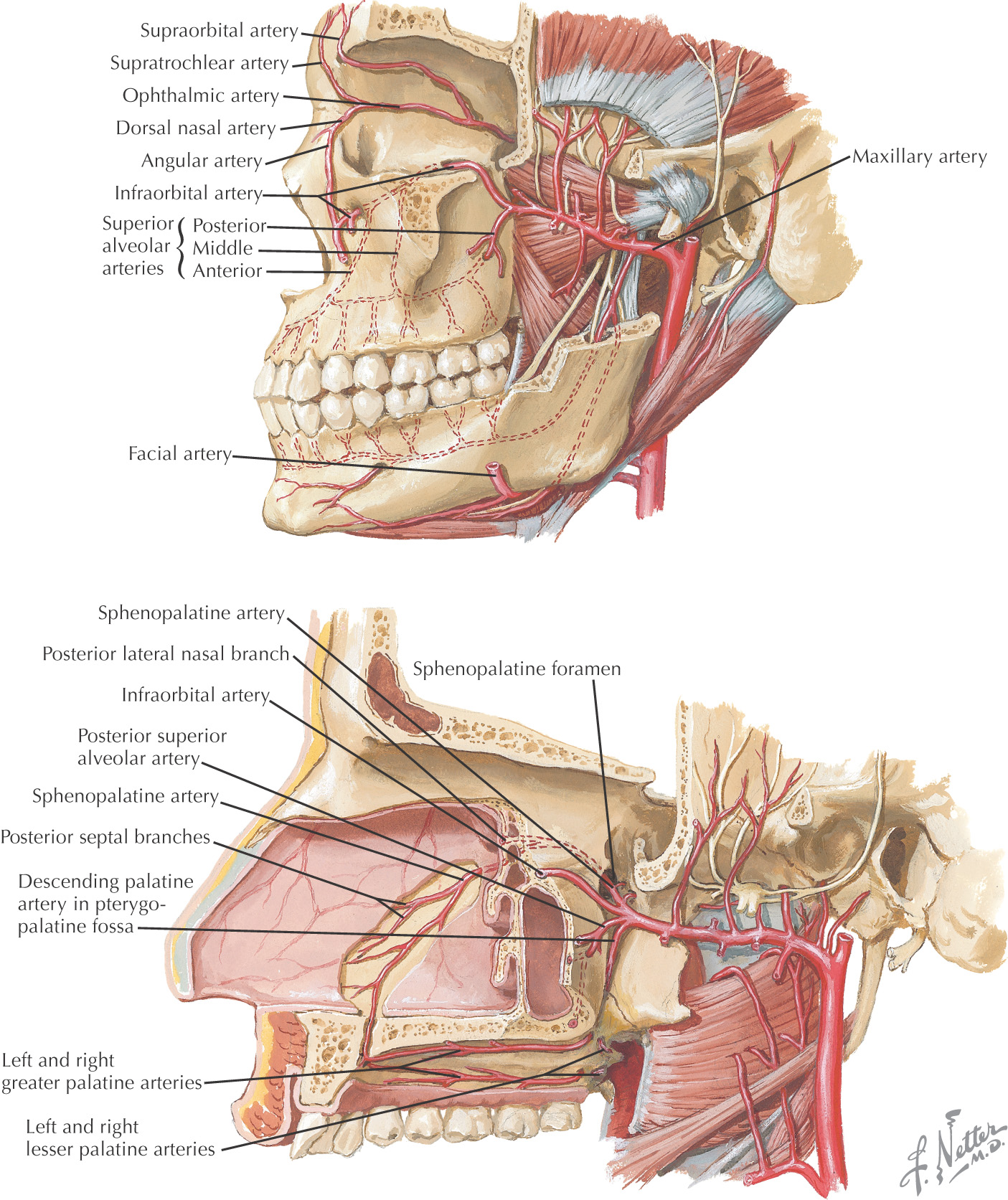
The blood supply to the nose arises from 3 major arteries:
These vessels are derived from the external and internal carotid arteries
These arteries anastomose along the nose
Many nosebleeds are due to trauma to the septal branch of the superior labial artery from the facial artery
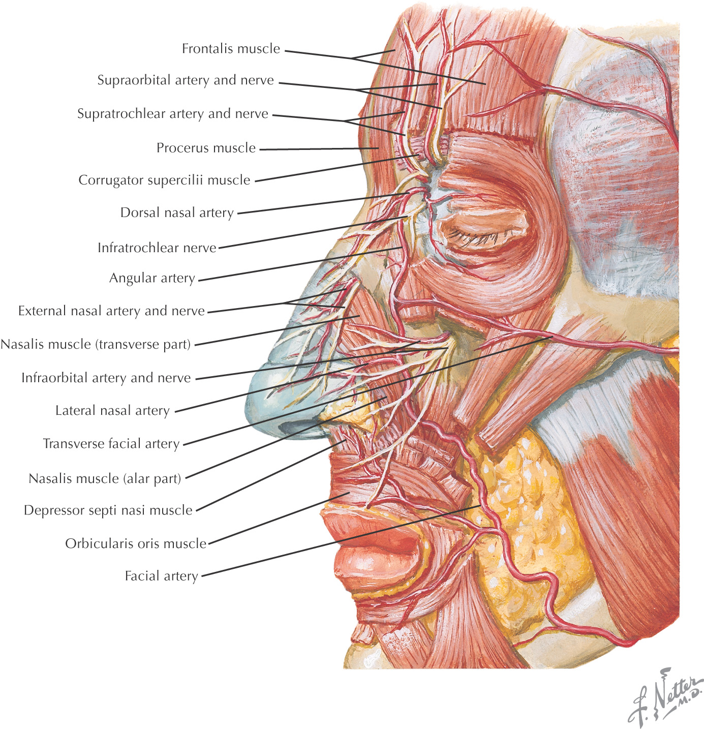

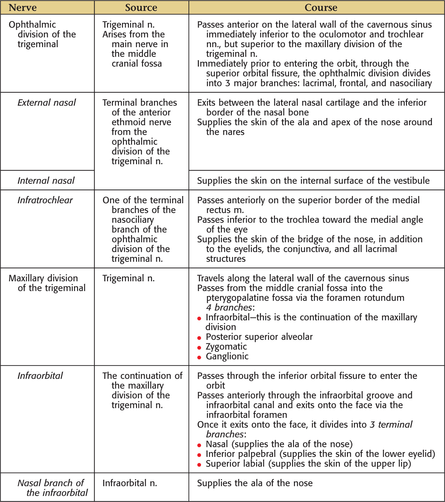
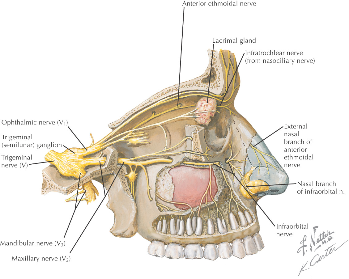
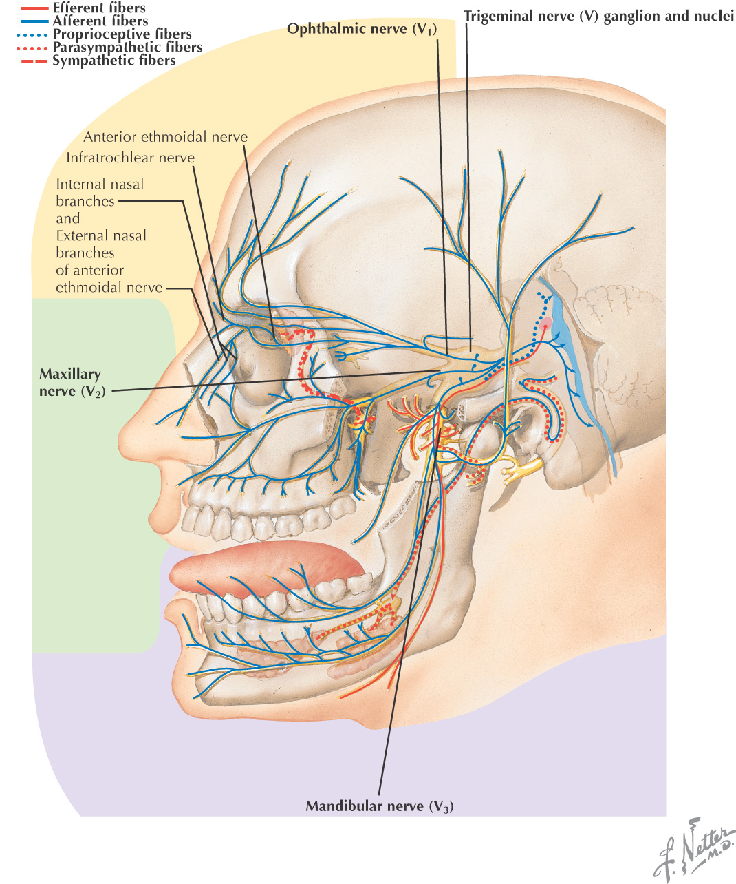
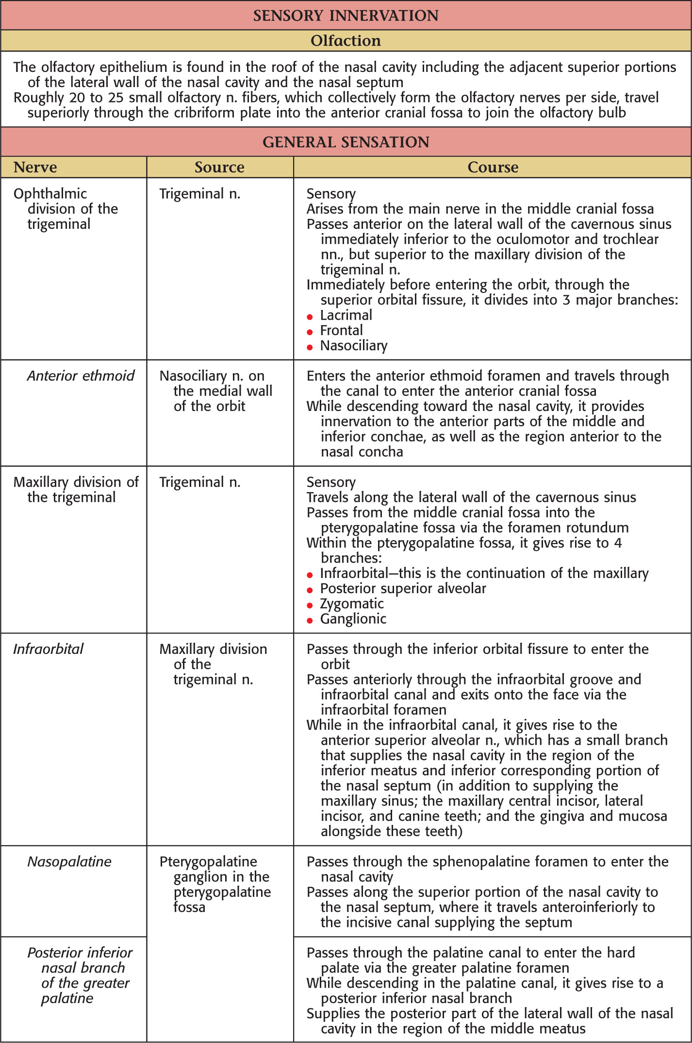
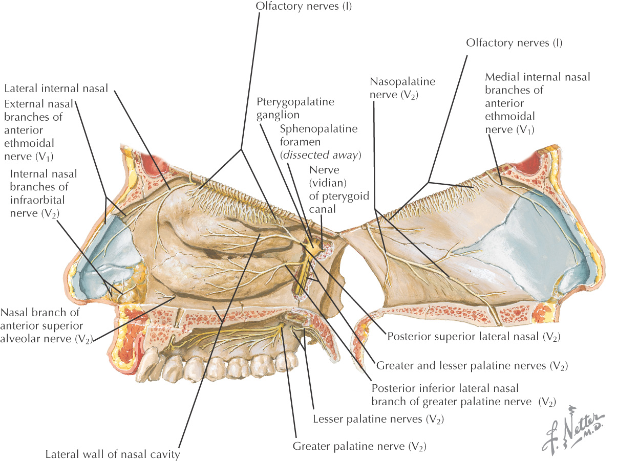
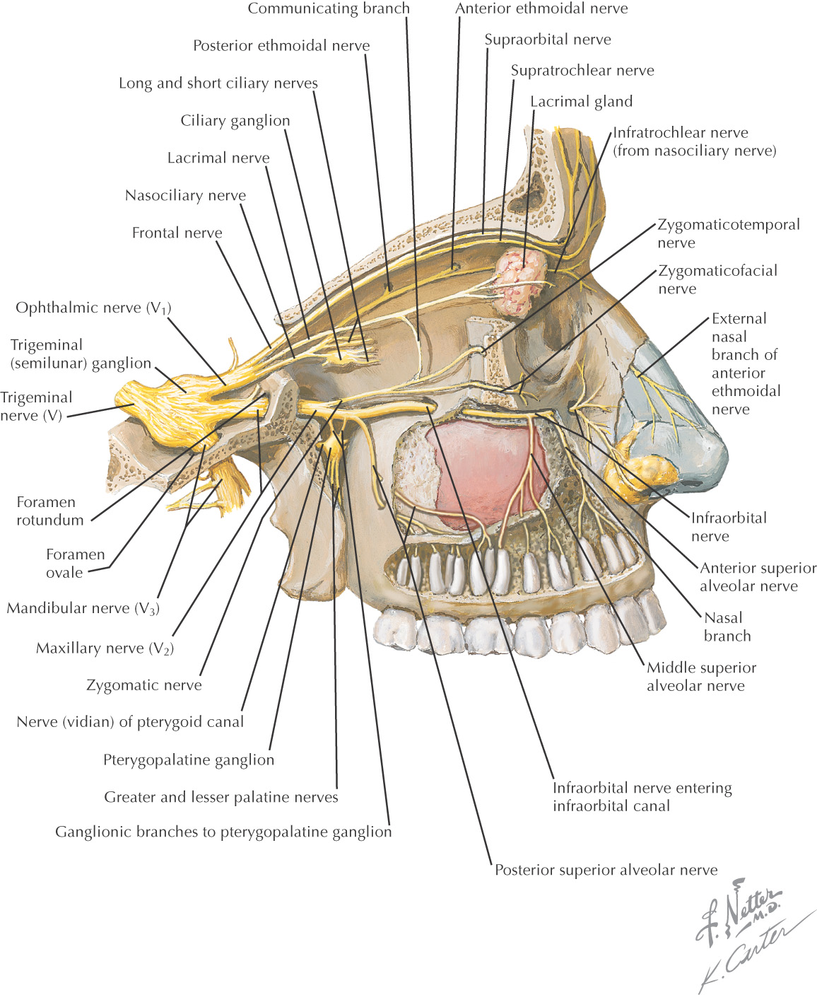
The sensory supply to the nose arises from branches of the ophthalmic and maxillary divisions of the trigeminal nerve
Lined by pseudostratified columnar epithelium with cilia
Inferior portion is larger than superior portion
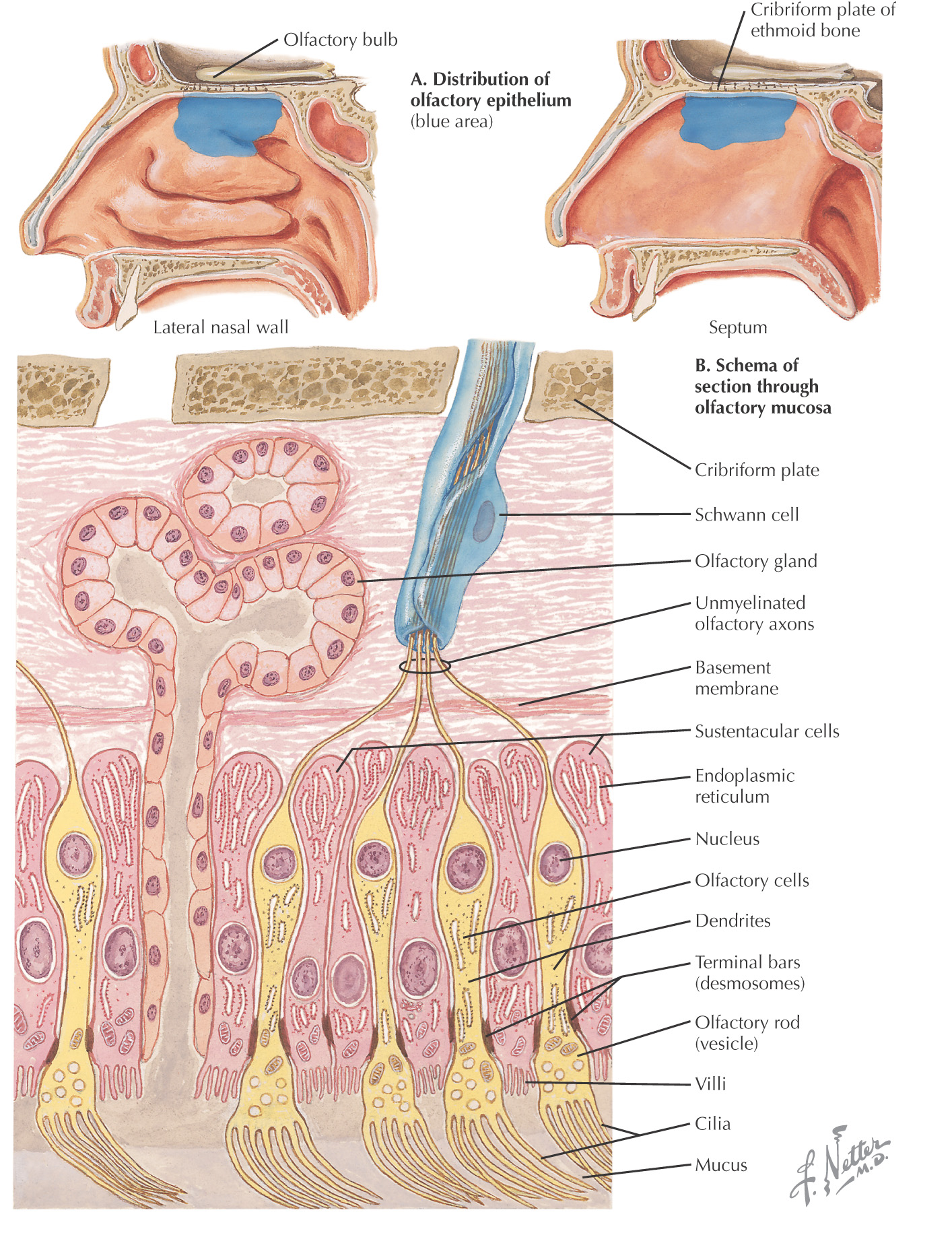
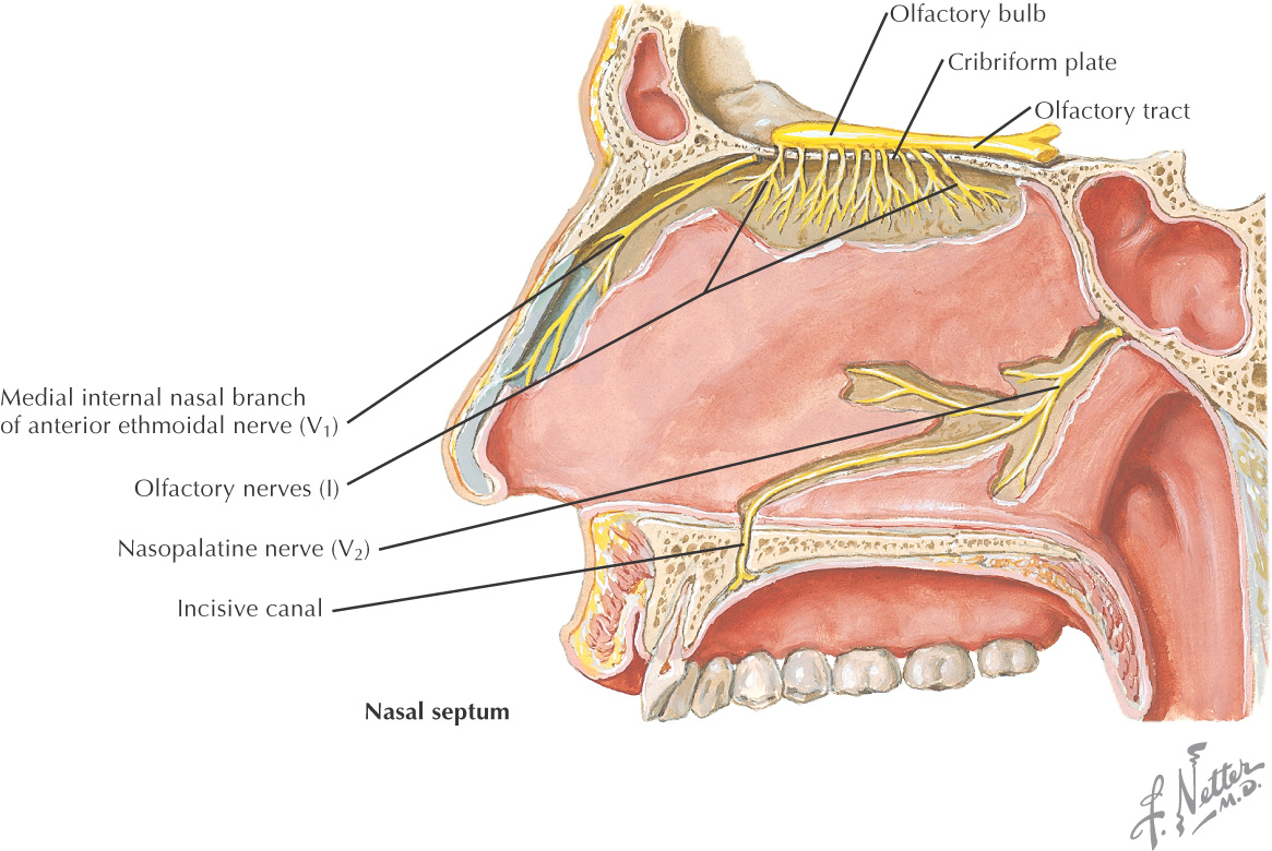
Olfactory epithelium is located at the superior part of the nasal cavity around the cribriform plate
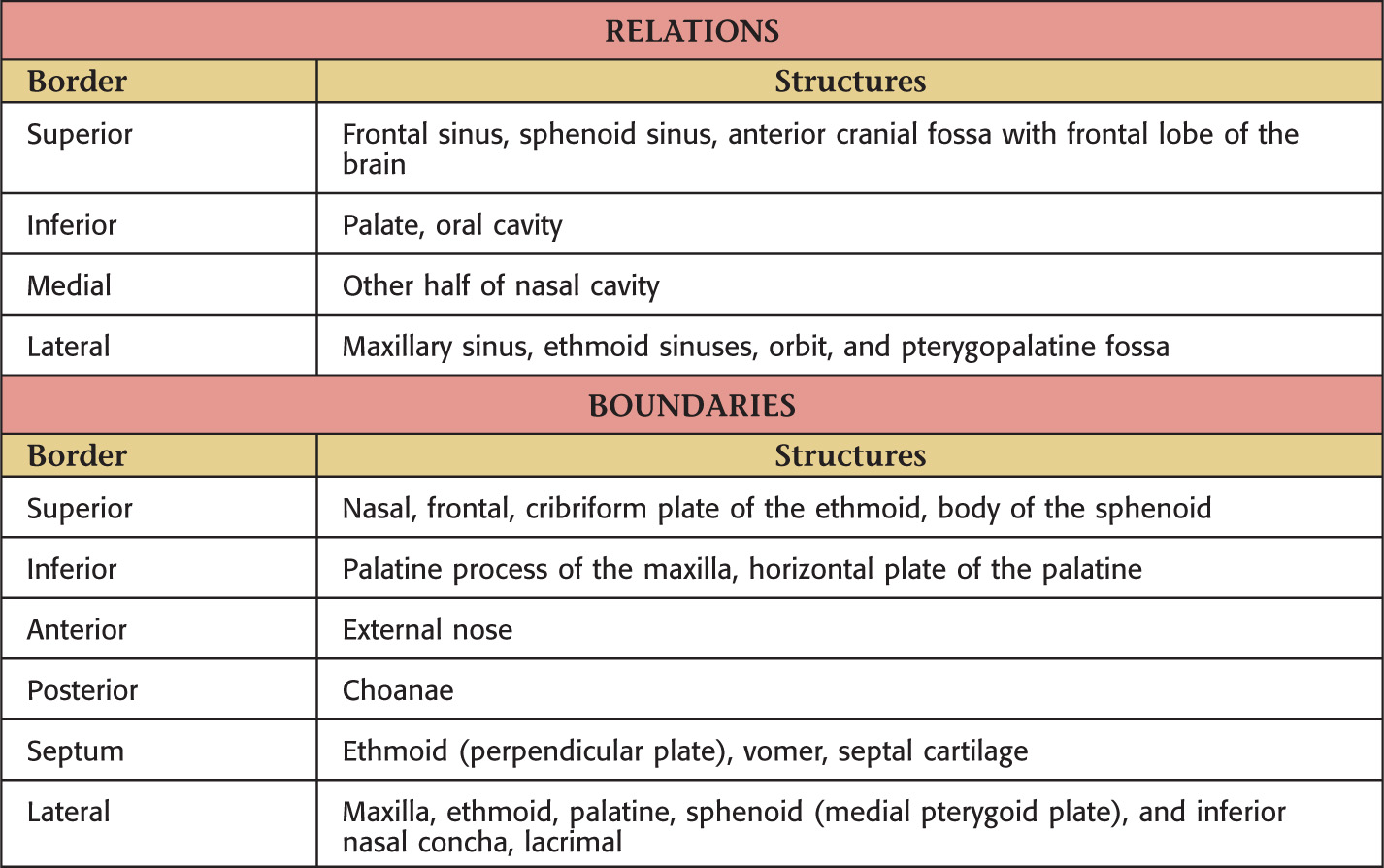
Anterior opening bounded by the nasal bones and maxilla
Frequently deviates to 1 side, giving rise to unequal chambers
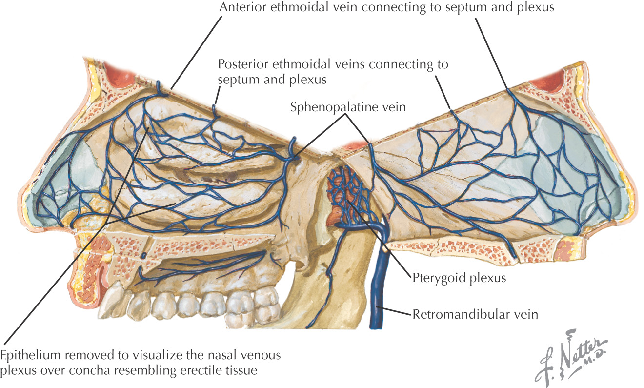
Composed of large venous plexuses that have the appearance of erectile tissue
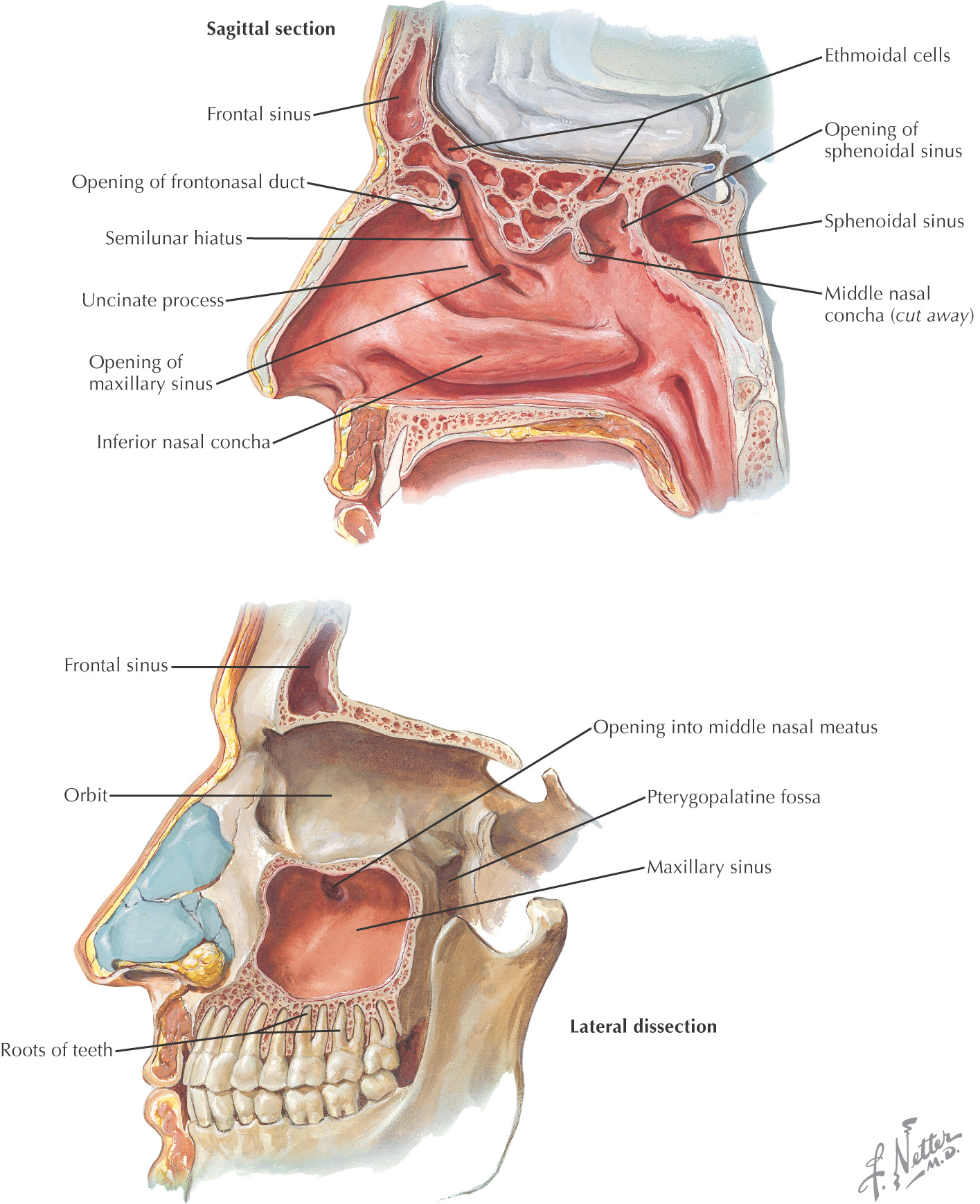
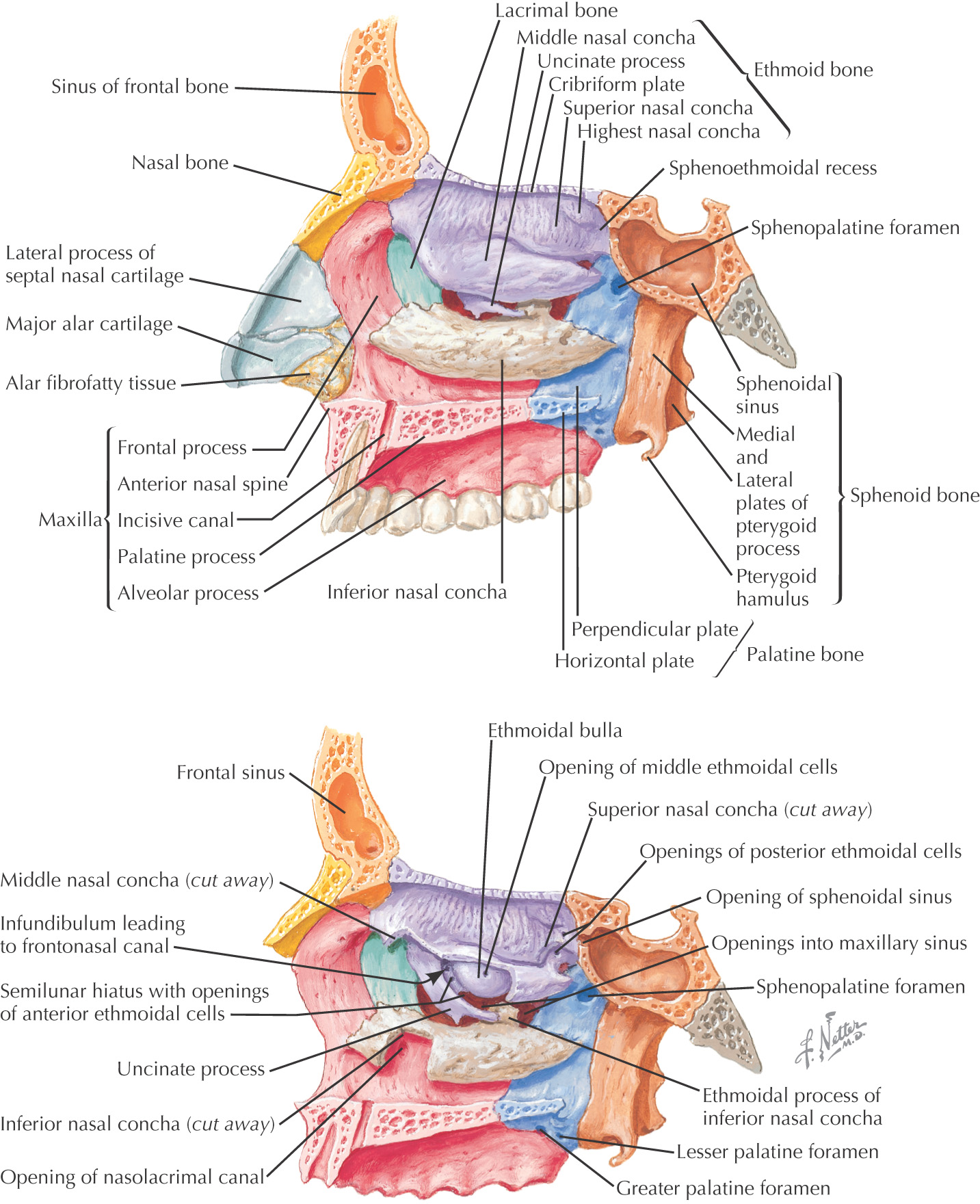
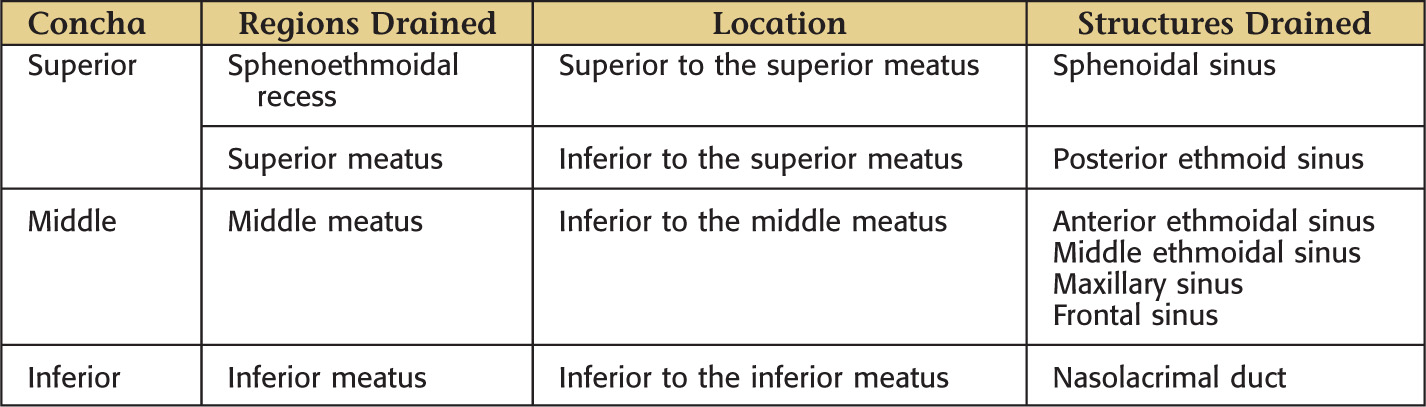
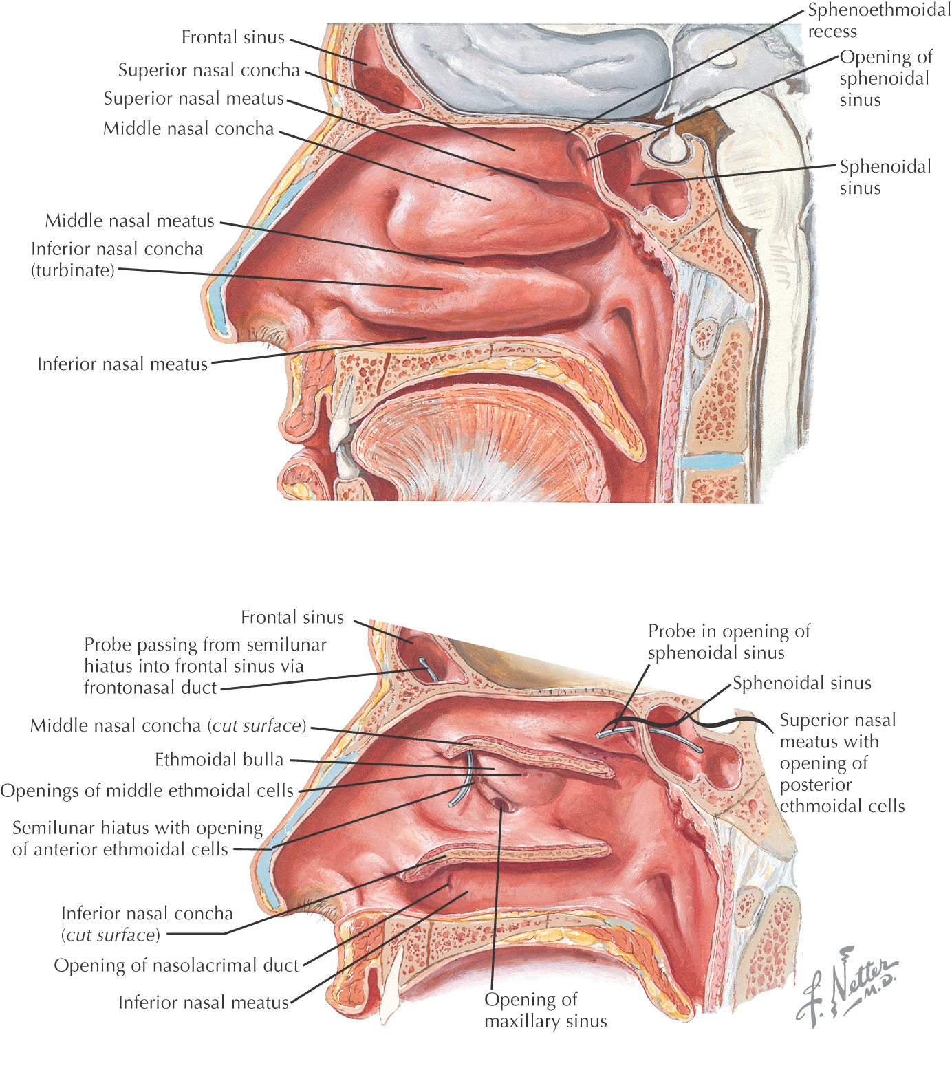
3 large elevations, known as conchae, protrude from the lateral wall
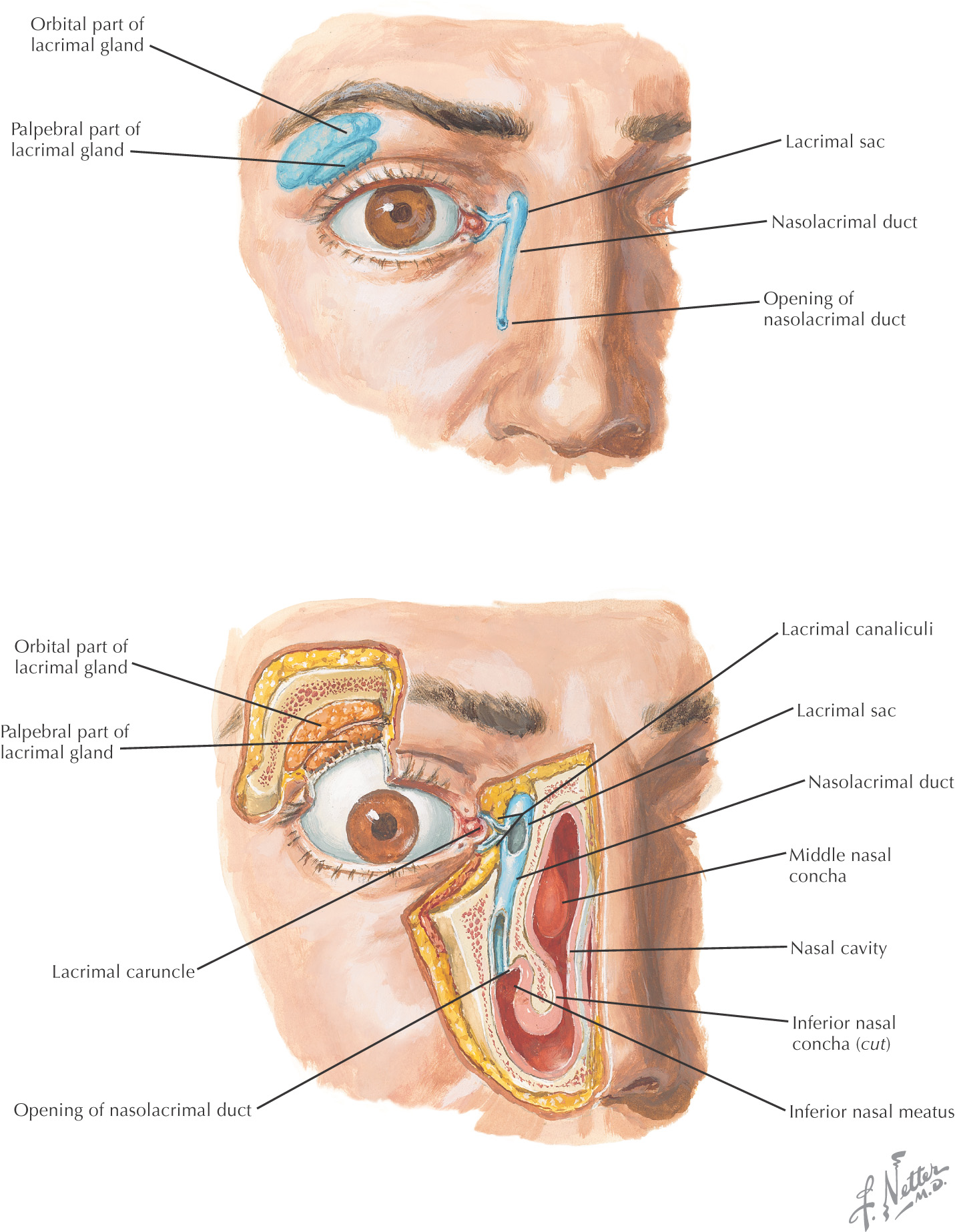
All of the paranasal sinuses and the nasolacrimal duct drain into the lateral walls of the nasal cavity
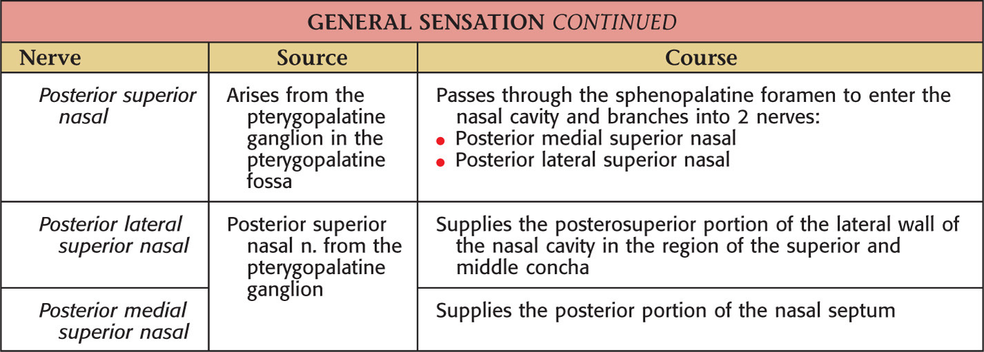
The sphenopalatine foramen, located in the posterior portion of the lateral walls, connects the nasal cavity to the pterygopalatine fossa
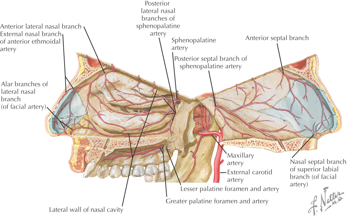
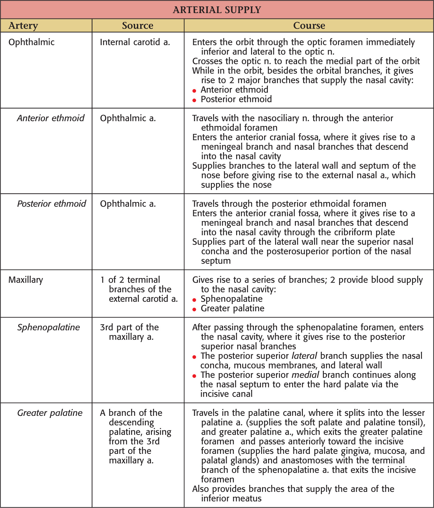
The blood supply to the nasal cavity arises from 3 major arteries:
These 3 vessels are derived from the external and internal carotid arteries and generally follow the paths of the nerves
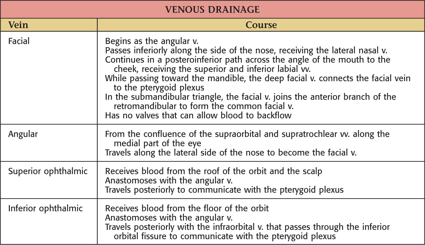
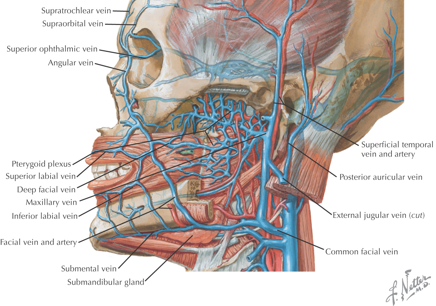
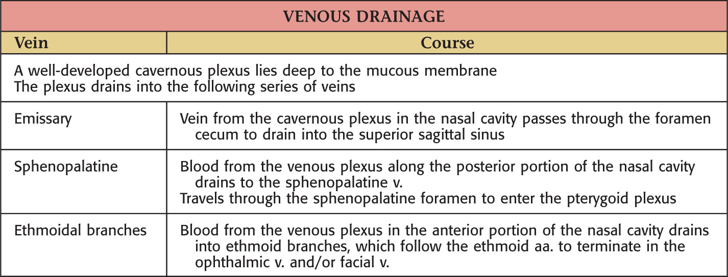
The veins generally correspond to the arteries
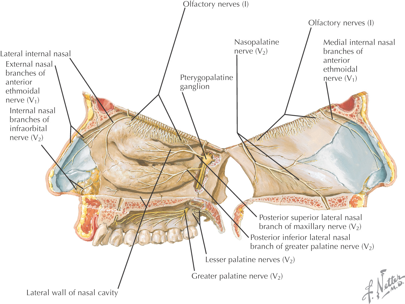
2 major types of sensory innervation to the nasal cavity:
• Olfaction (special visceral afferent) via the olfactory nerve
• General sensation (general somatic afferent) via ophthalmic and maxillary divisions of the trigeminal nerve
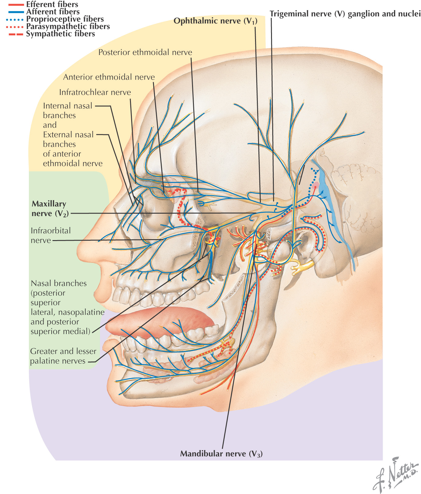
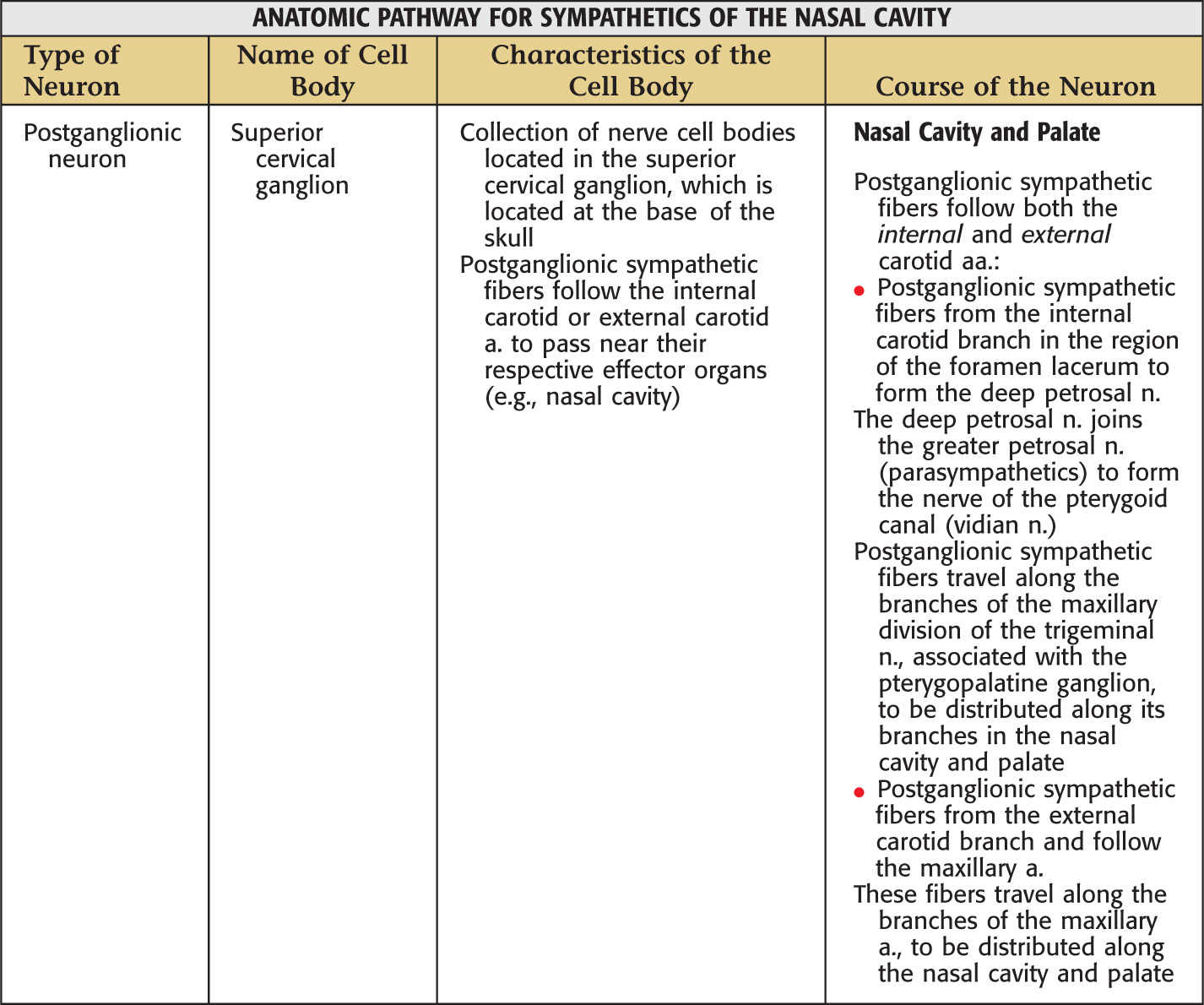
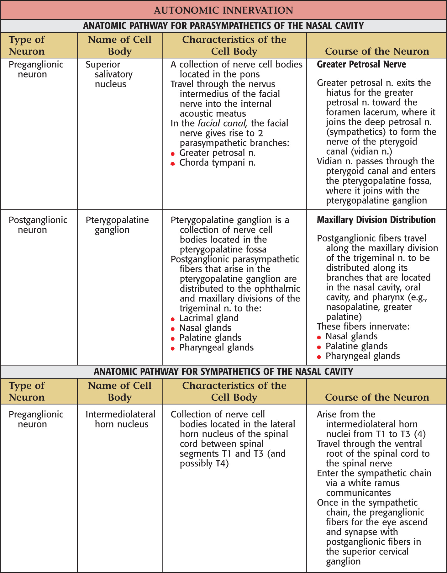
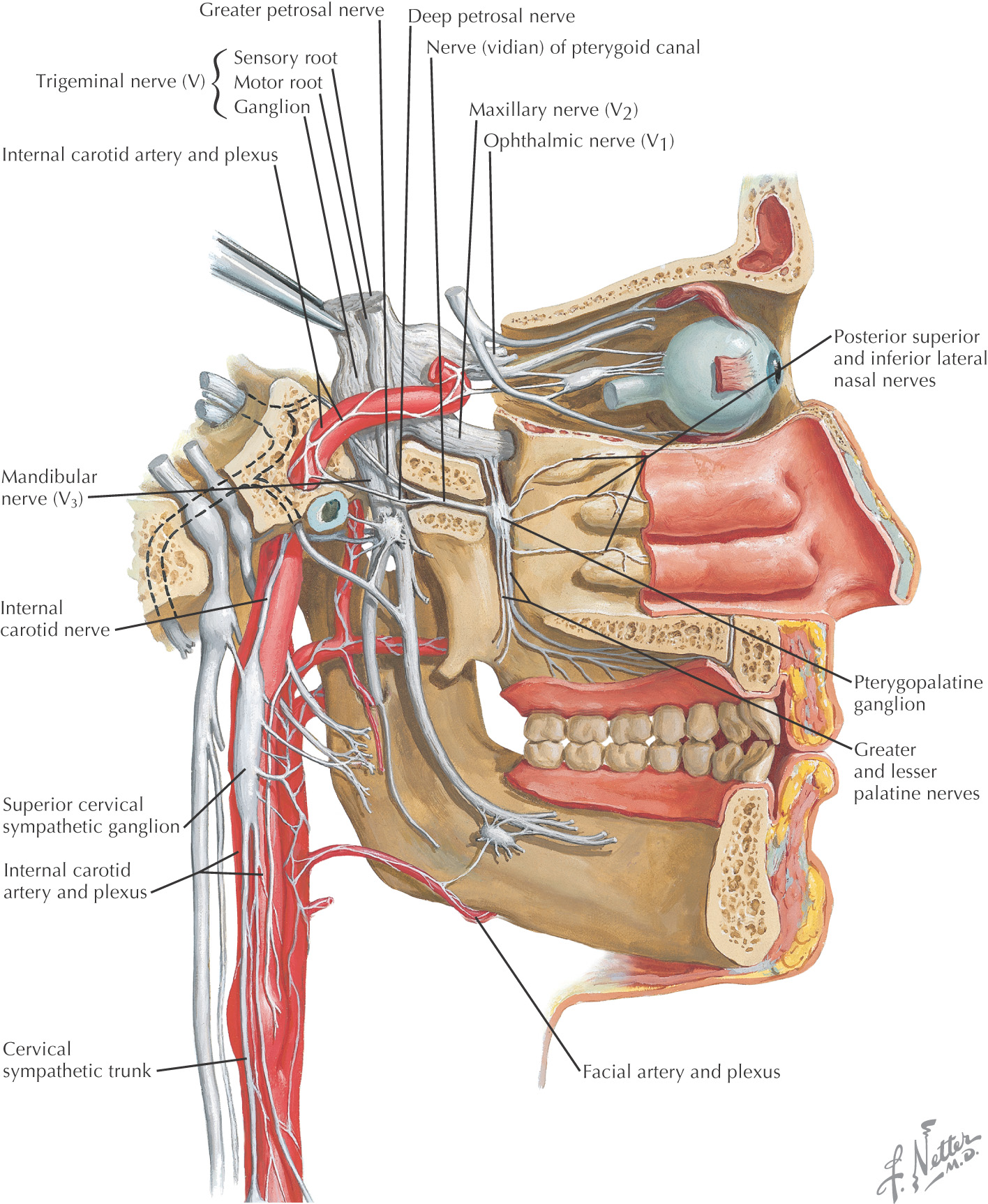
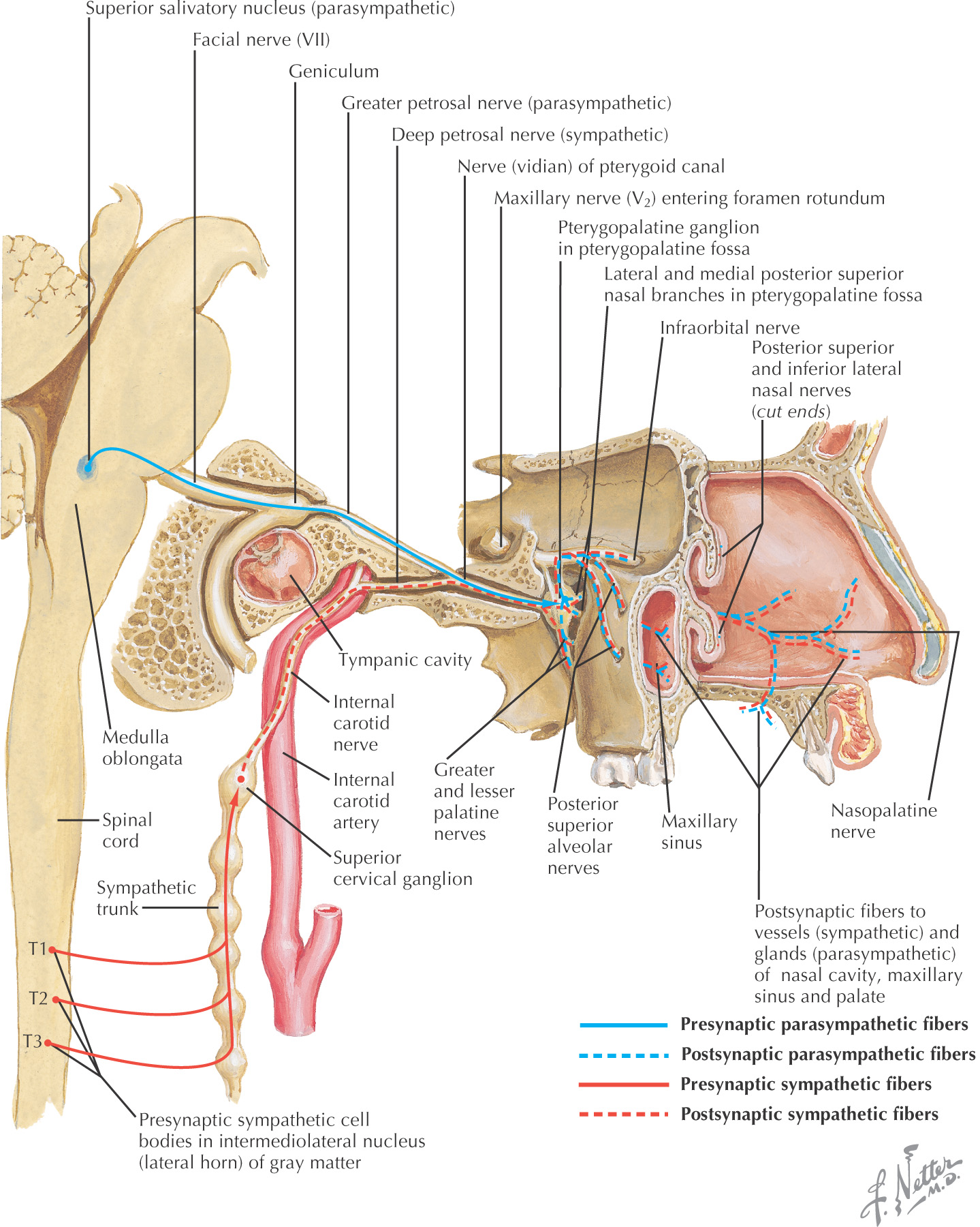
Autonomic fibers are distributed through the sensory branches of the maxillary division of the trigeminal nerve via the pterygopalatine ganglion (parasympathetics) and the superior cervical ganglion (sympathetics)
Autonomics travel to the glands and blood vessels of the nasal cavity
Epistaxis, or nosebleed, is a hemorrhage from the nasal cavity or nose
Classified by bleeding location:
• Trauma (blows to the face, fractures, nose picking)
The most common form (in about 90% of cases)
Usually found along the nasal septum and results from bleeding along Kiesselbach’s plexus
Many nosebleeds are due to trauma to the septal branch of the superior labial artery from the facial artery
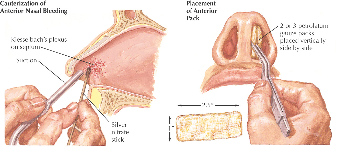
Typically managed with local pressure
May be controlled with cautery via a silver nitrate stick or anterior nasal packing if bleeding is persistent
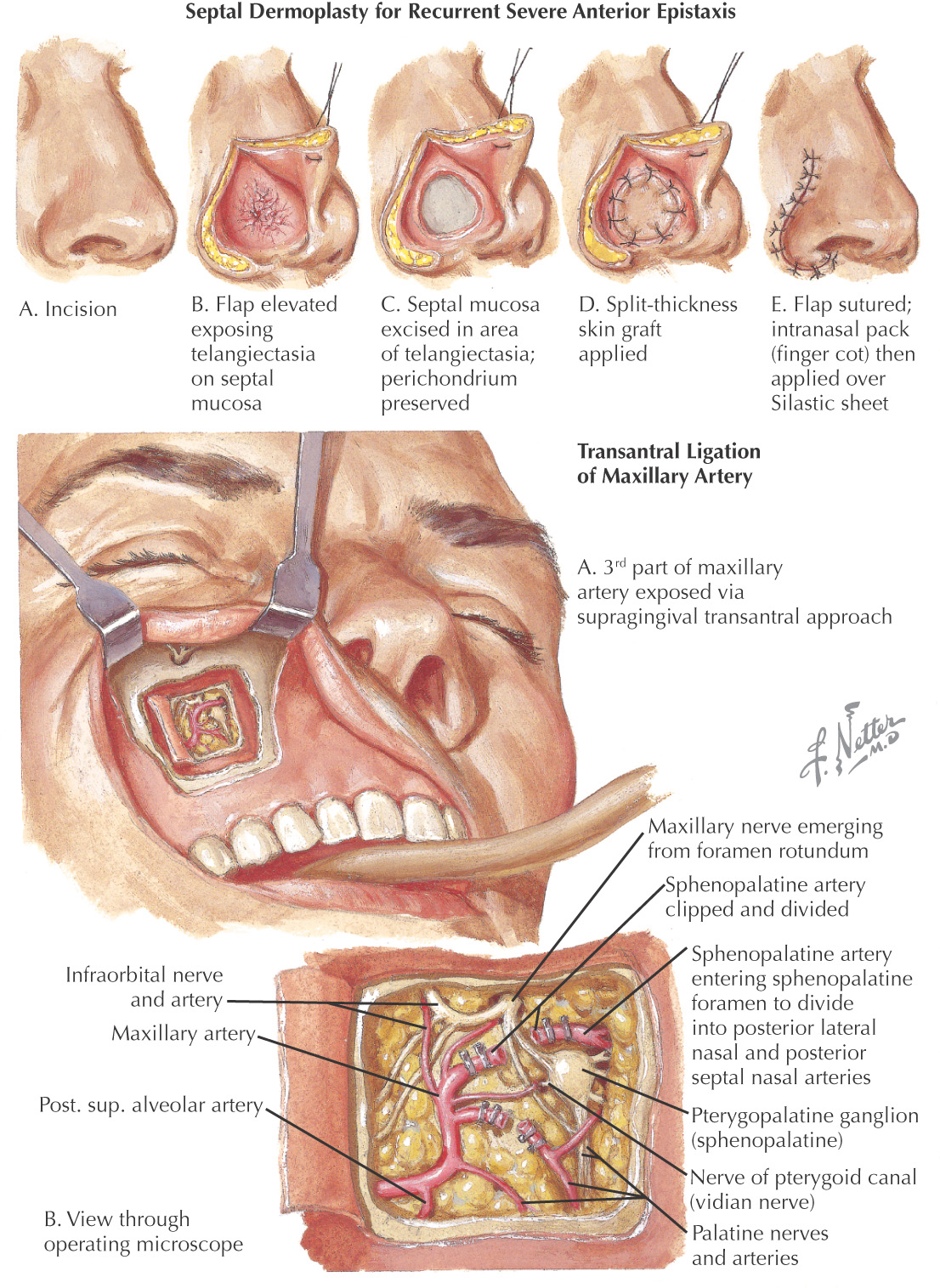
With anterior epistaxis, another treatment, although somewhat drastic, is septal dermoplasty
• The thin septal mucosa is replaced by a thicker graft of skin
• Often used to treat nosebleeds caused by hereditary hemorrhagic telangiectasia or septal perforations
Usually found along the posterior part of the nasal cavity
More difficult to treat and may be accomplished with posterior nasal packing or a balloon catheter
Severe posterior epistaxis may require ligation of the maxillary artery
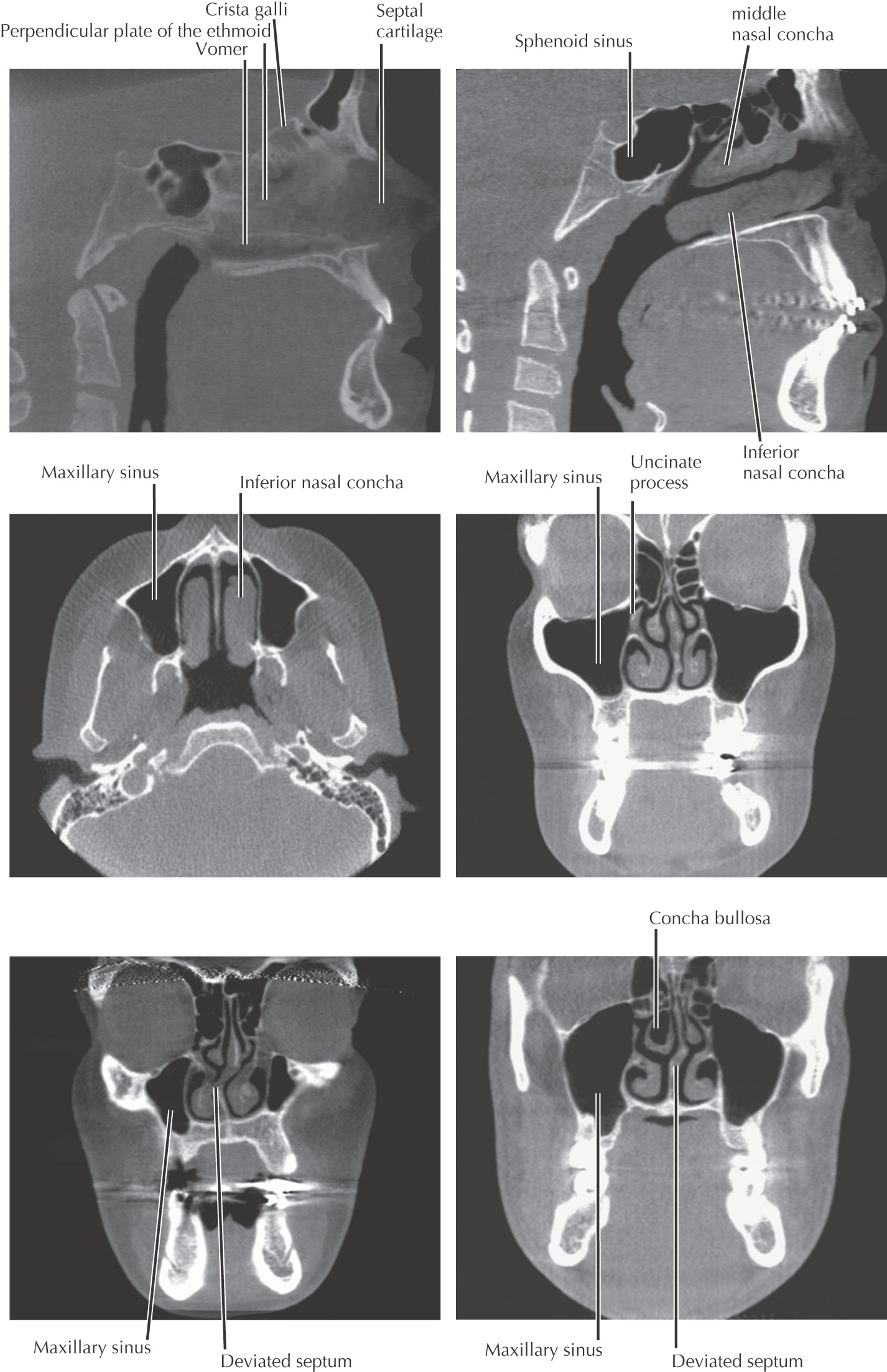
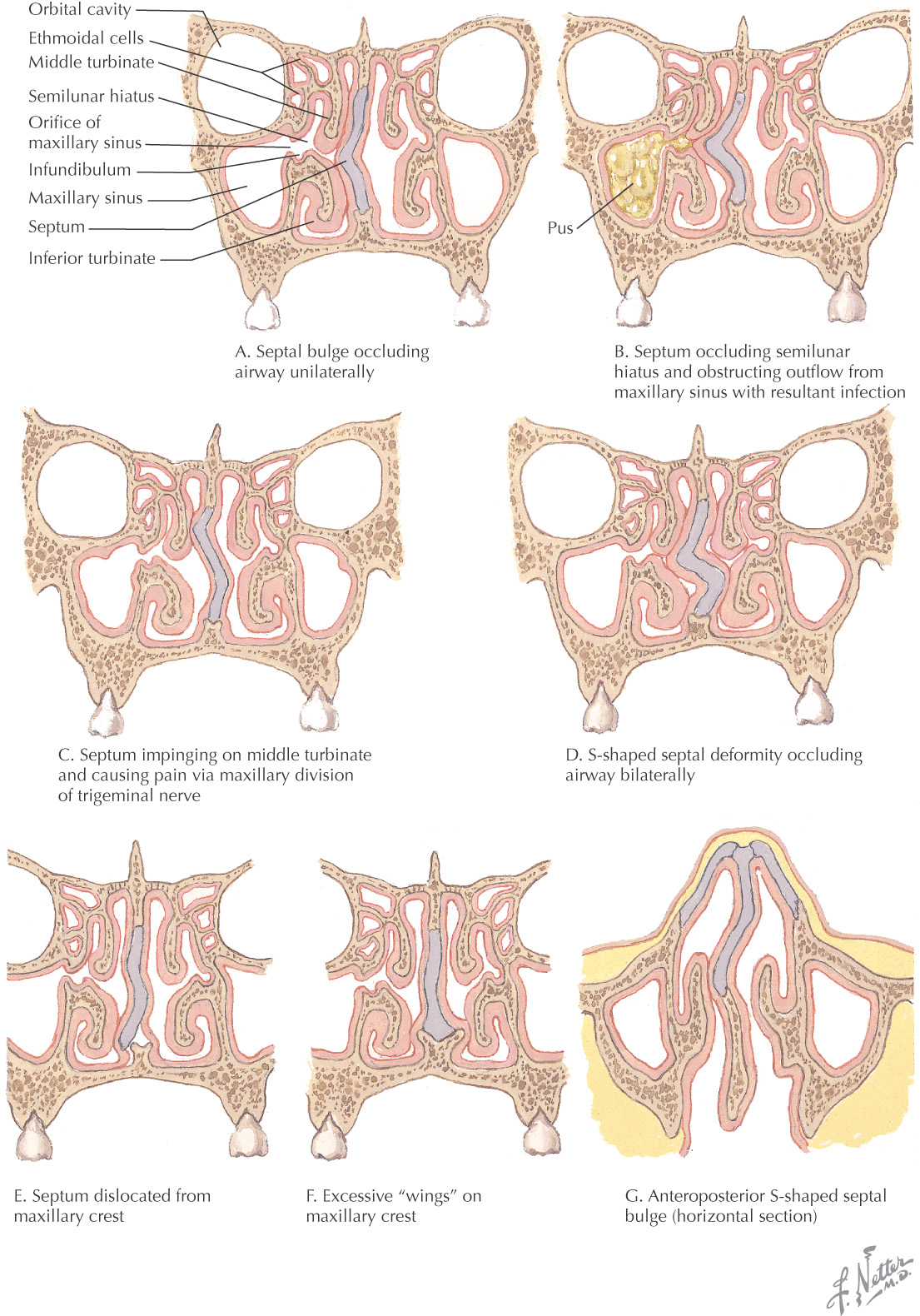
A severe shift of the nasal septum from the midline
Occlusion of one side, either partial or complete, producing difficulty in breathing or blocked air flow on that side
May also cause:
May be treated by septoplasty
An inflammation of the mucosa of the nasal cavity that results in:
May involve the eyes, ears, sinuses, and throat and cause headaches
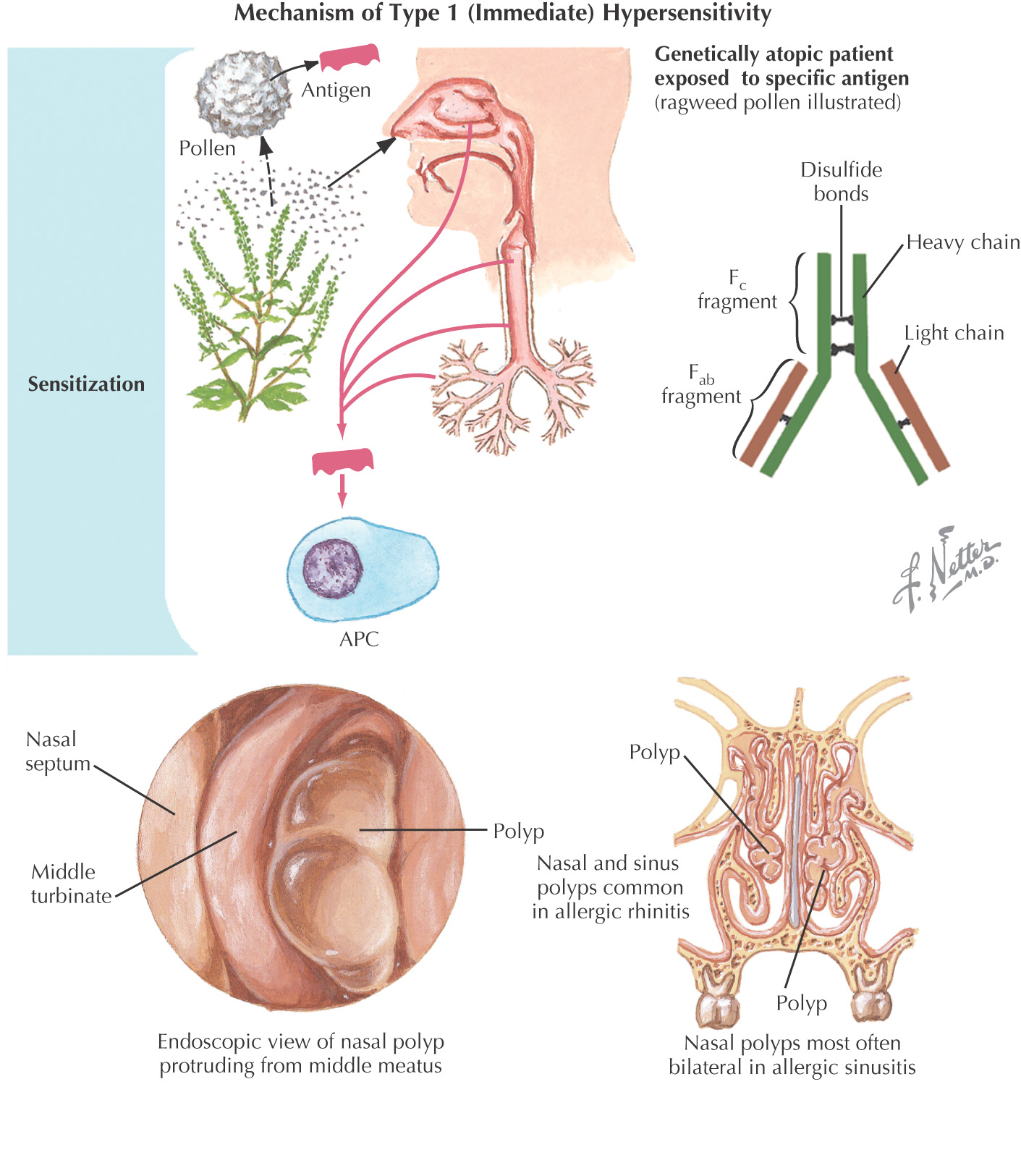
Most commonly caused by allergic rhinitis
Can be associated with nasal polyps, deviated septum, and asthma
Caused by an allergen inducing an immunoglobulin E (IgE)-mediated response on the mast cells
Because mast cells are located on the nasal mucosa, an allergen can bind to the mast cell, resulting in the release of histamines, prostaglandins, cytokines, and leukotrienes
Typically treated with decongestants, antihistamines, and steroids