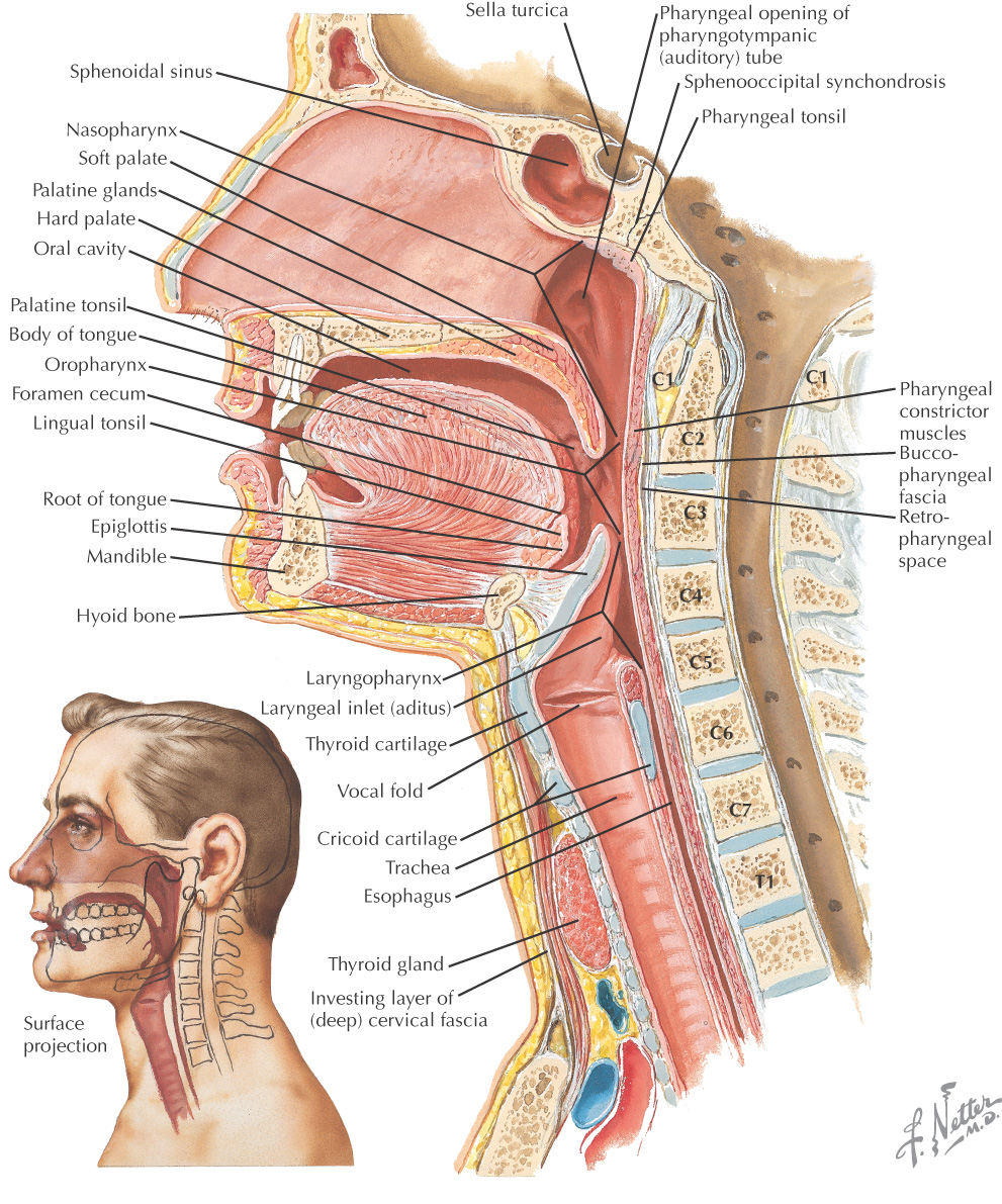
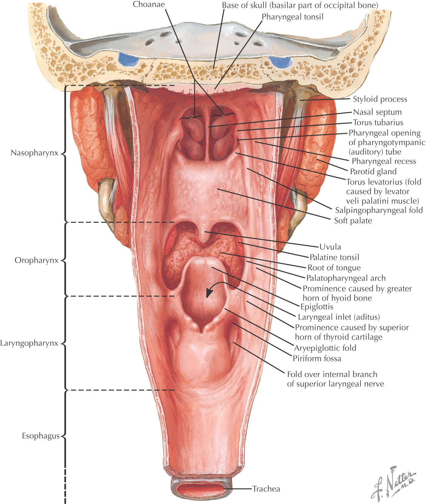
Overview and Topographic Anatomy
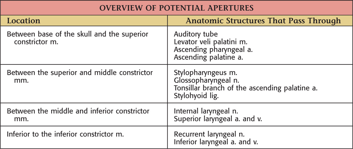
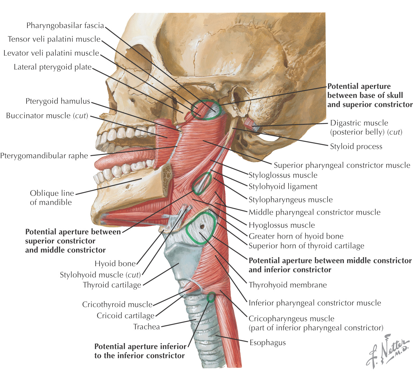
Potential Apertures in Pharyngeal Wall
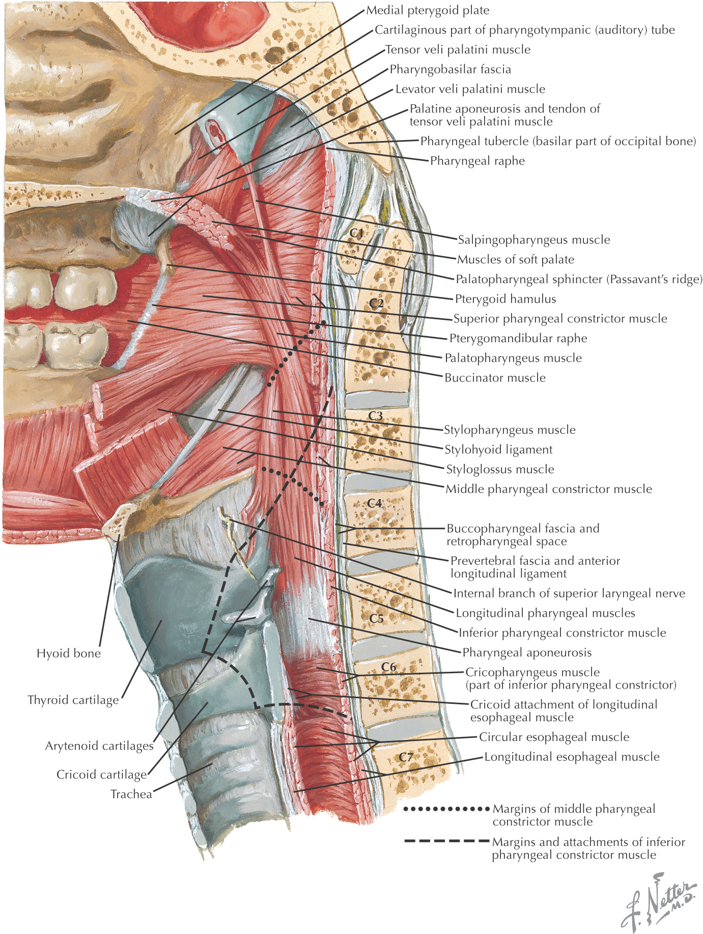
Pharynx: 5-inch muscular tube from base of the skull to the lower border of the cricoid cartilage (C6)
Posterior portion of the pharynx lies against the prevertebral fascia
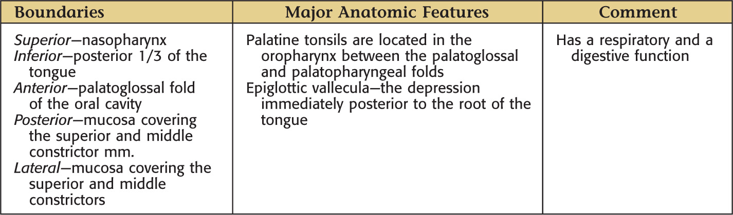
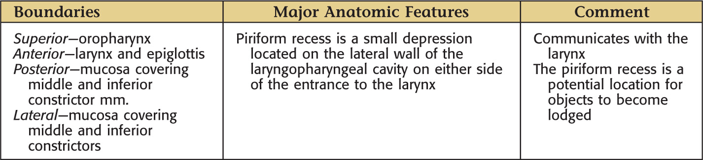
Lies posterior to the nasal and oral cavities and the larynx and thus is divided into 3 parts:
Responsible for properly conducting food to the esophagus and air to the lungs
Composed of:
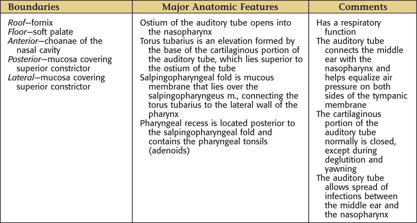
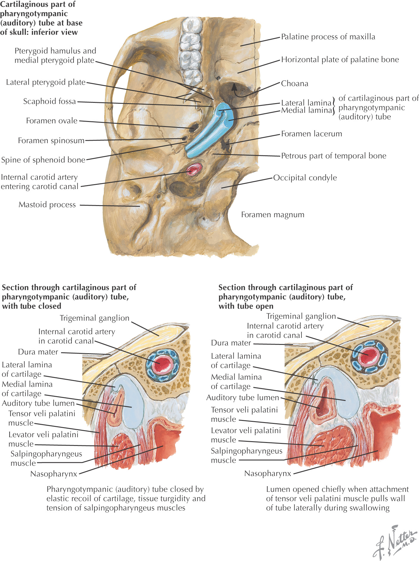
• Cartilaginous part of the pharyngotympanic tube
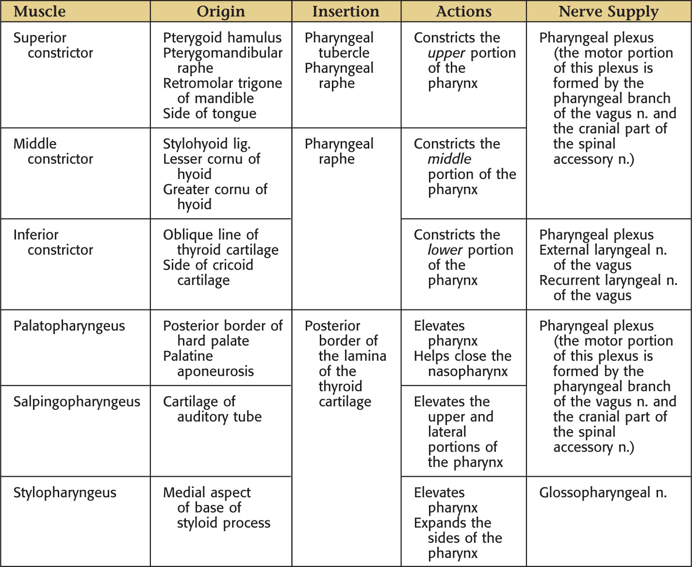
The wall of the pharynx has 5 layers:
• Mucous membrane—the innermost layer
• Pharyngobasilar fascia—the fibrous layer attached to the skull anchoring the pharynx
• Muscular—3 inner longitudinal and 3 outer circular (constrictor) muscles that overlap such that the superior constrictor is the innermost, whereas the inferior constrictor is the outermost muscle
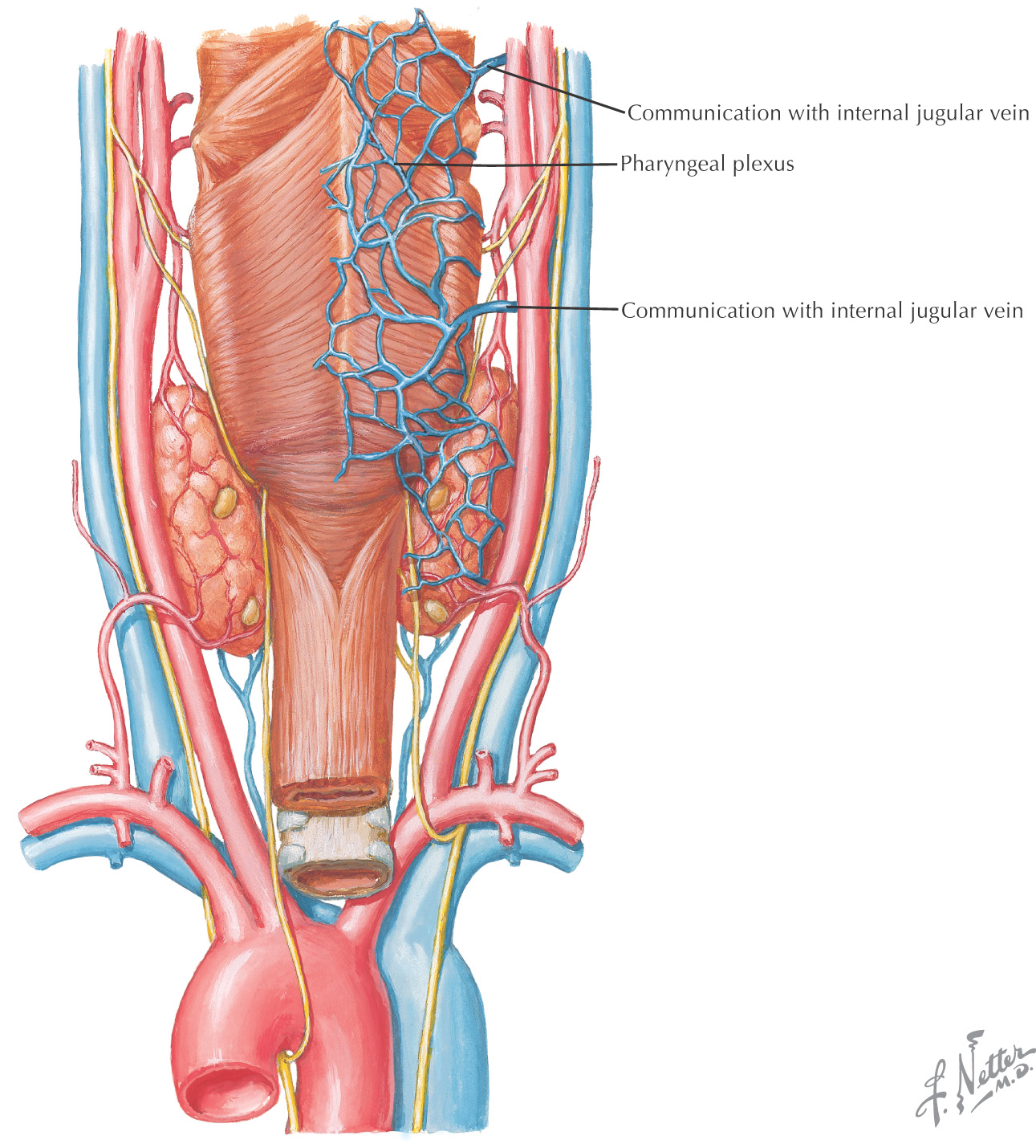
• Buccopharyngeal fascia—loose layer of connective tissue continuous with the fascia over the buccinator and pharyngeal muscles, and the location of the pharyngeal plexus of nerves and the pharyngeal plexus of veins
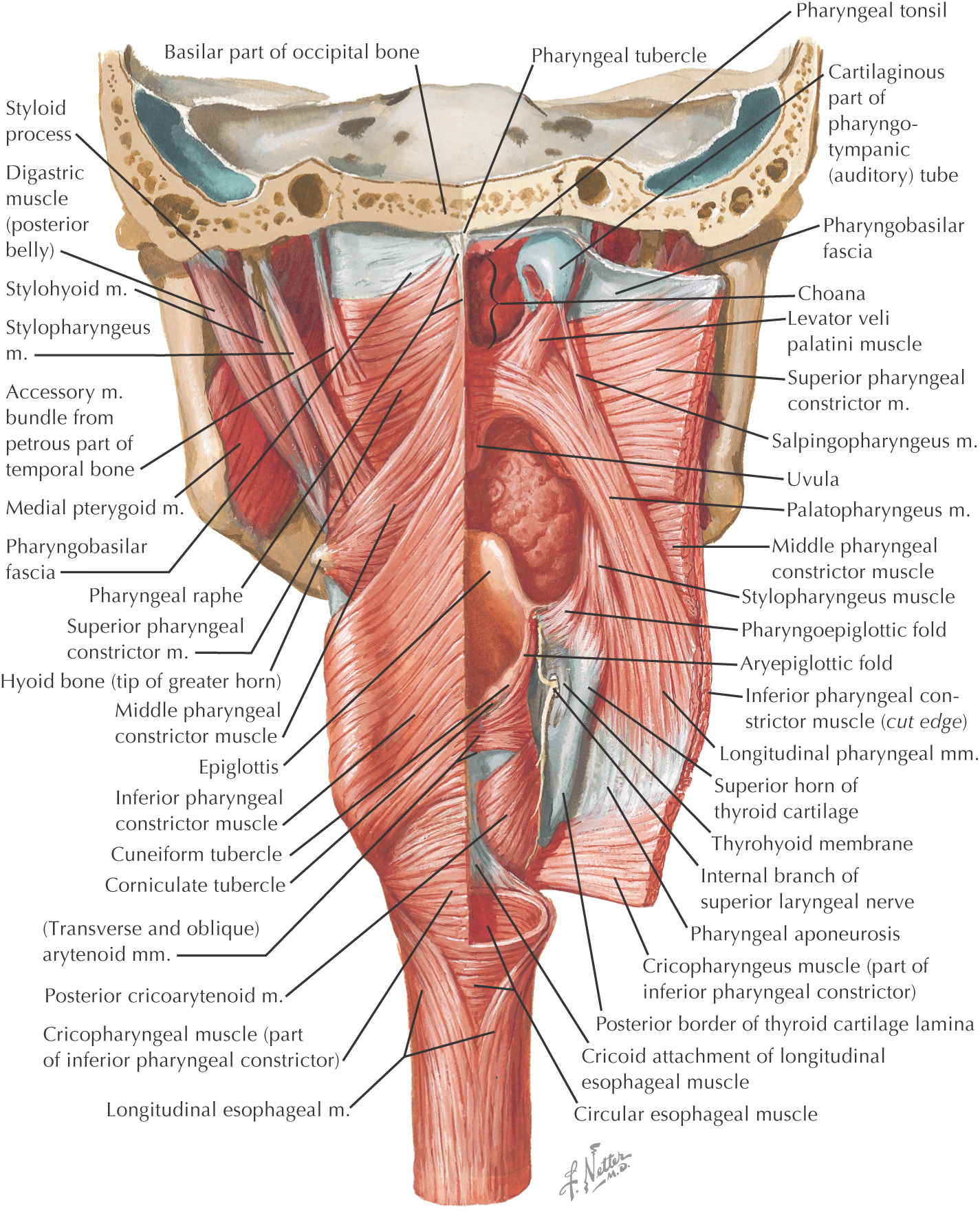
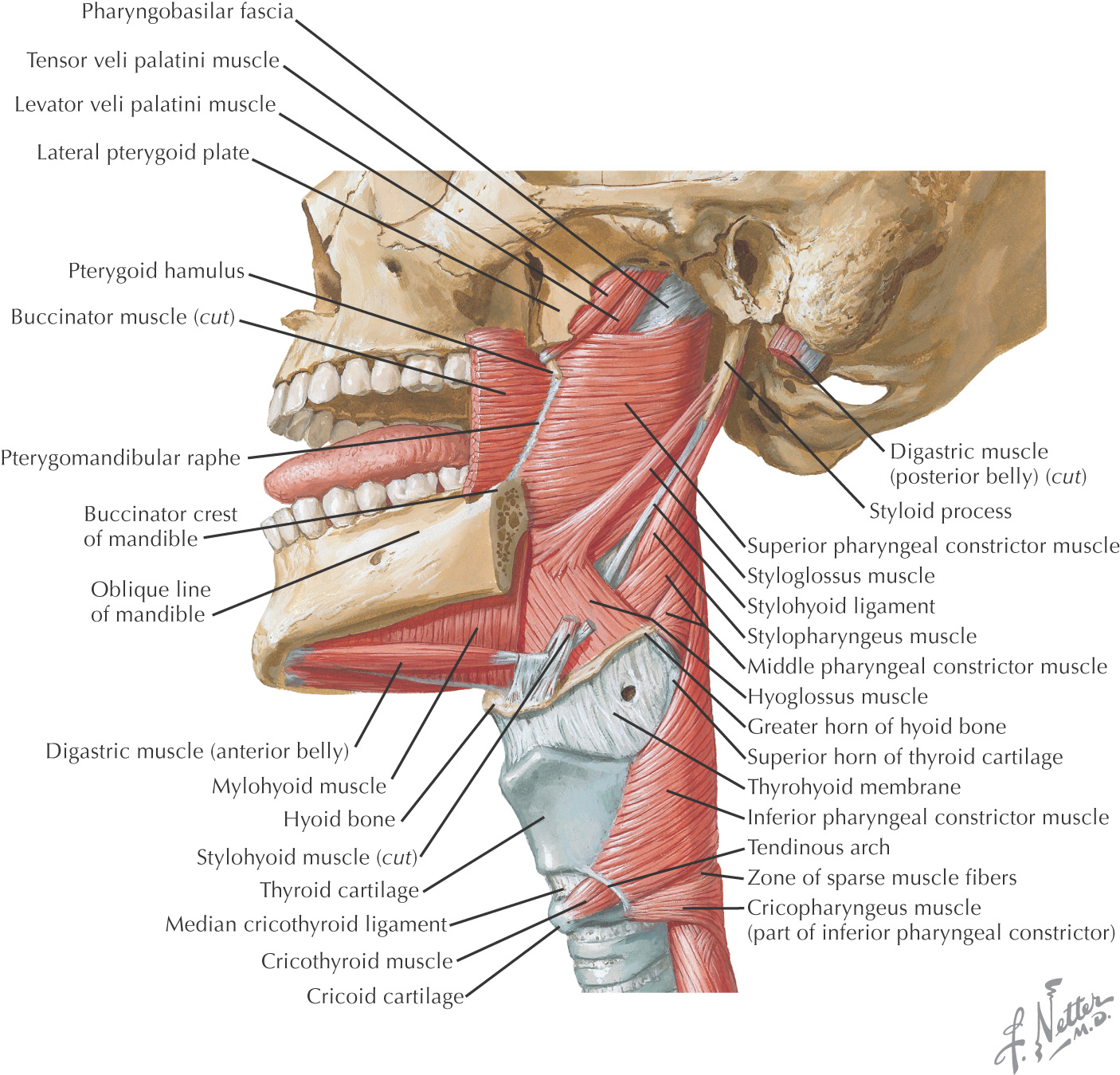
The overlapping arrangement of the 3 constrictor muscles leaves 4 potential apertures in the pharyngeal musculature
Anatomic structures enter and exit the pharynx through these potential apertures
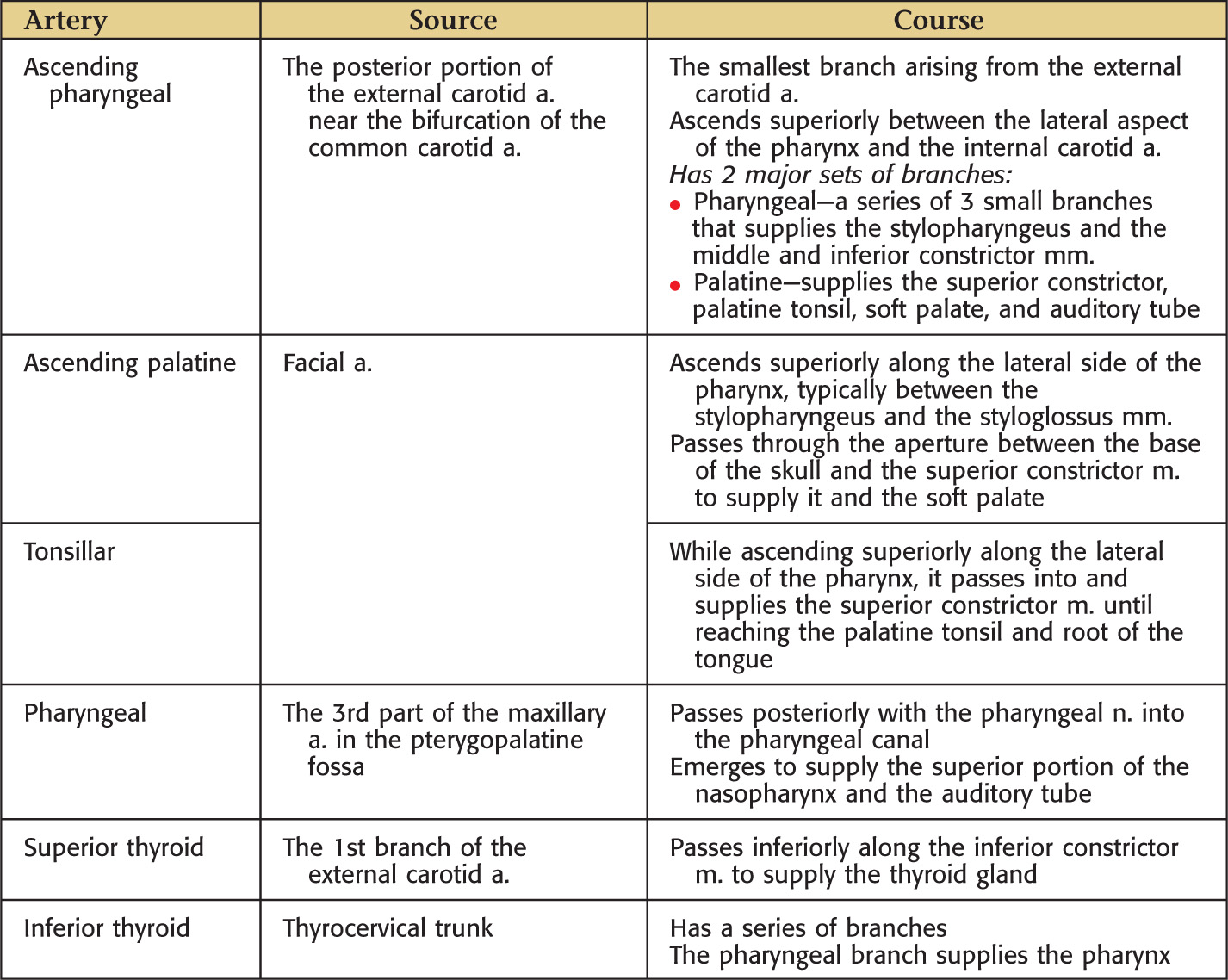
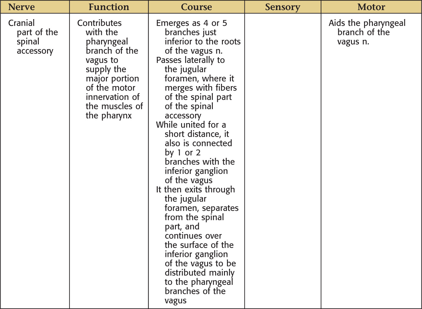
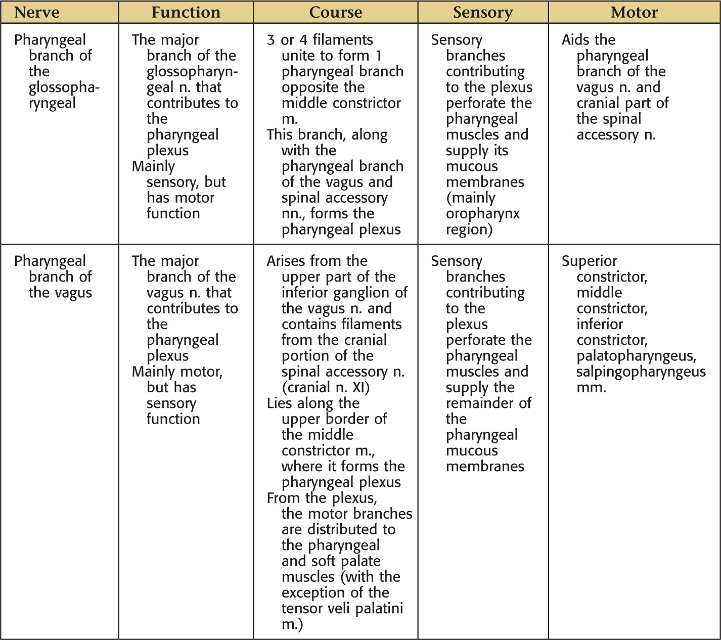
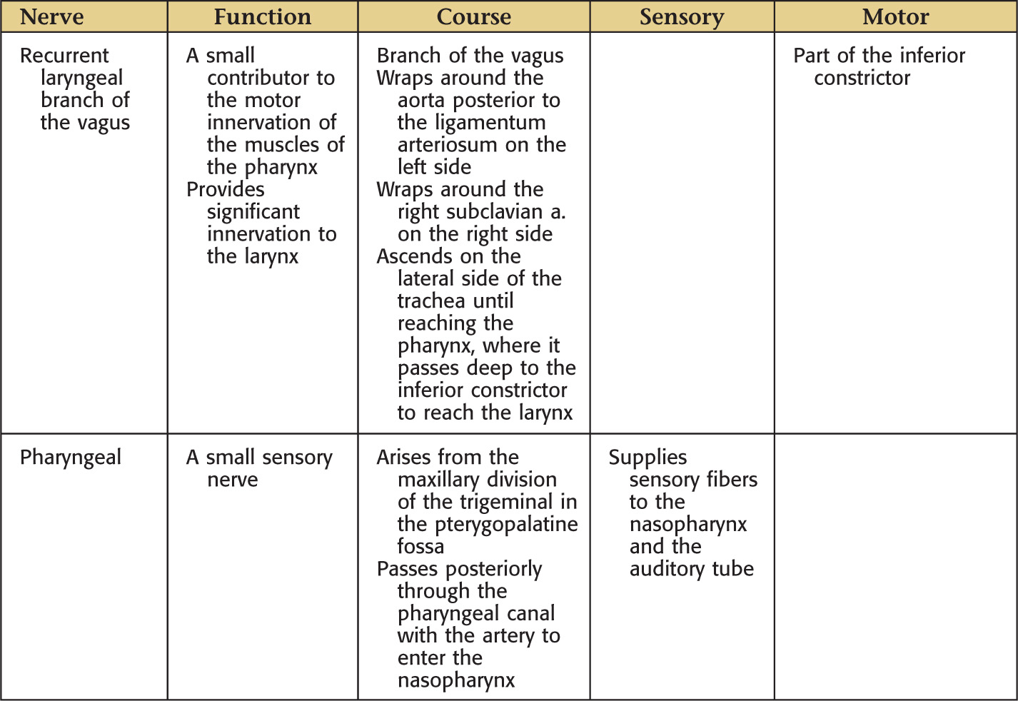
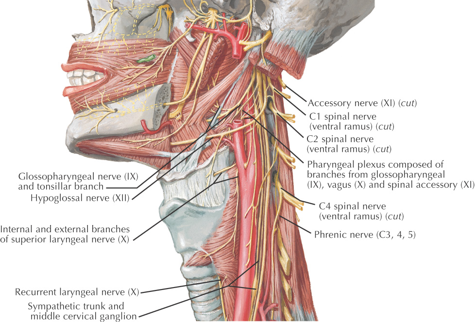
Supplies motor and sensory innervation to most of the pharynx
Composed of:
• Pharyngeal branch of the glossopharyngeal nerve
• Pharyngeal branch of the vagus nerve
• Cranial part of the spinal accessory nerve
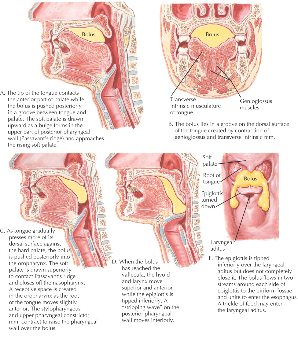
Deglutition, or swallowing, is a combination of voluntary and involuntary muscular contractions to move a bolus of food from the oral cavity to the esophagus
Deglutition begins when the tip of the tongue is placed into contact with the anterior portion of the palate and the bolus is pushed posteriorly
The soft palate begins to elevate, and Passavant’s ridge starts to form in the posterior wall of the pharynx and moves closer to the soft palate
As more of the tongue is pushed against the hard palate, the bolus is moved into the oropharynx, and the soft palate makes contact with Passavant’s ridge to close off the nasopharynx from the oropharynx
Once the bolus reaches the epiglottic vallecula, the hyoid and larynx are elevated and the tip of the epiglottis is tipped down slightly over the laryngeal aditus
A “stripping wave” is created on the posterior wall of the pharynx to help move the bolus
Bolus splits into 2 streams that flow on either side of the epiglottis and unite to enter the esophagus
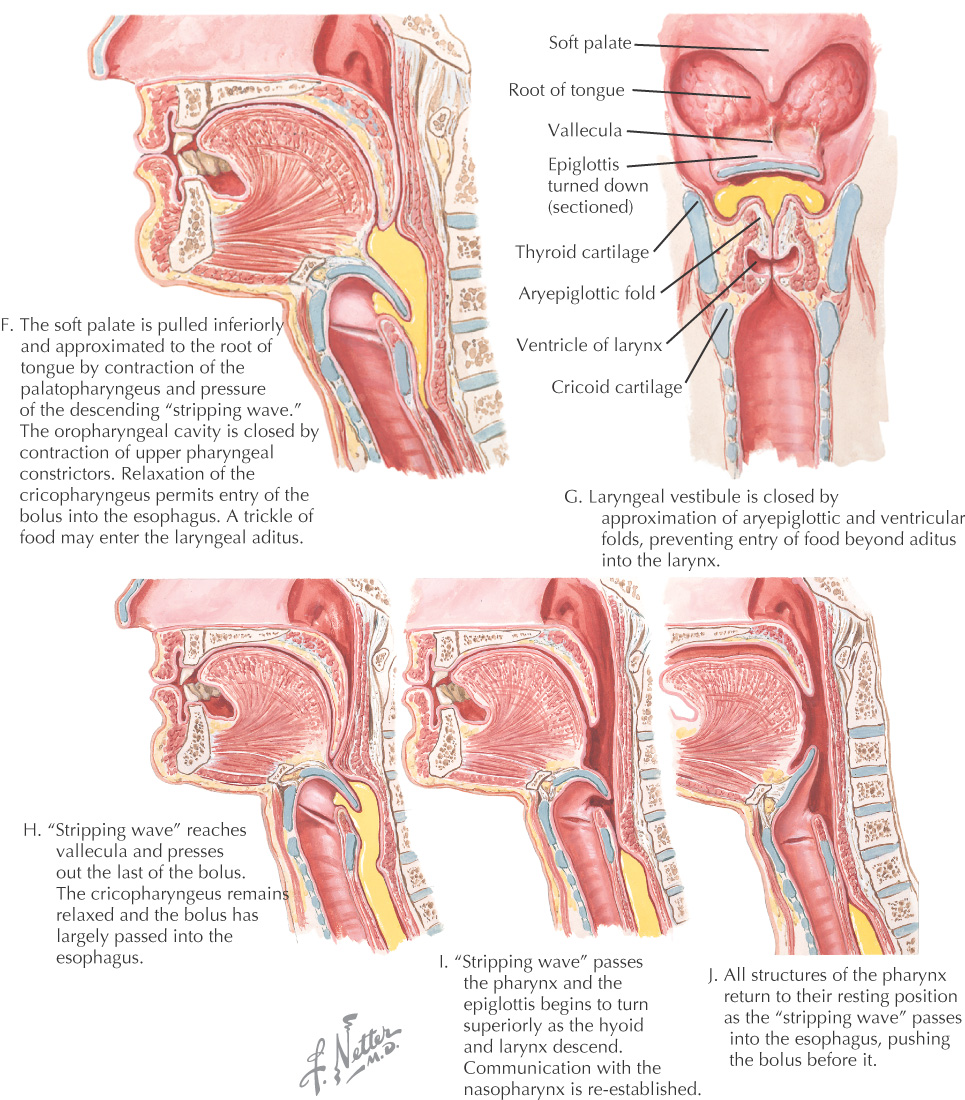
The soft palate is pulled down by the palatopharyngeus muscles and the pressure of the wave from the movement of the bolus, while the stripping wave continues to help move the bolus from the oropharynx
The cricopharyngeal portion of the inferior constrictor relaxes to help the bolus enter the esophagus
Laryngeal vestibule and rima glottidis are closed to prevent the bolus from entering the larynx
Stripping wave empties the last of the bolus from the epiglottic vallecula, and the major portion of the bolus is already in the esophagus
All structures return to their initial position as the stripping wave moves into the esophagus