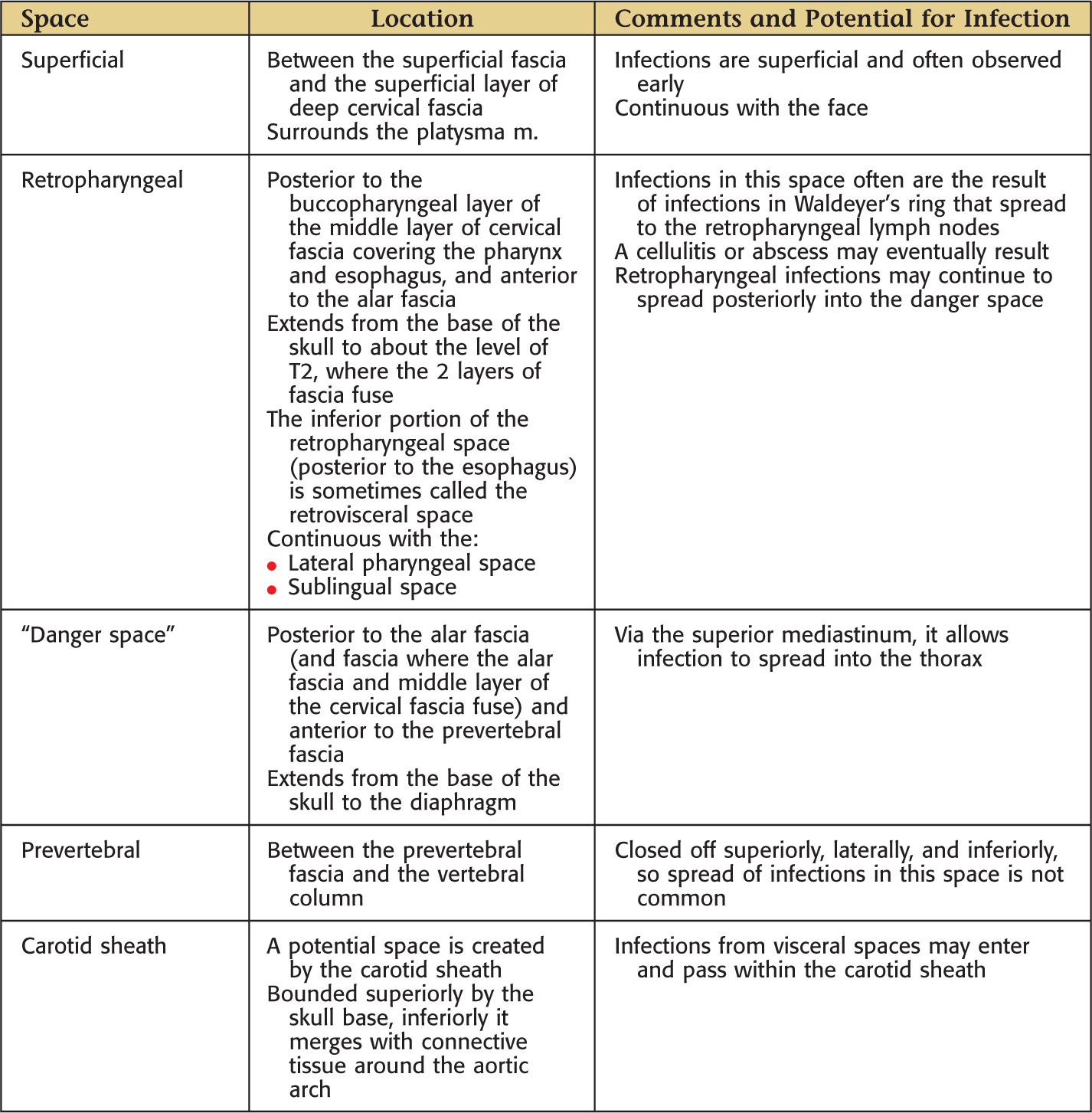
Overview and Topographic Anatomy
Fascia: a band of connective tissue that surrounds structures (such as enveloping muscles), giving rise to potential tissue spaces and pathways that allow infection to spread
Immediately deep to the skin
Contains fat
Deep to the superficial fascia
Aids muscle movements
Provides passageways for nerves and vessels
Provides attachment for some muscles
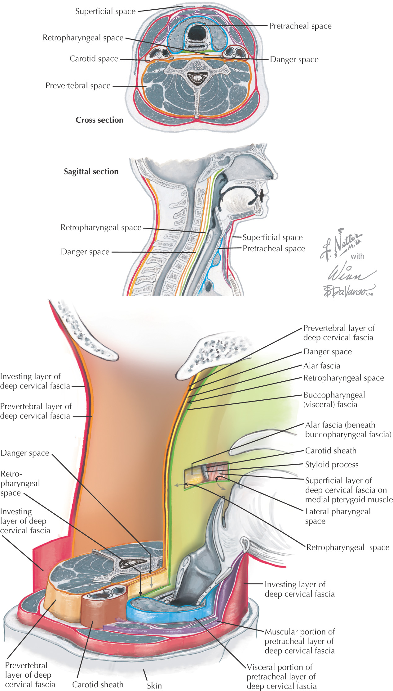
In the neck, it is divided into 4 regions:
• 2 neurovascular compartments
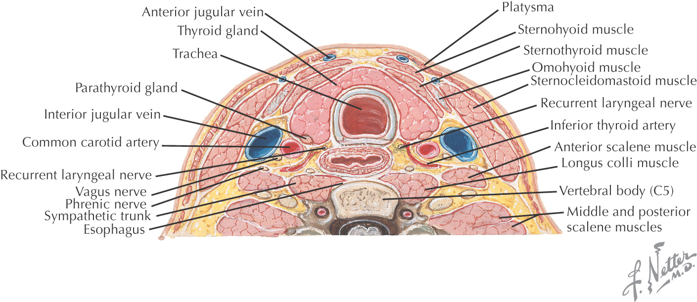
Also divided into 4 layers:
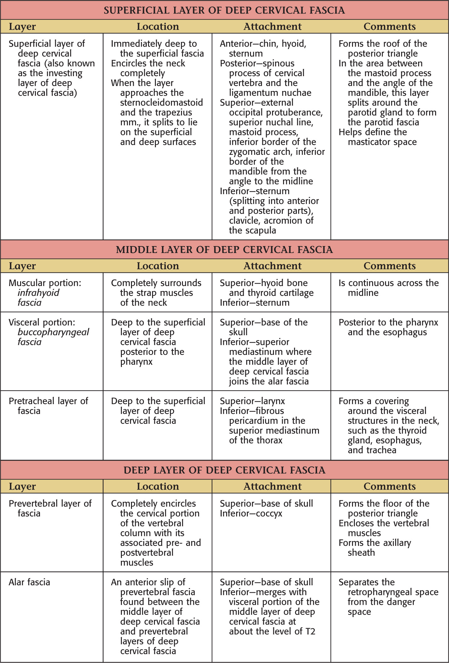
• Superficial layer of deep cervical fascia (investing layer of deep cervical fascia)
• Middle layer of deep cervical fascia
• Deep layer of deep cervical fascia
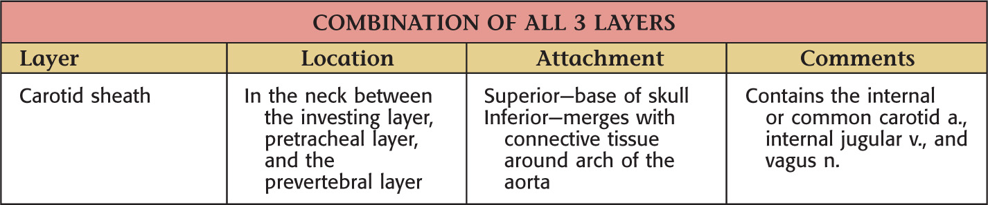
• Carotid sheath (composed by the contribution of all 3 layers of deep cervical fascia)
There is no deep fascia in the face, which allows free spread of fluid
Superficial fascia lies deep to the skin and contains the cutaneous vessels and nerves
In the neck, the platysma muscle lies within the superficial fascia
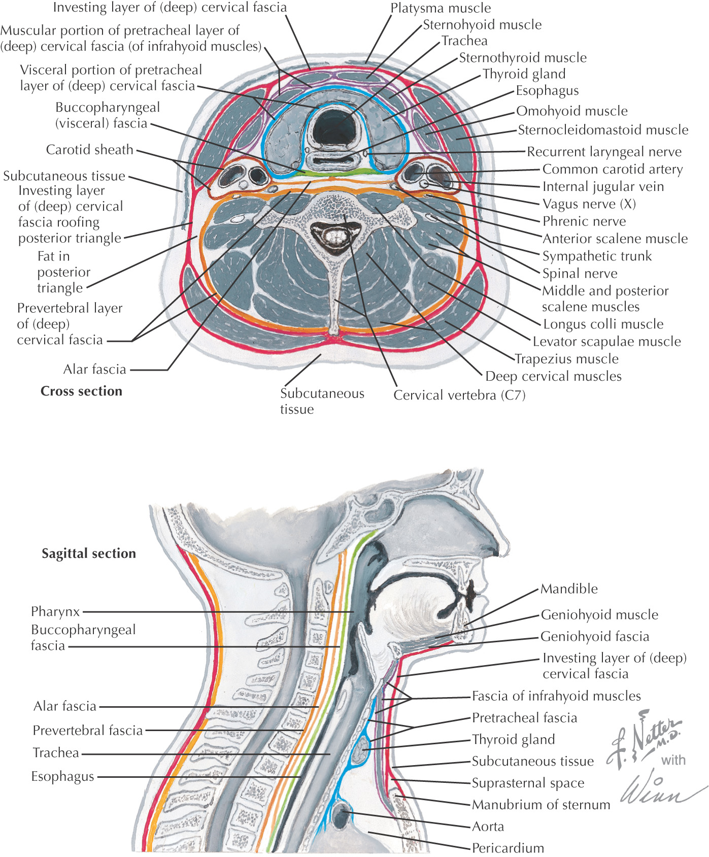
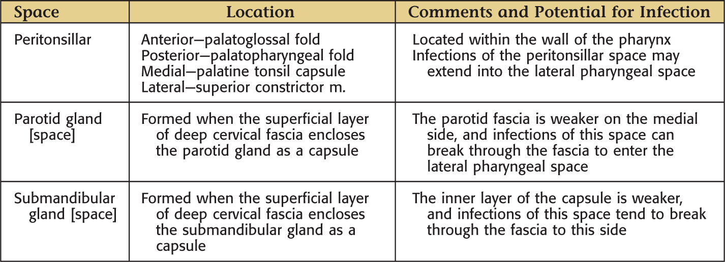
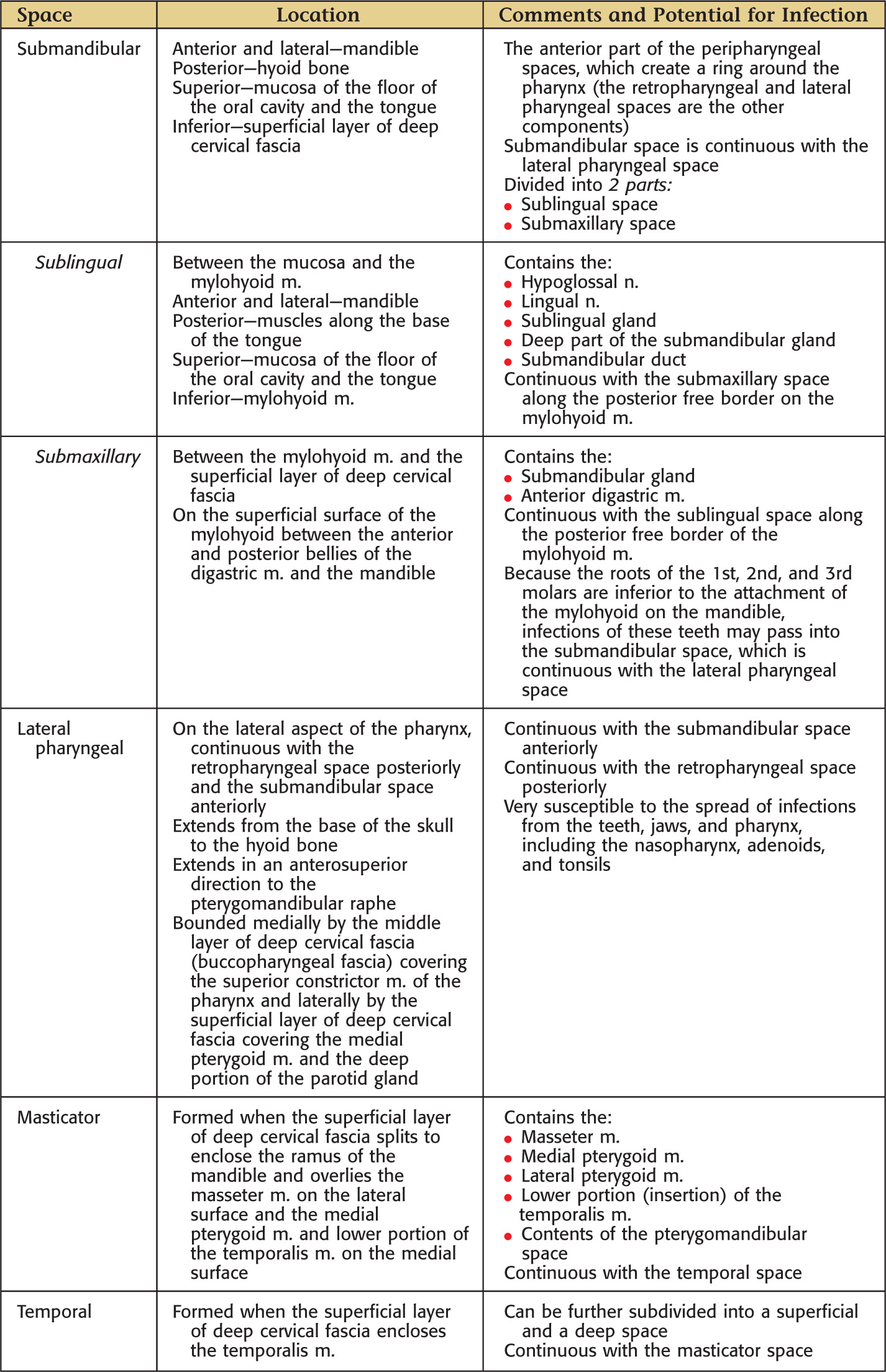
Layers of fascia “create” potential fascial spaces
All are filled by loose areolar connective tissue
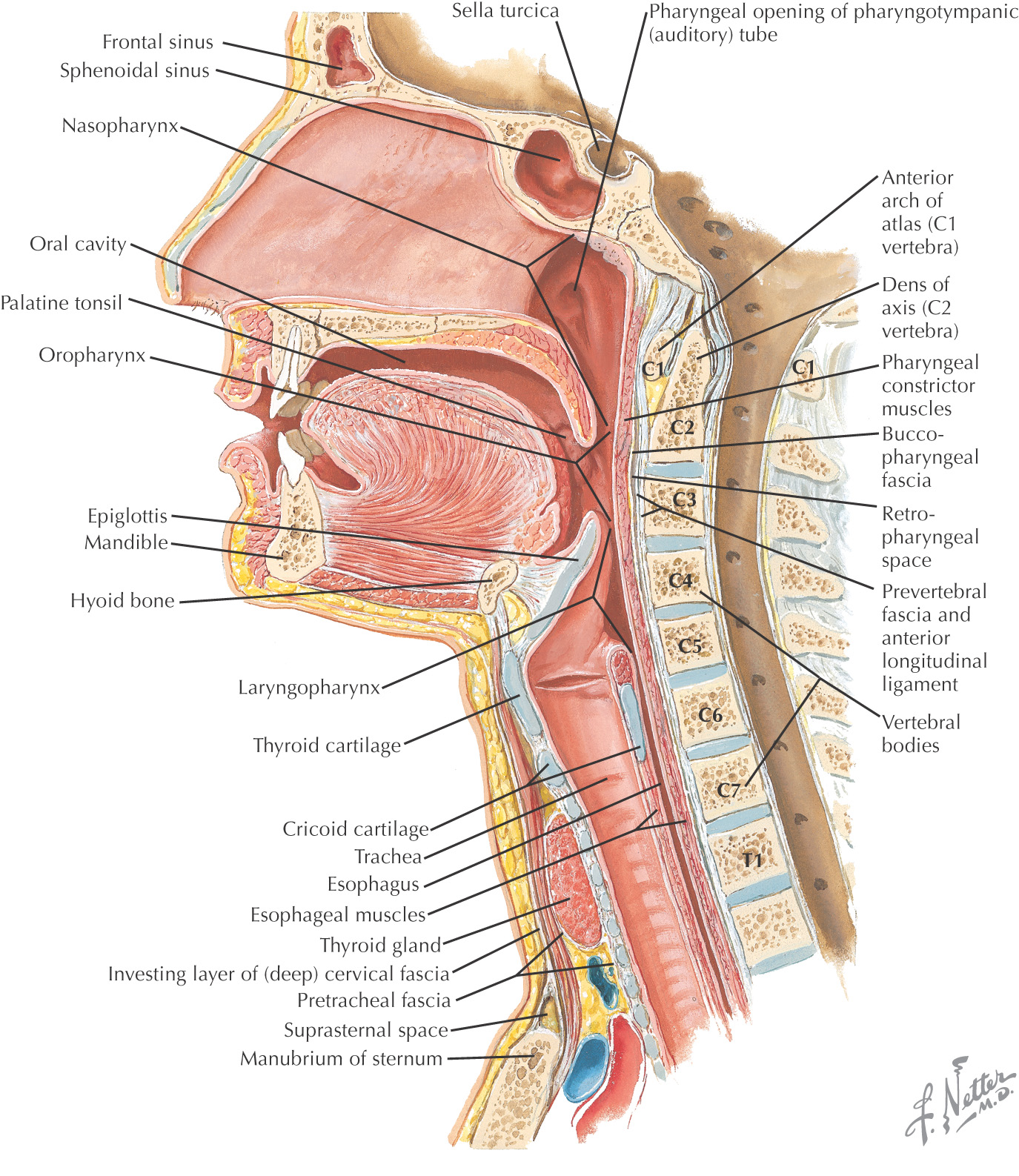
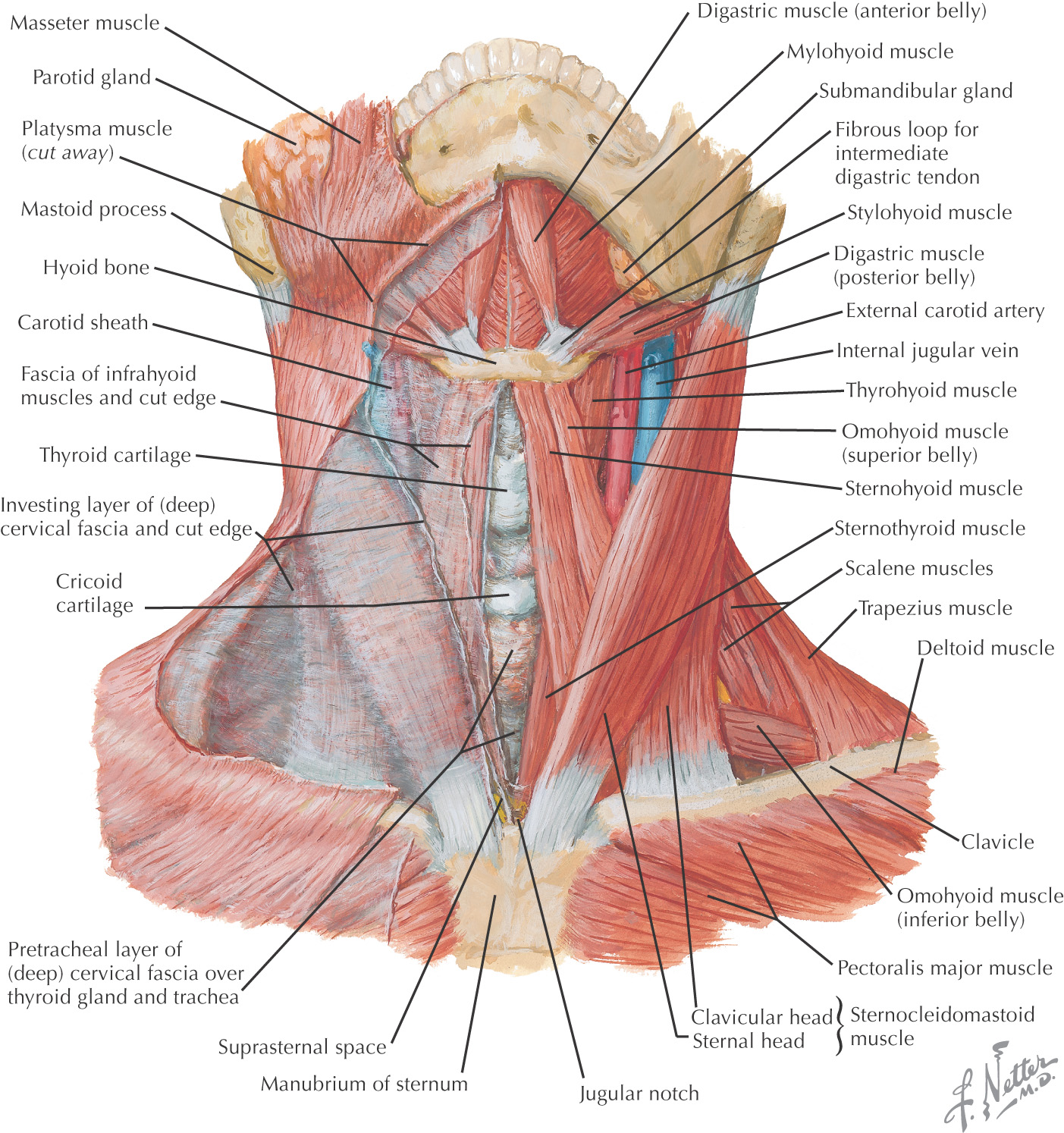
The hyoid bone is the most important anatomic structure in the neck that limits the spread of infection
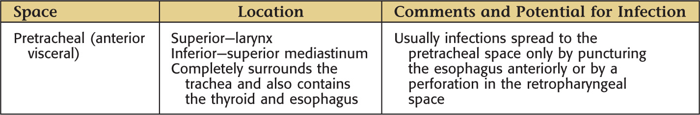
Most are divided into spaces in relation to the hyoid bone:
Infections or other inflammatory conditions spread by the path of least resistance to reach the fascial spaces
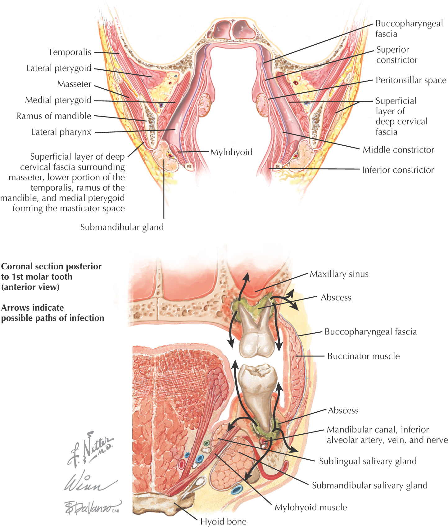
A severe cellulitis due to bacterial infection (usually from Streptococcus, Actinomyces, Prevotella, Fusobacterium, or Staphylococcus) in the floor of the oral cavity under the tongue
Often begins in the sublingual and submandibular spaces after infection of the premolar teeth or, more commonly, molar teeth (such as an abscess of a mandibular molar) because their roots extend inferior to the mylohyoid line of the mandible
May follow the planes of the fascial spaces to spread in the neck
May cause sufficient neck swelling to block the airway
More common in children
Antibiotic therapy, incision of the neck to drain the infection, and excision of the infected tooth are the possible treatments.
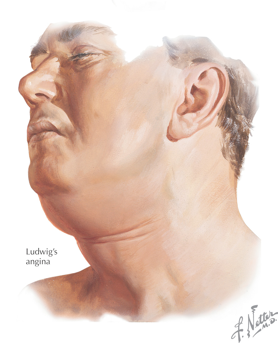
May spread via the fascial planes of the neck to become more serious, such as in Ludwig’s angina
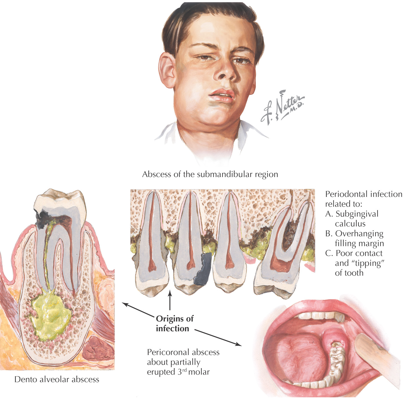
An acute lesion characterized by localization of pus in the structures surrounding the apex of a tooth
May originate in the dental pulp and be secondary to dental caries with erosion of enamel and dentin, or to traumatic injury to tooth, allowing bacteria to invade the dental pulp
Resulting pulpitis can progress to necrosis as bacteria invade the surrounding alveolar bone, causing formation of a local abscess
Typically involves the supporting structures of the teeth, such as the periodontal ligaments and alveolar bone, leading to formation of a local abscess
An inflammation around the crown of a tooth from an infection of the gingiva, leading to formation of an abscess
Most commonly affected tooth is a partially erupted 3rd mandibular molar
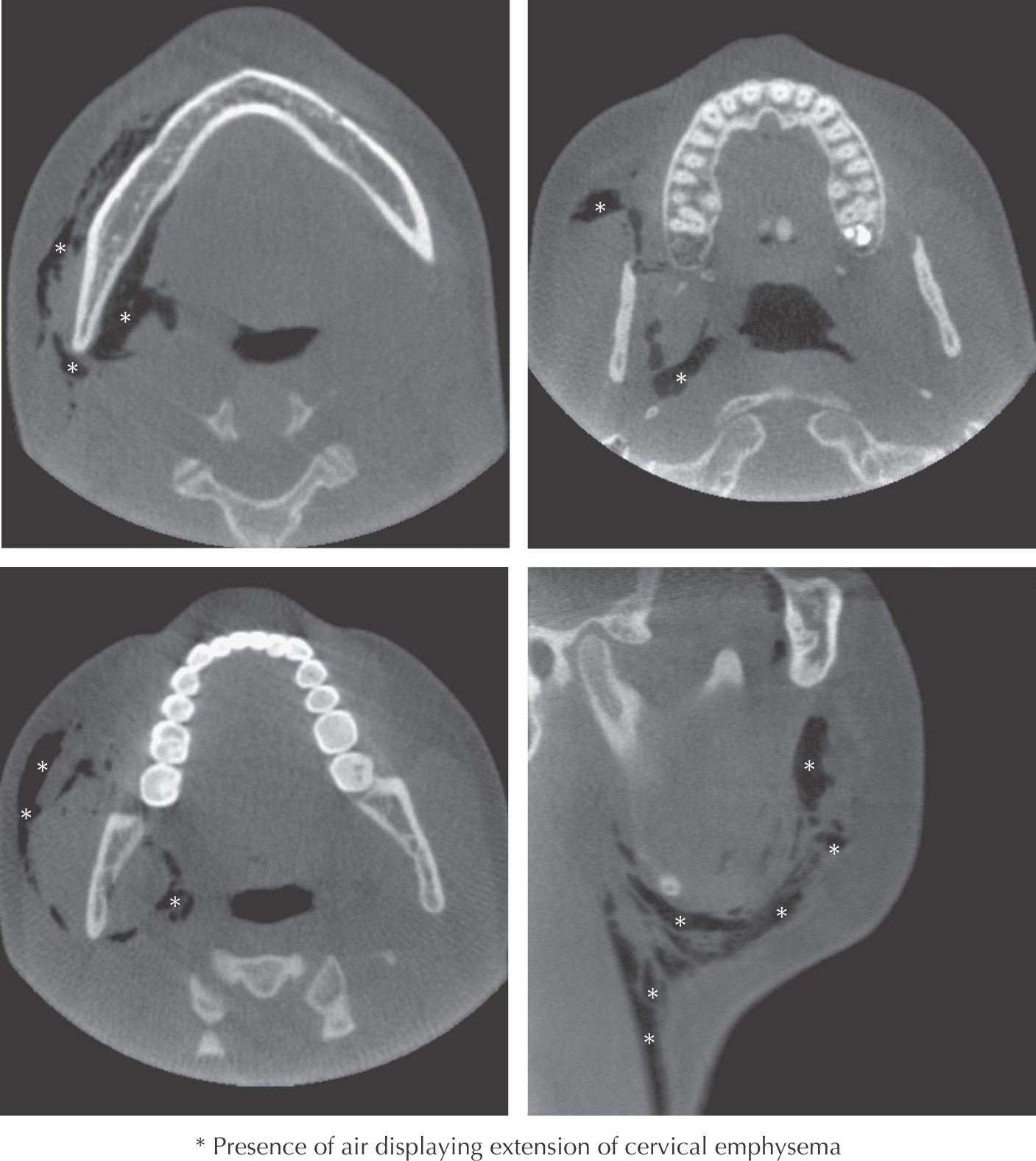
Introduction of gas deep to the skin which may be due to trauma, iatrogenic, or infection.
Some causes include fractures of the head and neck, introduction of air from a high speed dental drill, and surgical procedures such as root canals and extractions of mandibular 3rd molars
In the head and neck, cervical emphysema can spread via the fascial planes
May be benign or fatal, depending on the spread