Overview and Topographic Anatomy
Dual functions:
• Maintains the balance of the body (vestibular)
3 divisions:
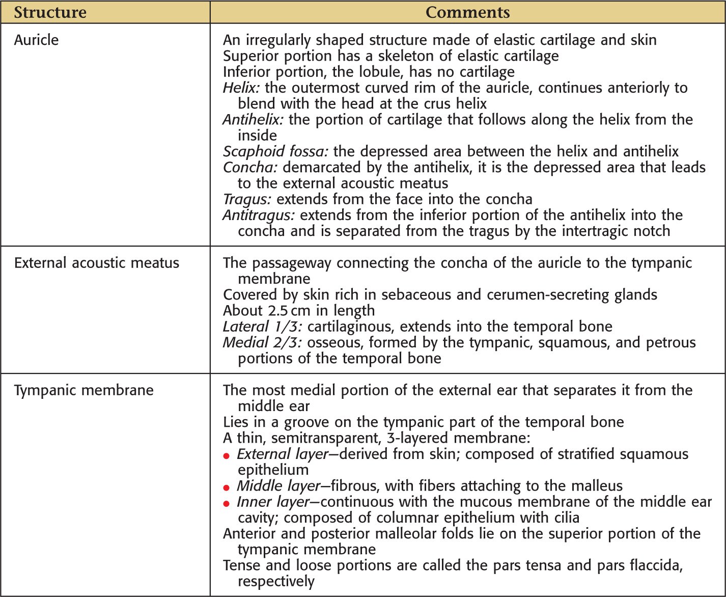
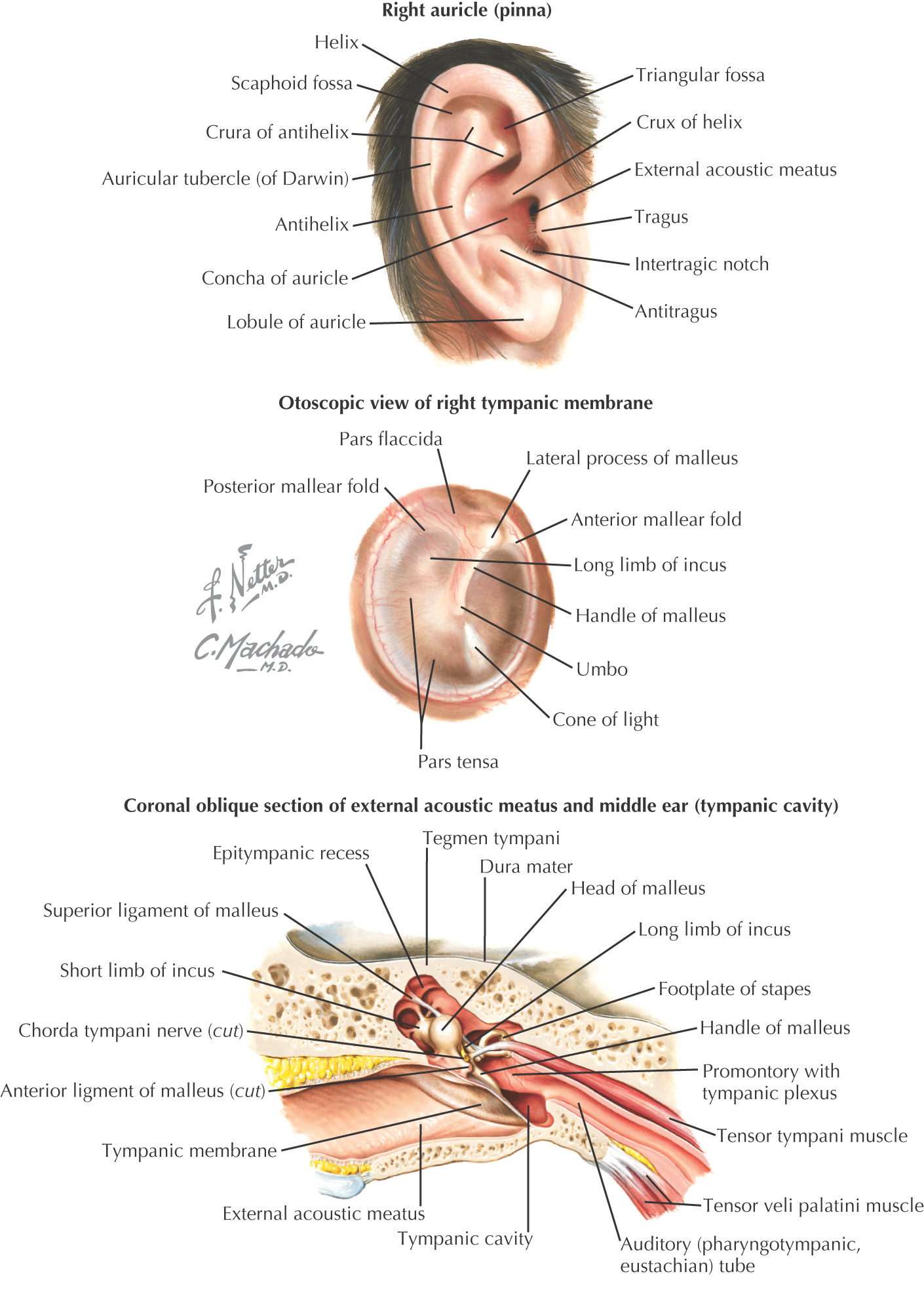
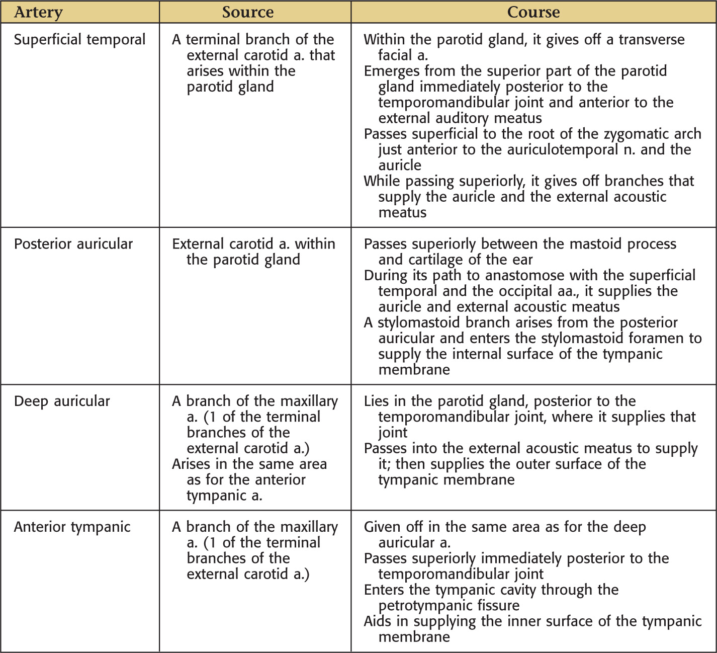
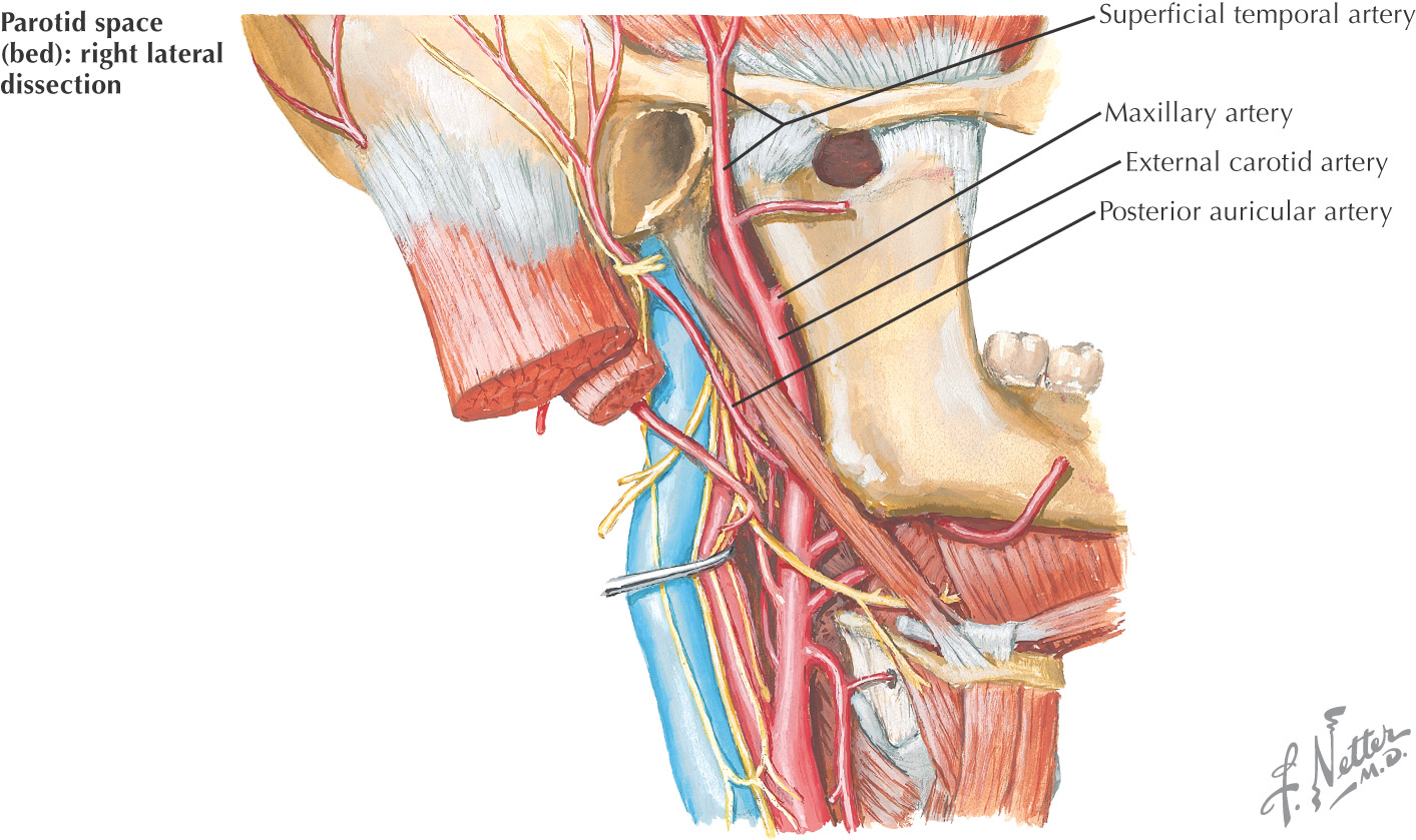
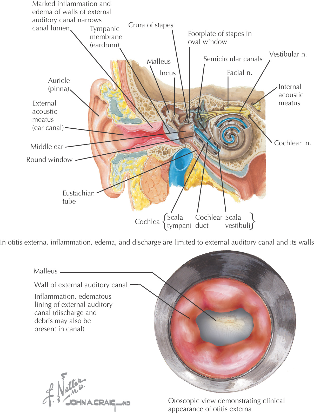
The most superficial portion of the ear, the external ear includes the auricle, external acoustic meatus, and the tympanic membrane
Helps gather sound and direct it to the tympanic membrane
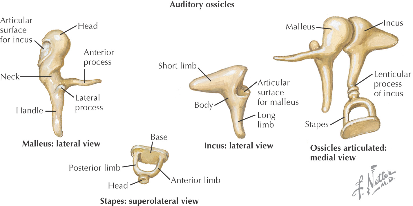
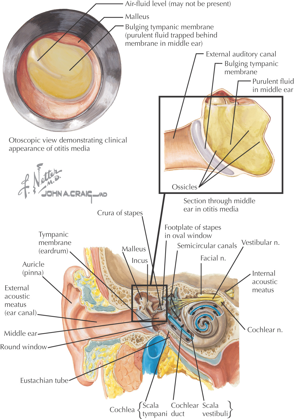
Transmits sound vibrations from the tympanic membrane to the inner ear via the ear ossicles: malleus, incus, and stapes
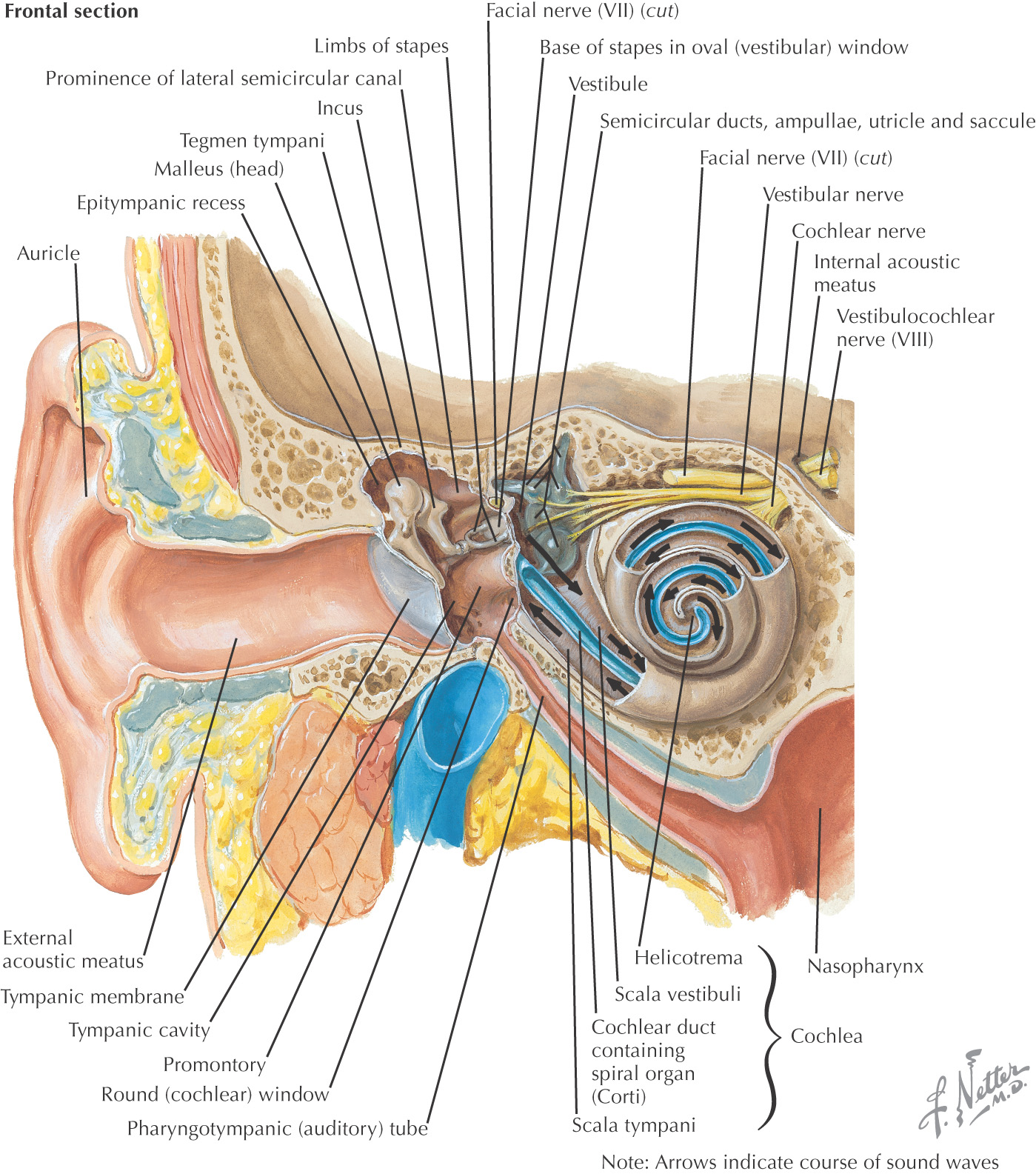
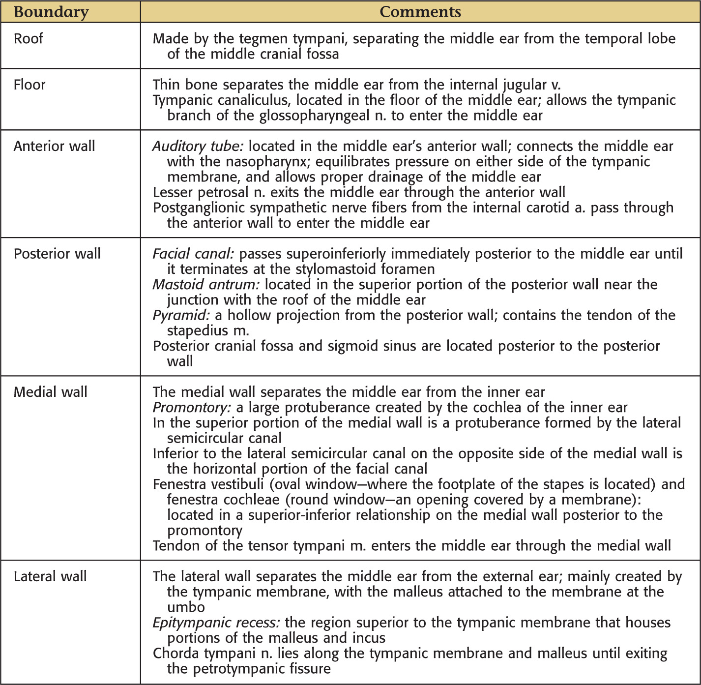
Mainly within the petrous portion of the temporal bone
General shape resembles a biconcave lens
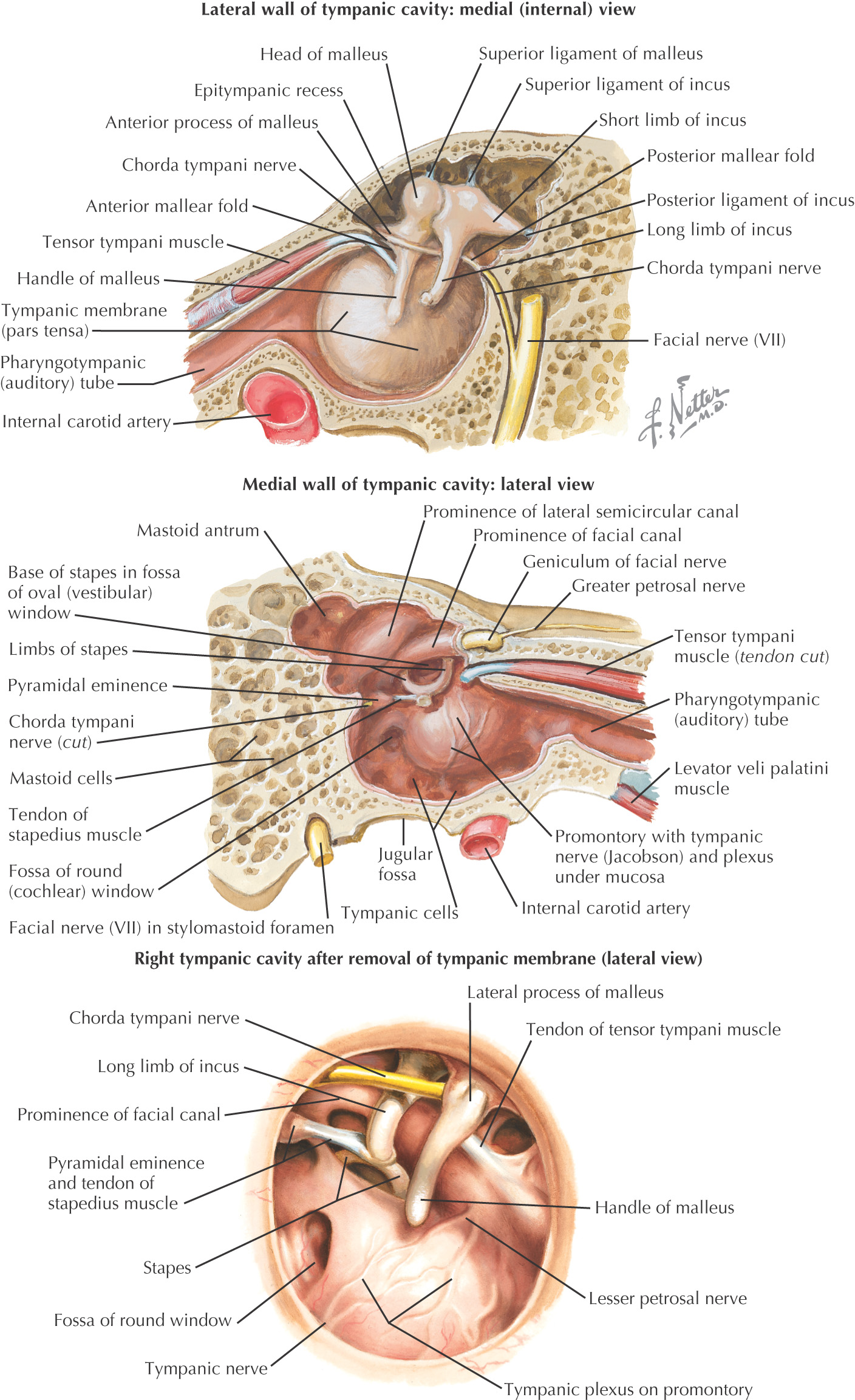
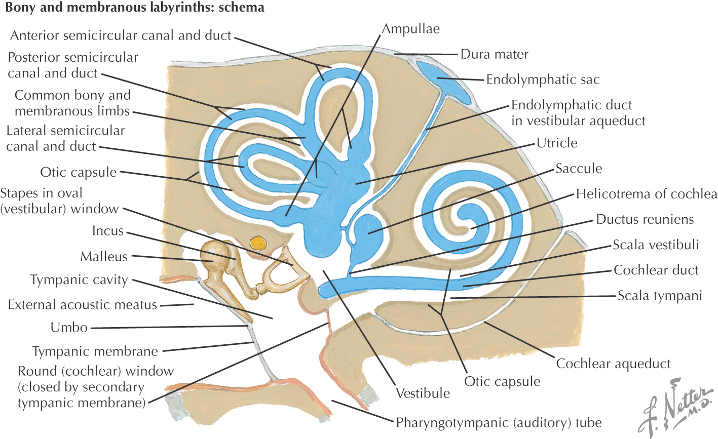
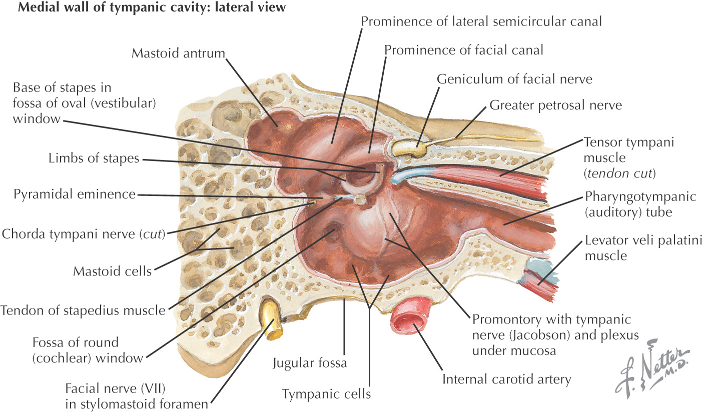
Composed of the tympanic cavity that connects anteriorly with the nasopharynx via the auditory tube and the mastoid air cells posteriorly
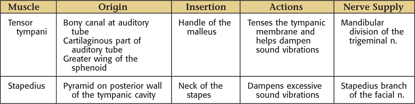
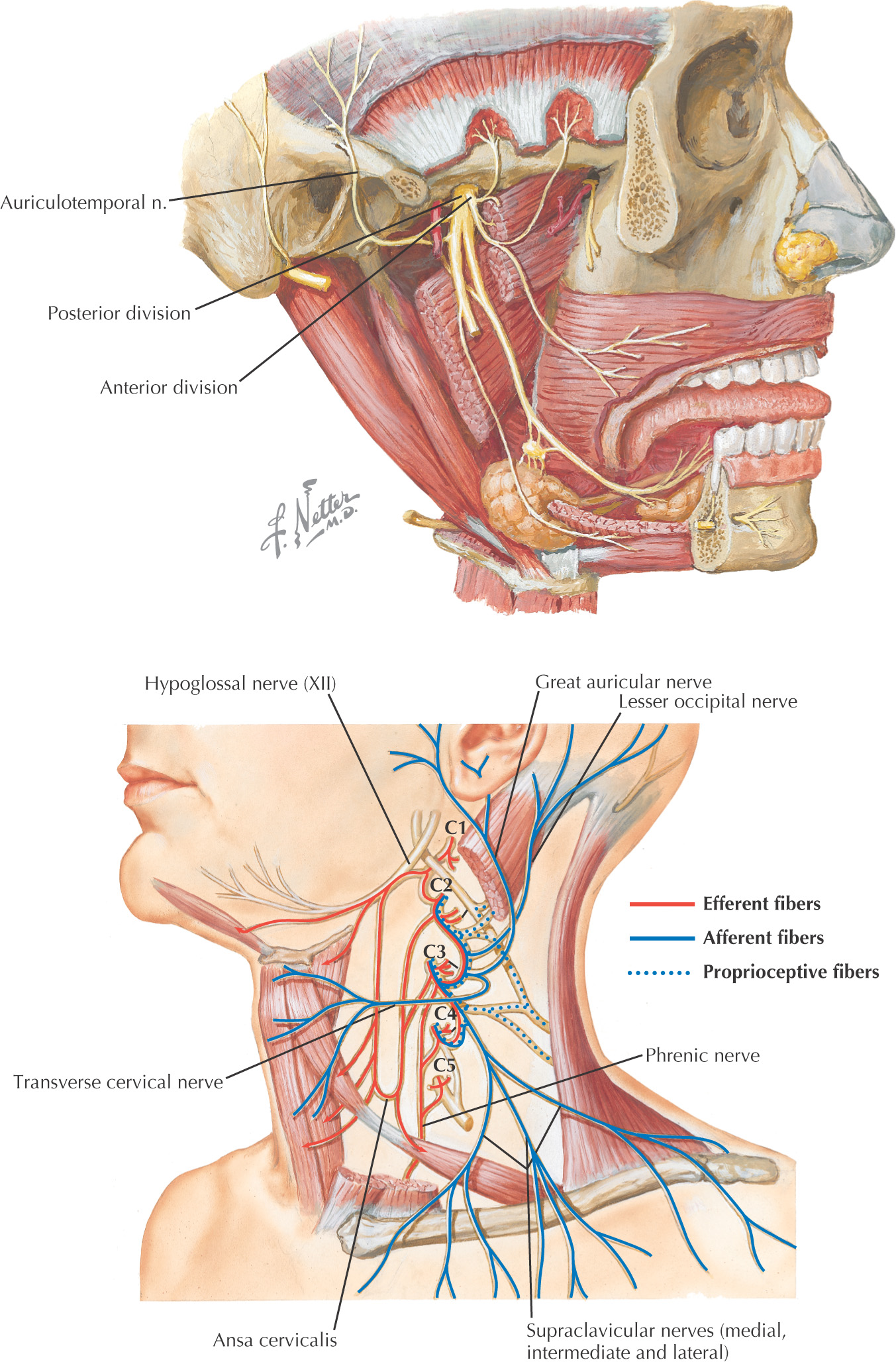
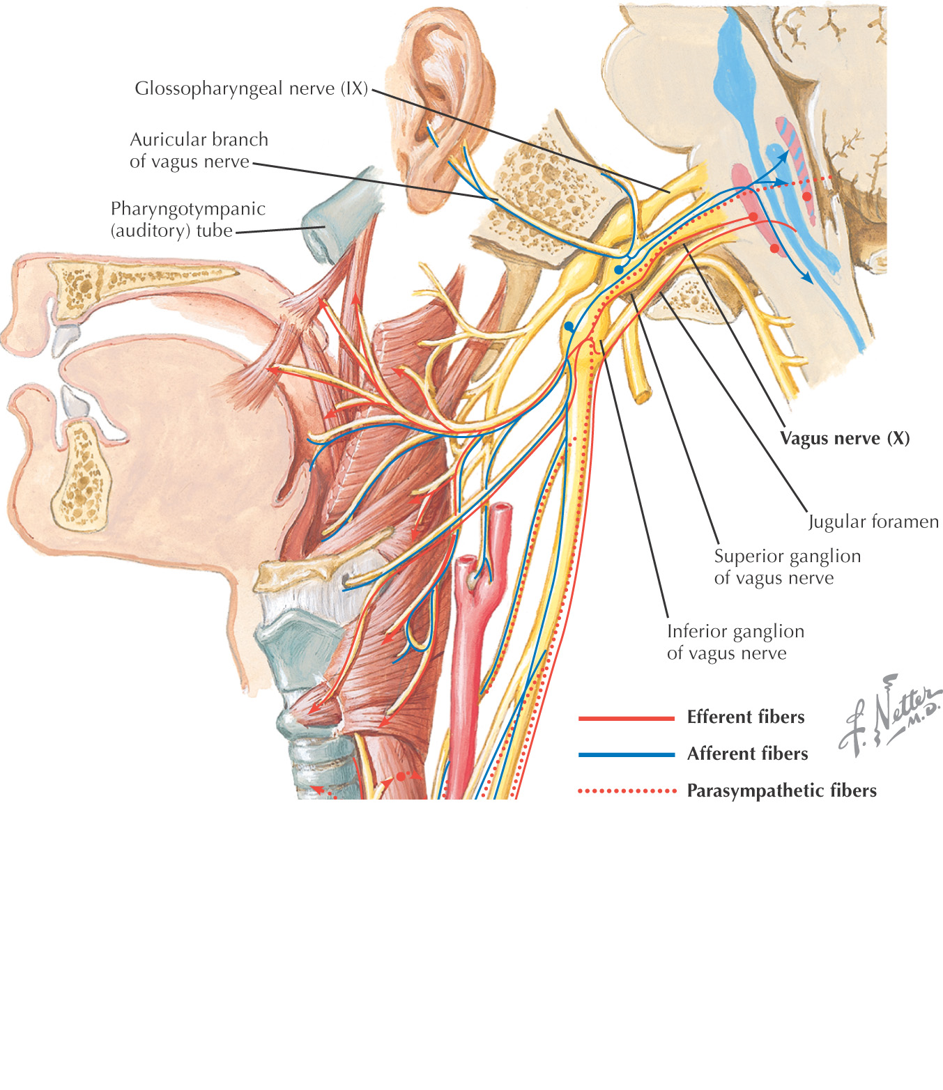
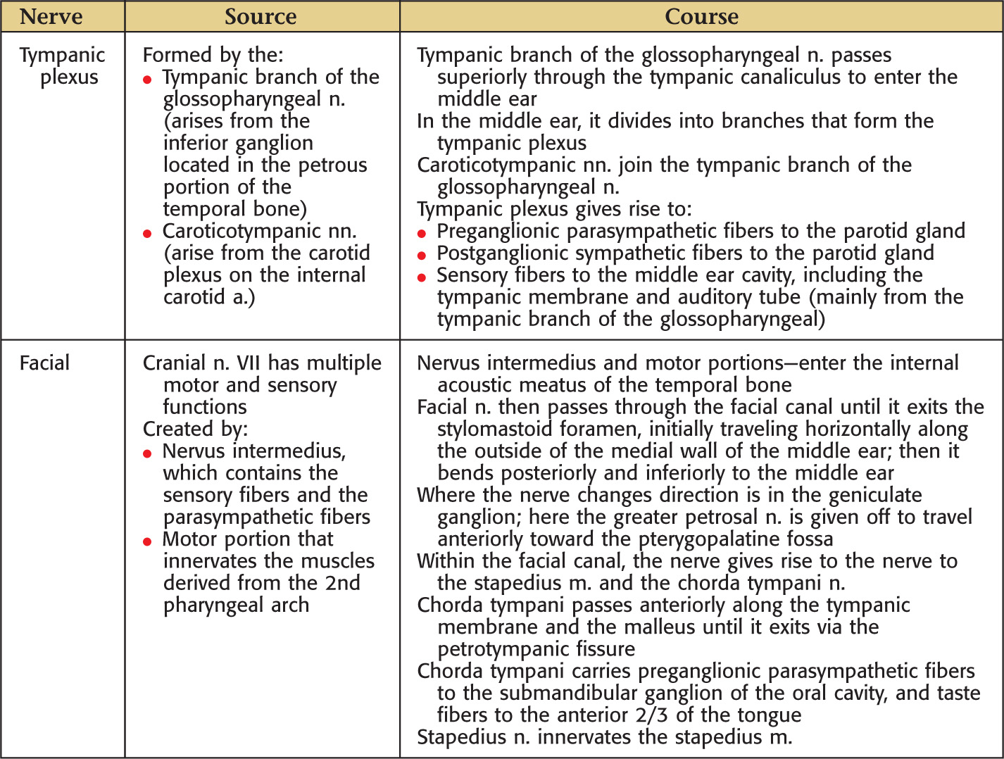
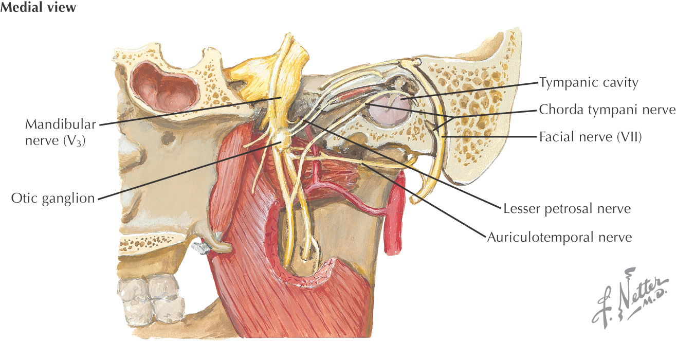
Tympanic cavity contains the ear ossicles (malleus, incus, and stapes), muscles (tensor tympani and stapedius muscles), nerves (chorda tympani, tympanic branch of the glossopharyngeal nerve, and lesser petrosal nerve), and tympanic plexus (parasympathetics from the glosspharyngeal nerve plus sympathetics from the superior cervical ganglion via the carotid plexus)
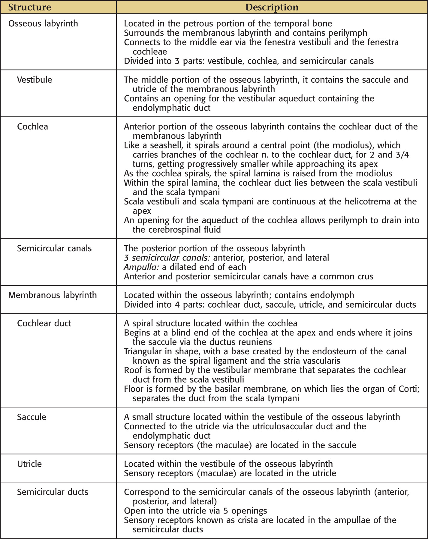
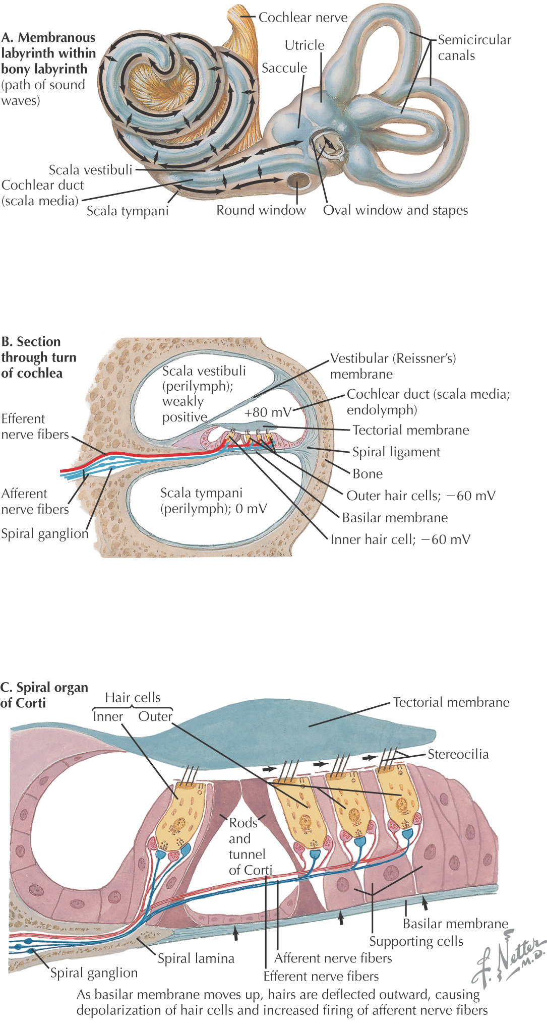
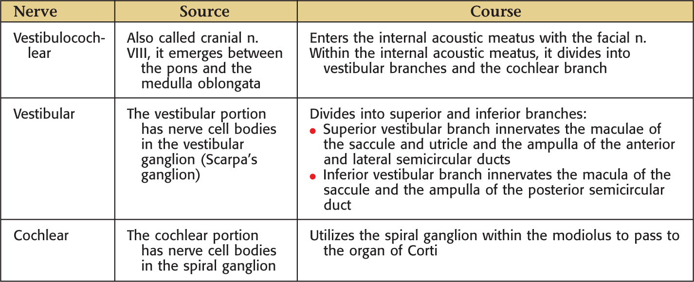
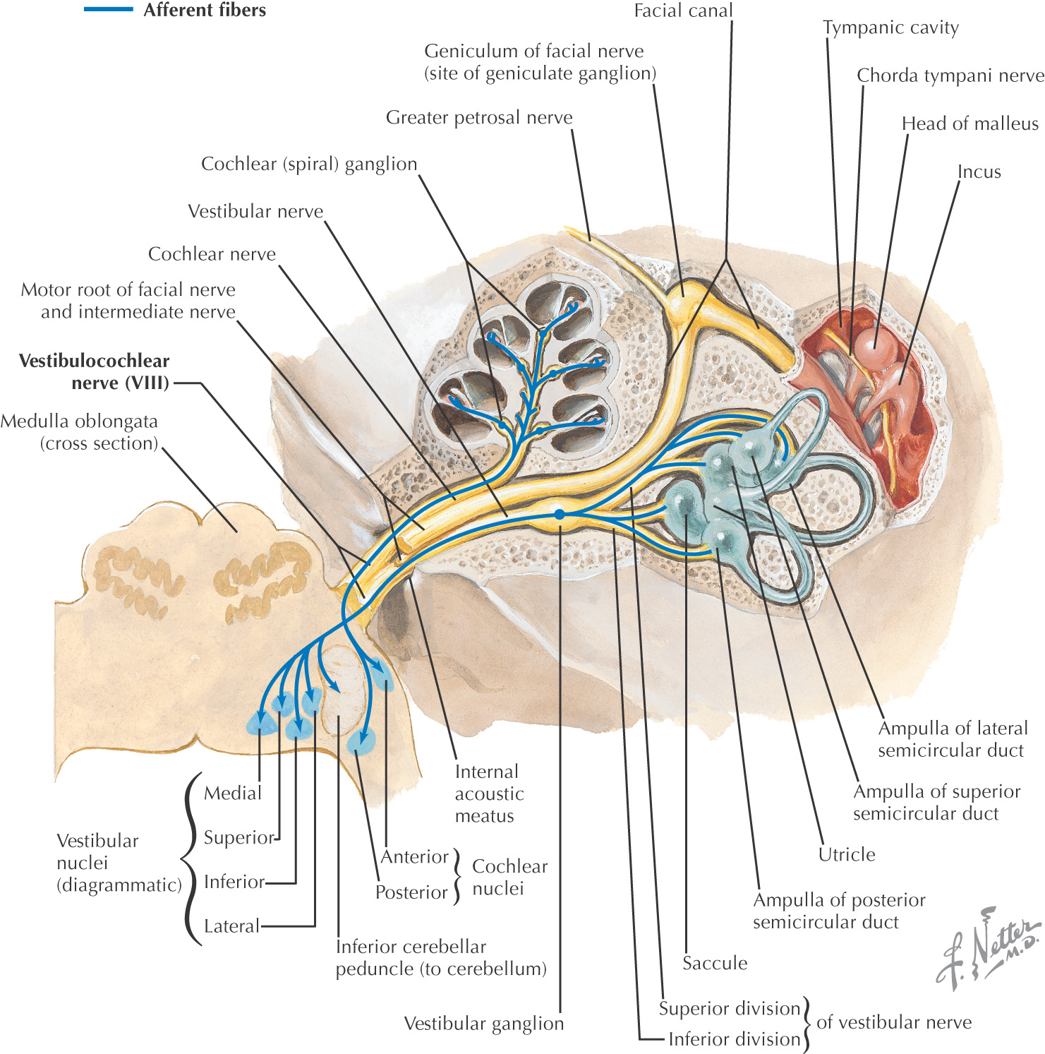
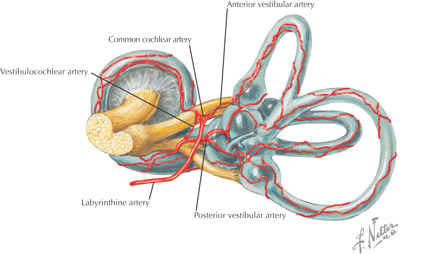
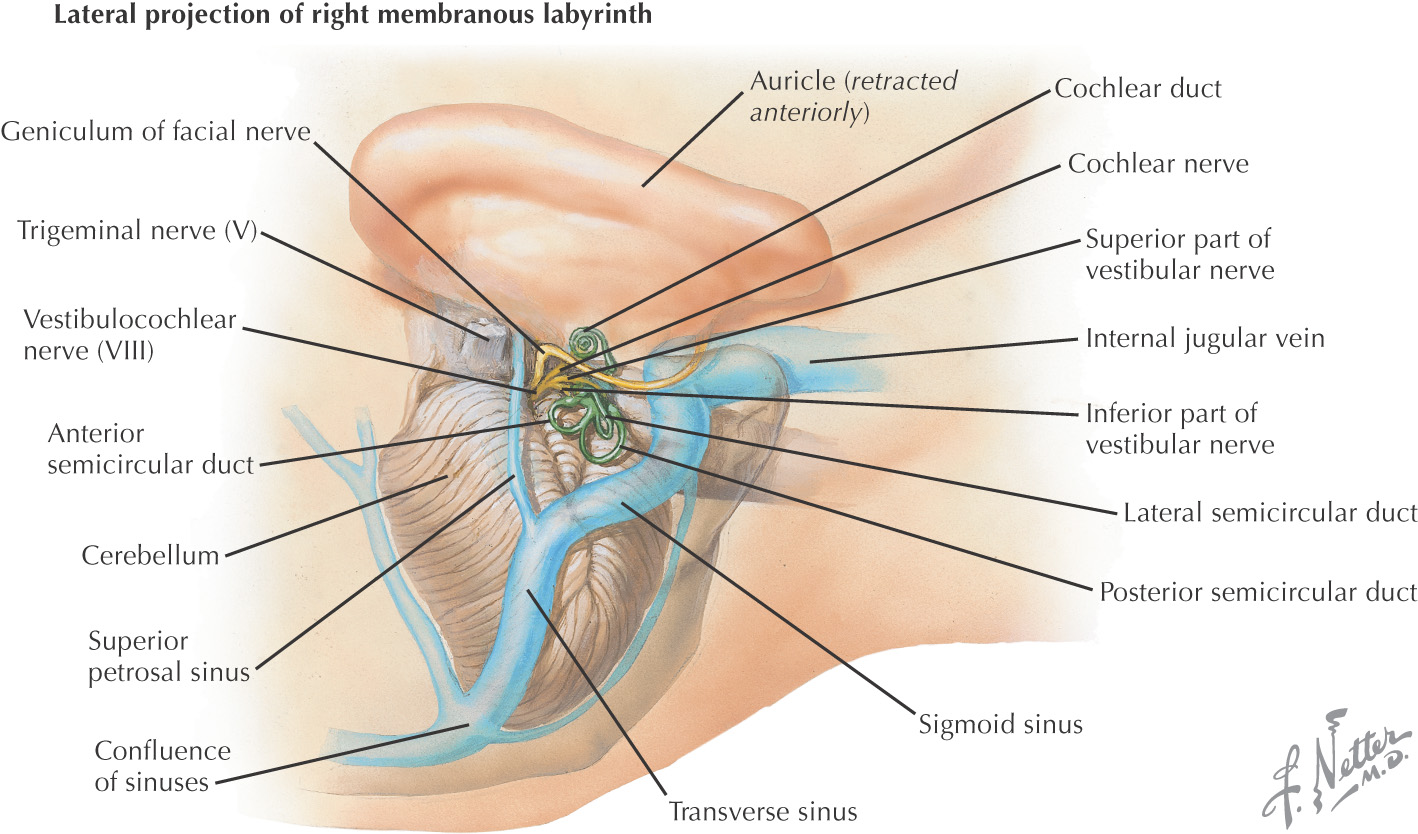
Vestibular and auditory structures, which are filled with fluid, make up the inner ear:
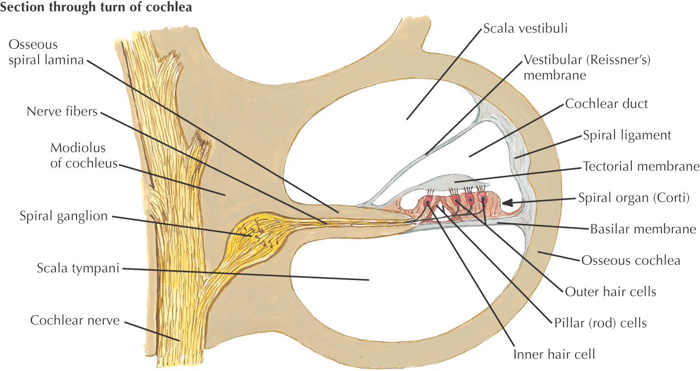
• Auditory portion (cochlea) is stimulated by the movement of the fluid
• Vestibular portion (utricle, saccule, and semicircular canals) is stimulated by fluid movement within these chambers
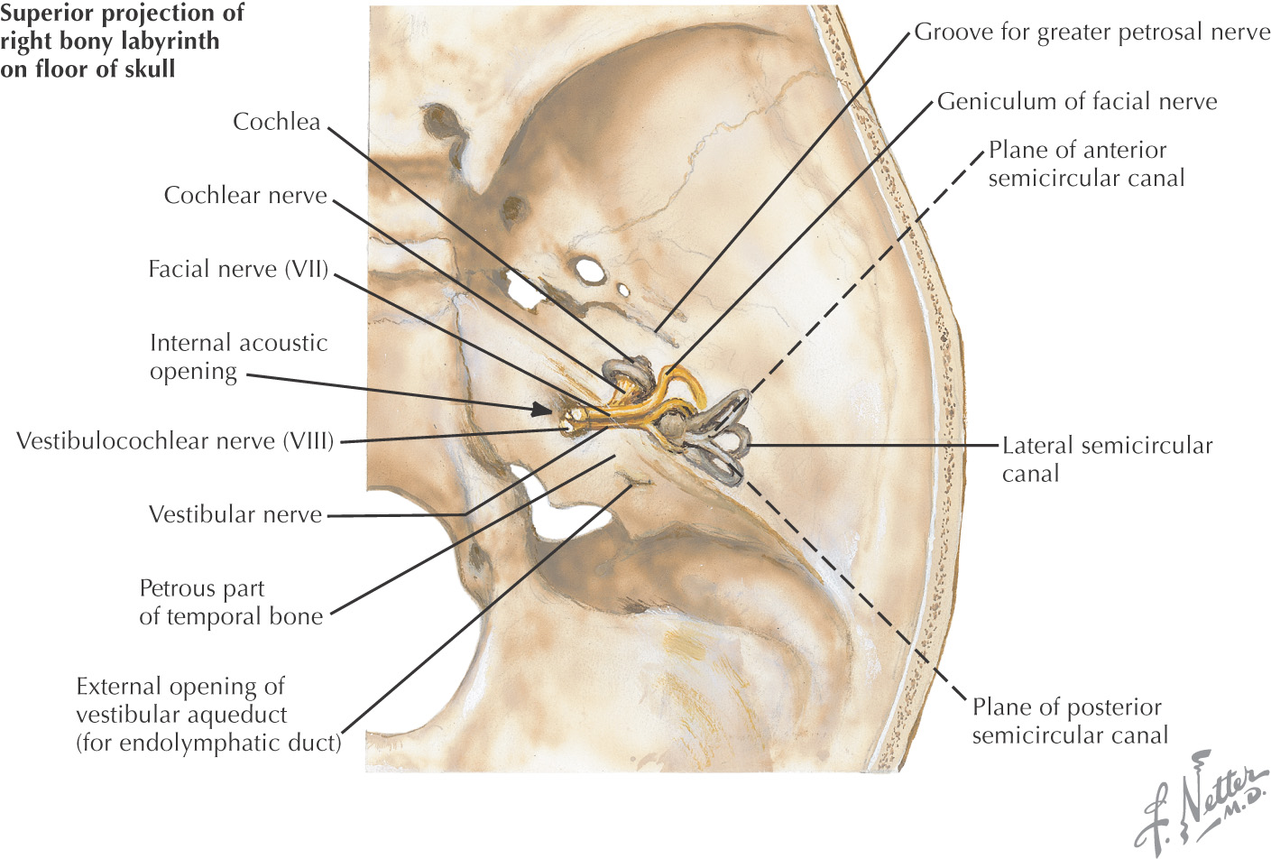
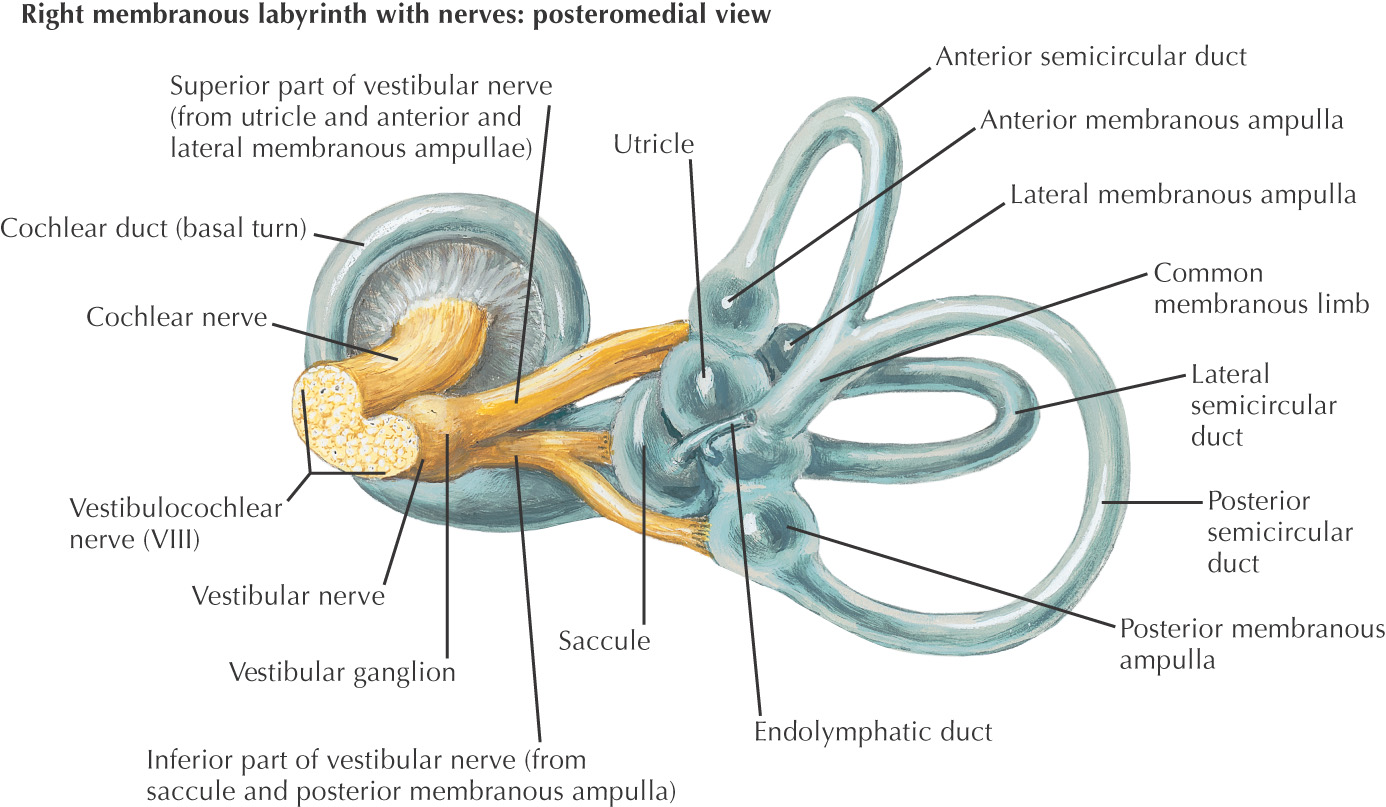
Consists of a membranous labyrinth that lies within an osseous labyrinth
The receptors for auditory and vestibular function are located within the membranous labyrinth
Fluids located in the membranous labyrinth (endolymph) and osseous labyrinth (perilymph) stimulate the auditory and vestibular receptors
The vestibulocochlear nerve enters the internal ear via the internal acoustic meatus

Infection or inflammation of the auricle and external auditory canal located in the external ear, causing ear pain (otalgia)
Also called “swimmer’s ear”
2 major bacteria are involved: Staphylococcus aureus and Pseudomonas aeruginosa
Excess water from swimming removes some of the ceruminous wax that lines the external auditory canal
Because the wax helps maintain a healthy canal, loss of the wax predisposes the canal to bacterial infections
An inflammation of the middle ear cavity
More common in children
2 major bacteria are involved: Streptococcus pneumoniae and Haemophilus influenzae
Often results from auditory tube dysfunction
Because the auditory tube allows drainage from the tympanic cavity into the nasopharynx, any blockage leads to a buildup of fluid in the tympanic cavity
When the fluid sits in the tympanic cavity, it predisposes the region to a bacterial infection
The resulting inflammation leads to ear pain (otalgia) and often diminished hearing
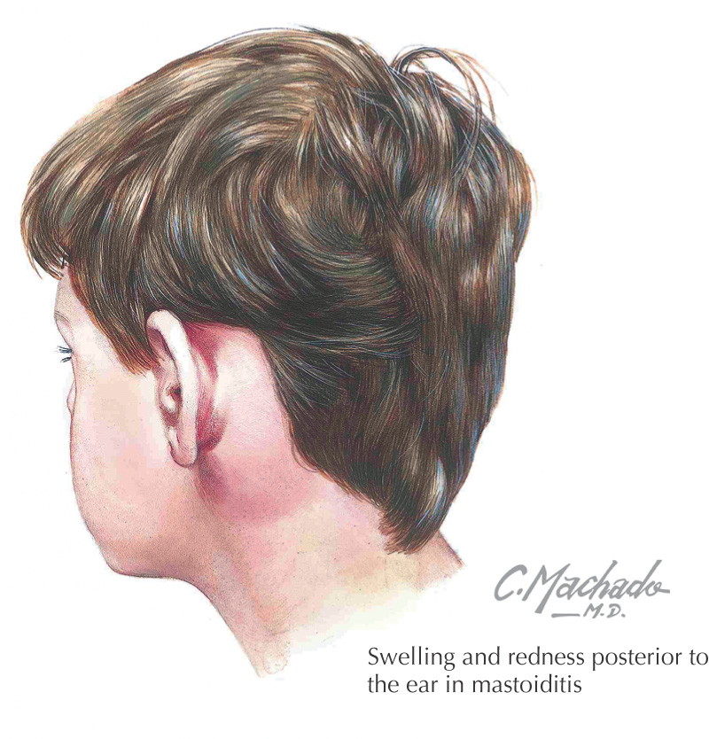
A bacterial infection of the mastoid air cells
More common in children than in adults
Although less common since the advent of antibiotics, formerly it often occurred as a complication of acute otitis media, when infection spread from the middle ear cavity to the mastoid air cells
Once within the mastoid air cells, the infection can lead to inflammation and destruction of the mastoid bone
Because of the infection’s location, it may lead to partial (or total) hearing loss, damage to the mastoid bone, or formation of an epidural abscess, or it may spread to involve the brain
Can be difficult because medications cannot readily reach the mastoid air cells
In some cases, a mastoidectomy may be performed to drain the mastoid if antibiotic therapy is not successful
A myringotomy (creating an opening in the middle ear cavity through the tympanic membrane) is performed to drain the ear in acute otitis media