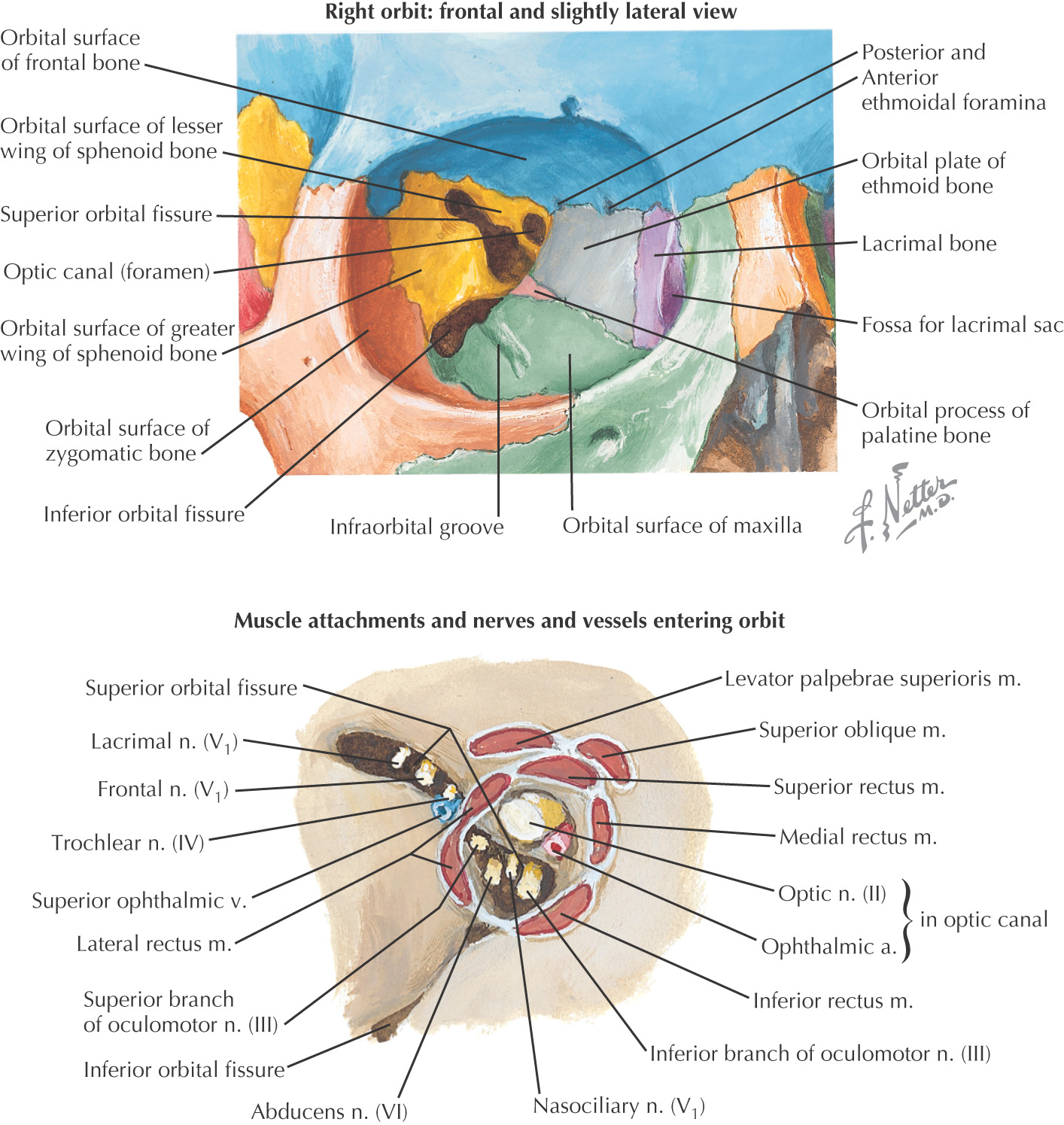
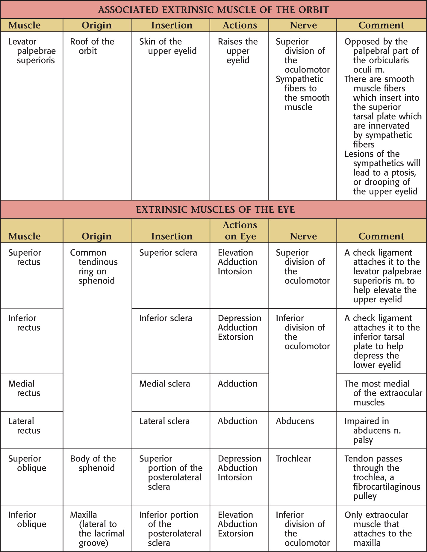
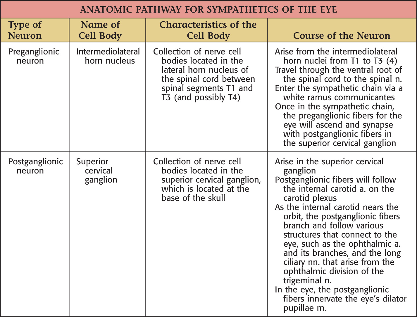
Overview and Topographic Anatomy of the Orbit
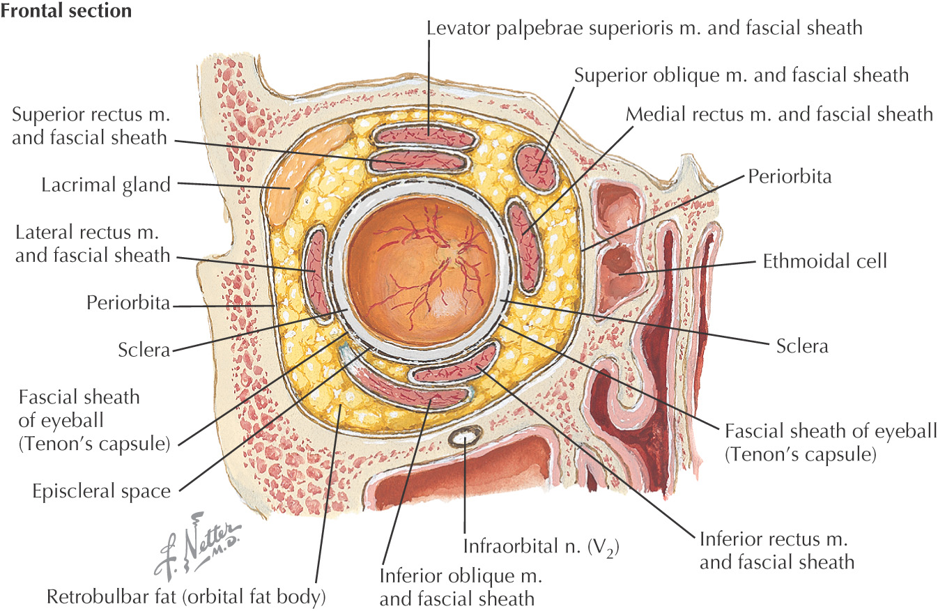
Orbit: a pyramid-shaped bony recess in the anterior part of the skull, lined by periosteum called the periorbital fascia
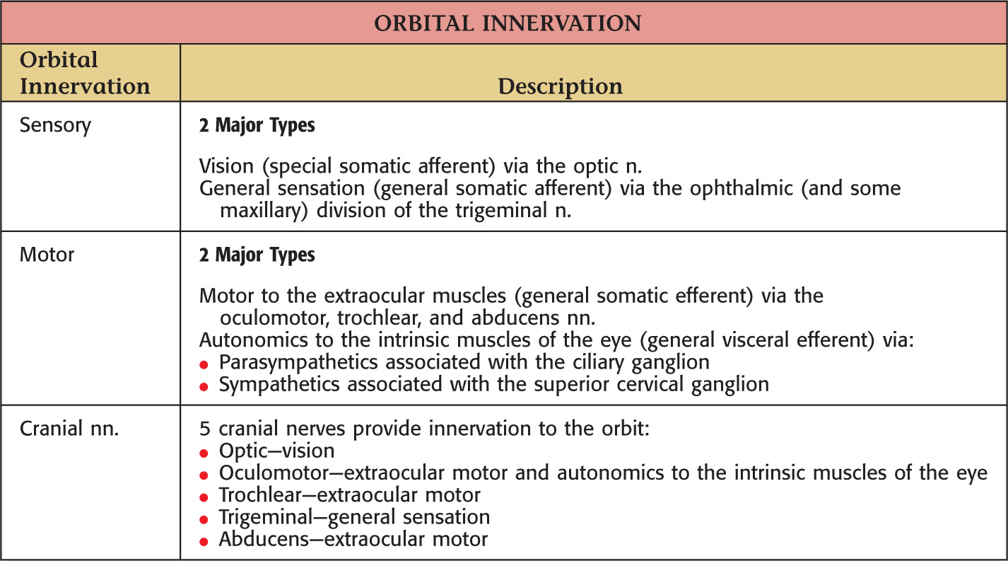
Contents include:
• Eye—organ associated with vision
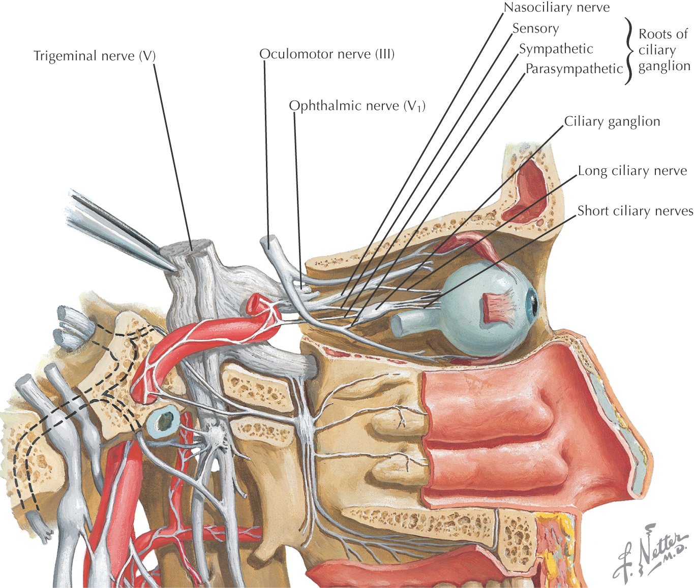
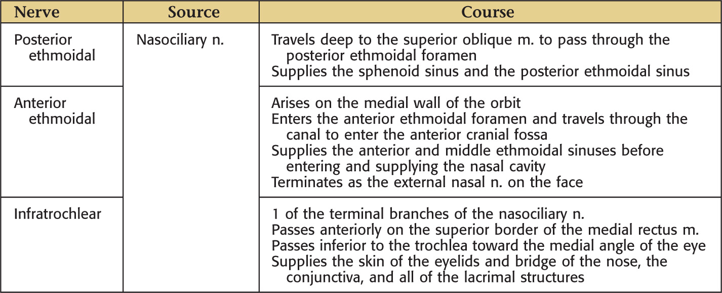
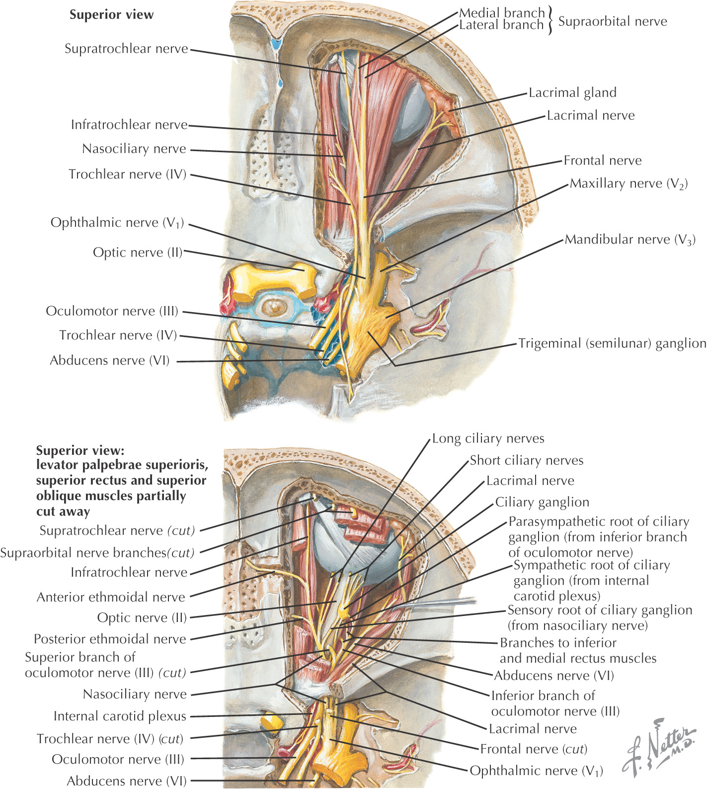
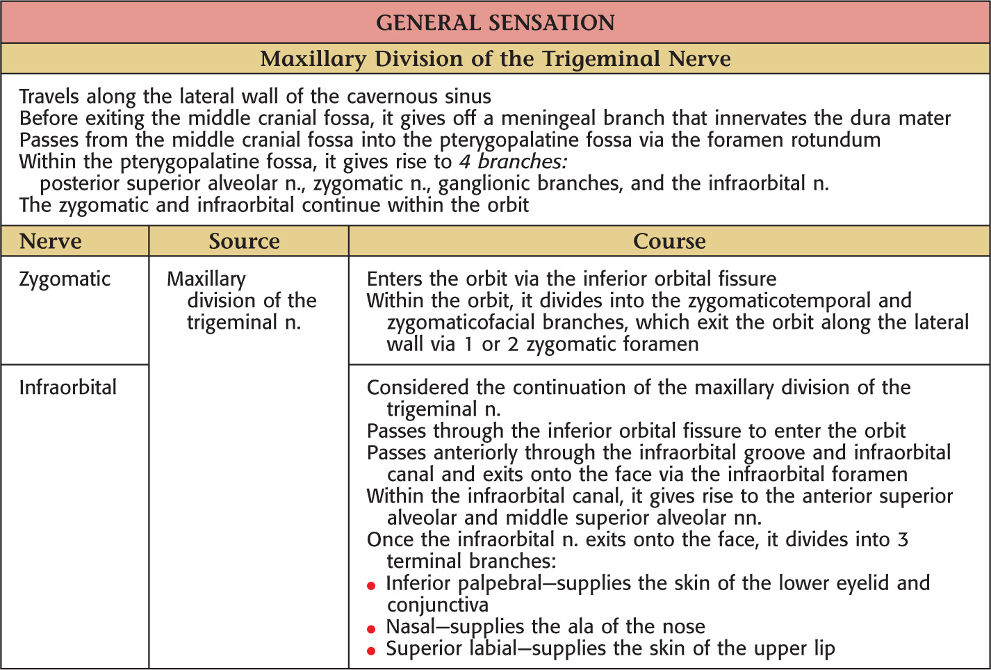
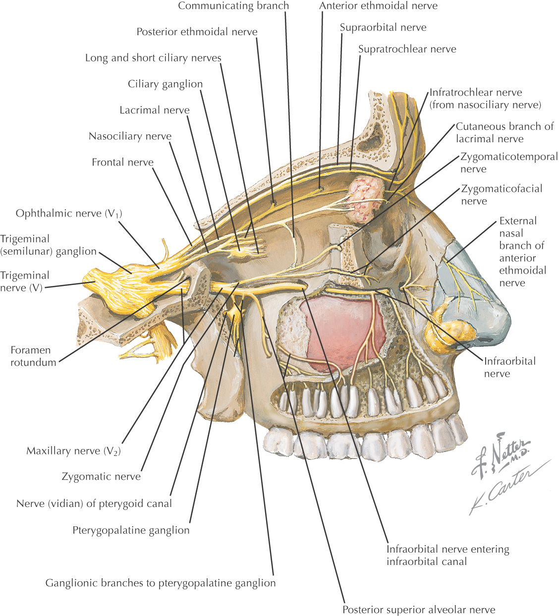
• Ophthalmic division of the trigeminal nerve
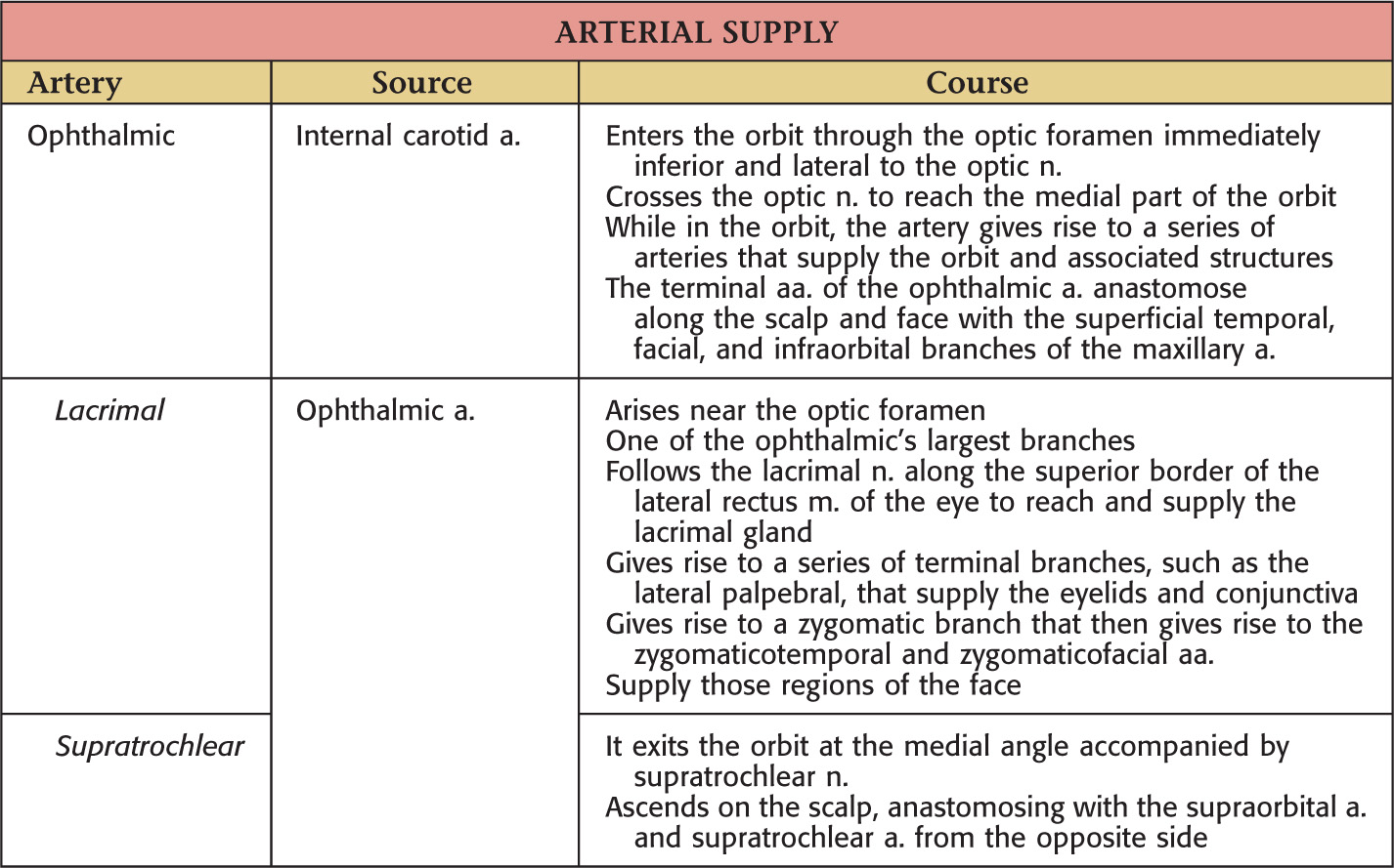
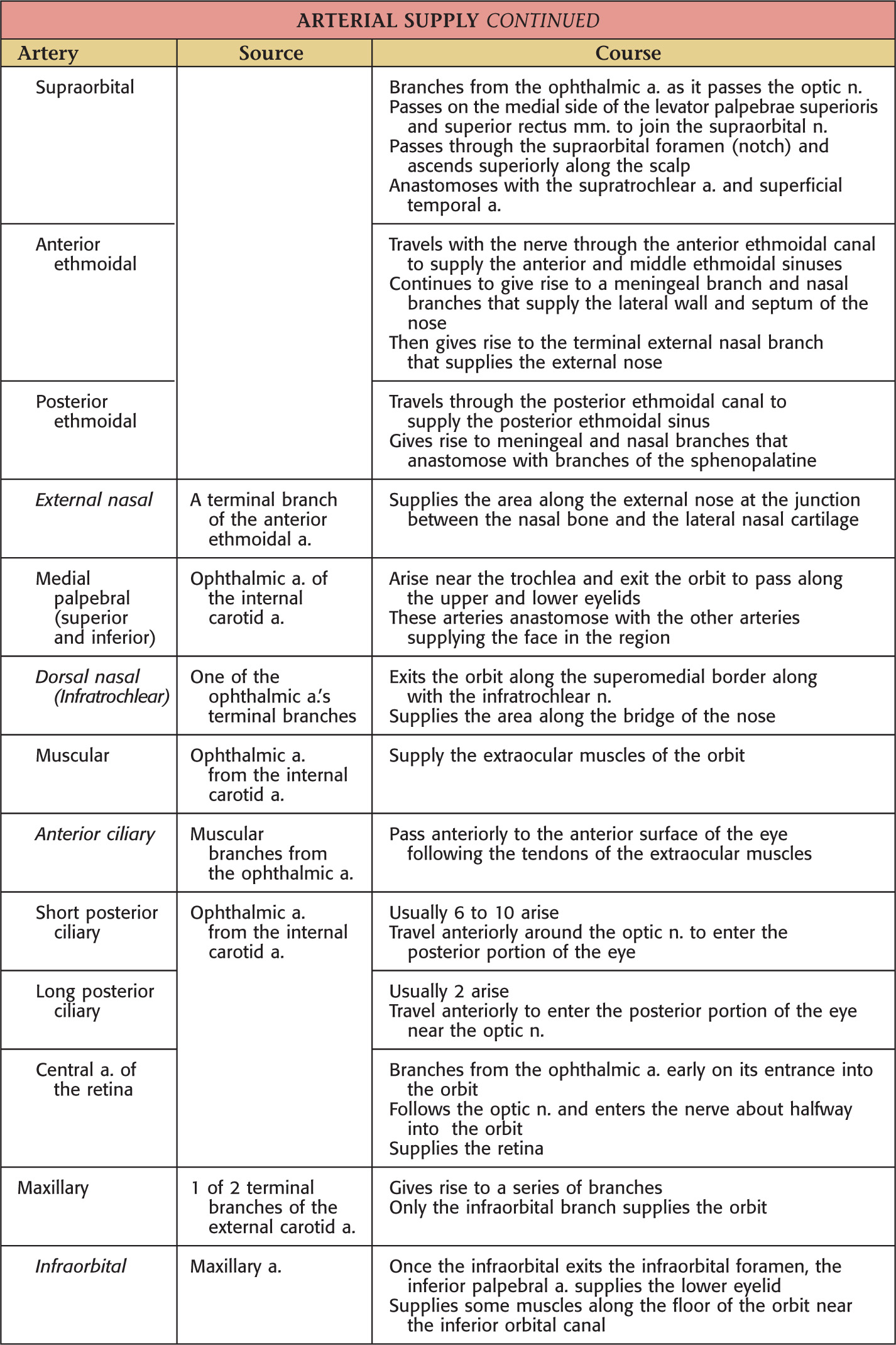
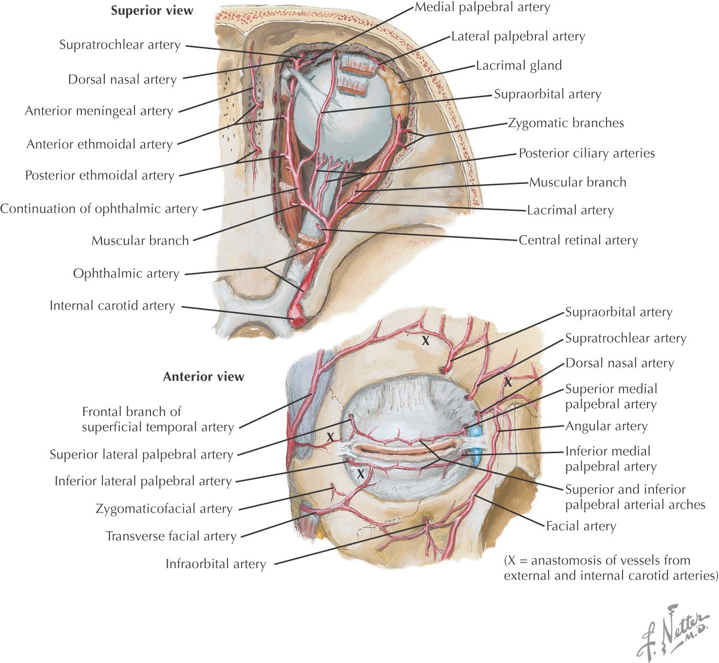
• Ophthalmic artery and branches
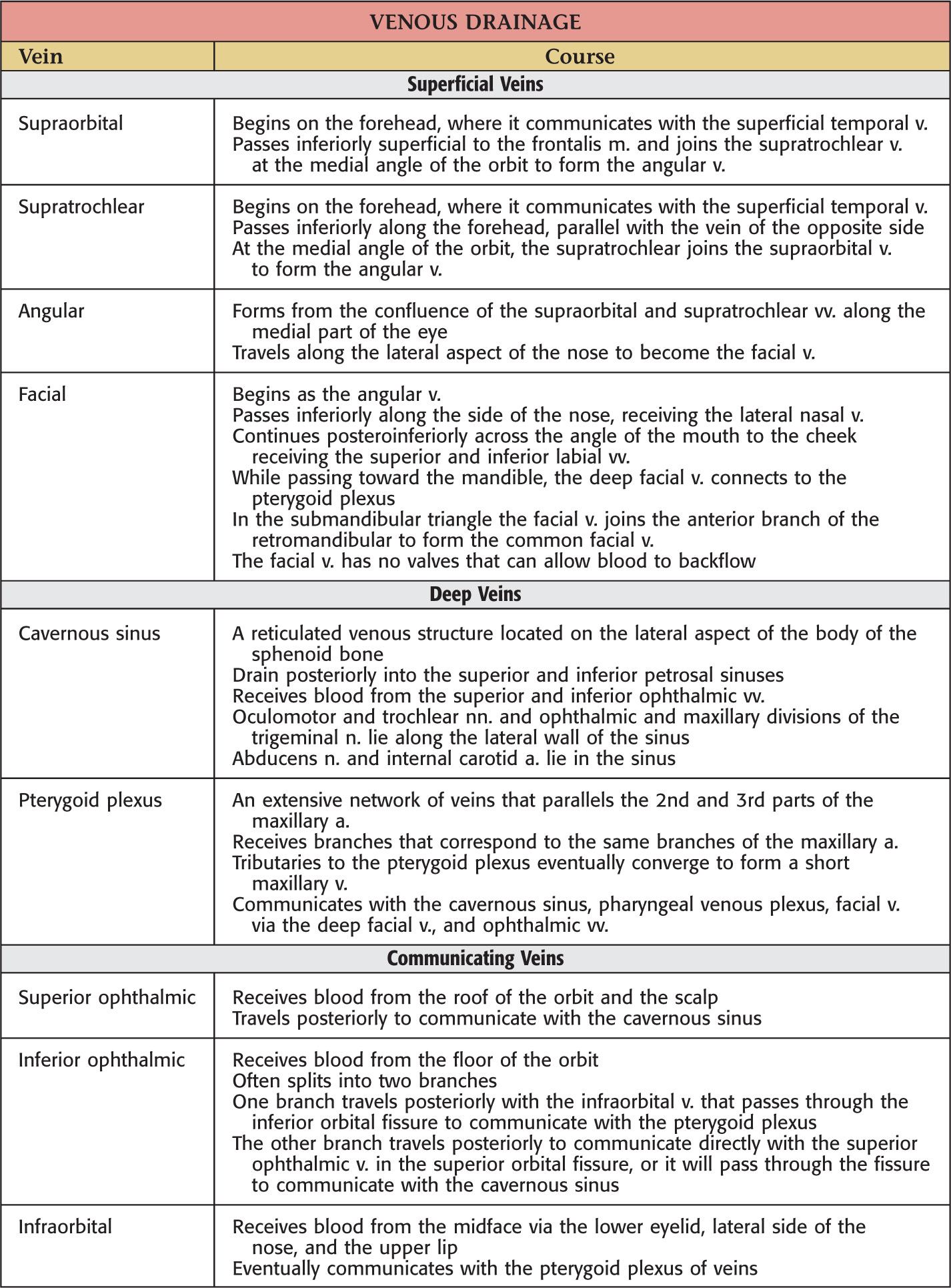
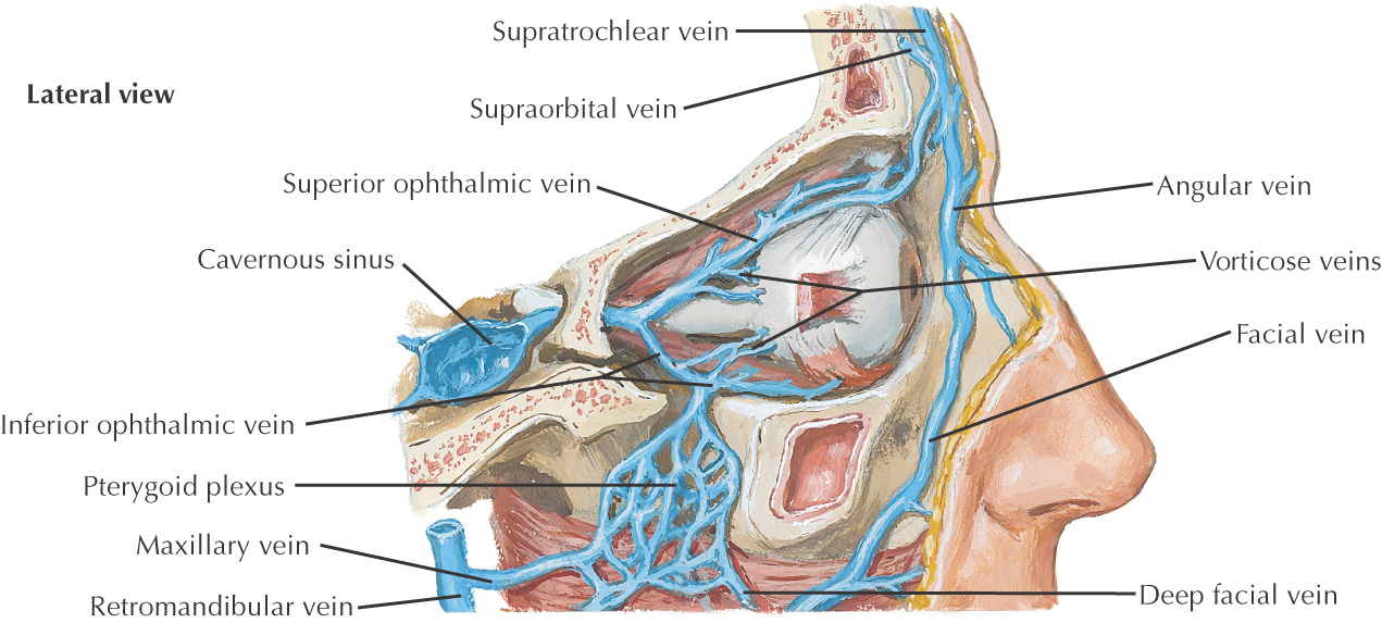
• Superior and inferior ophthalmic veins
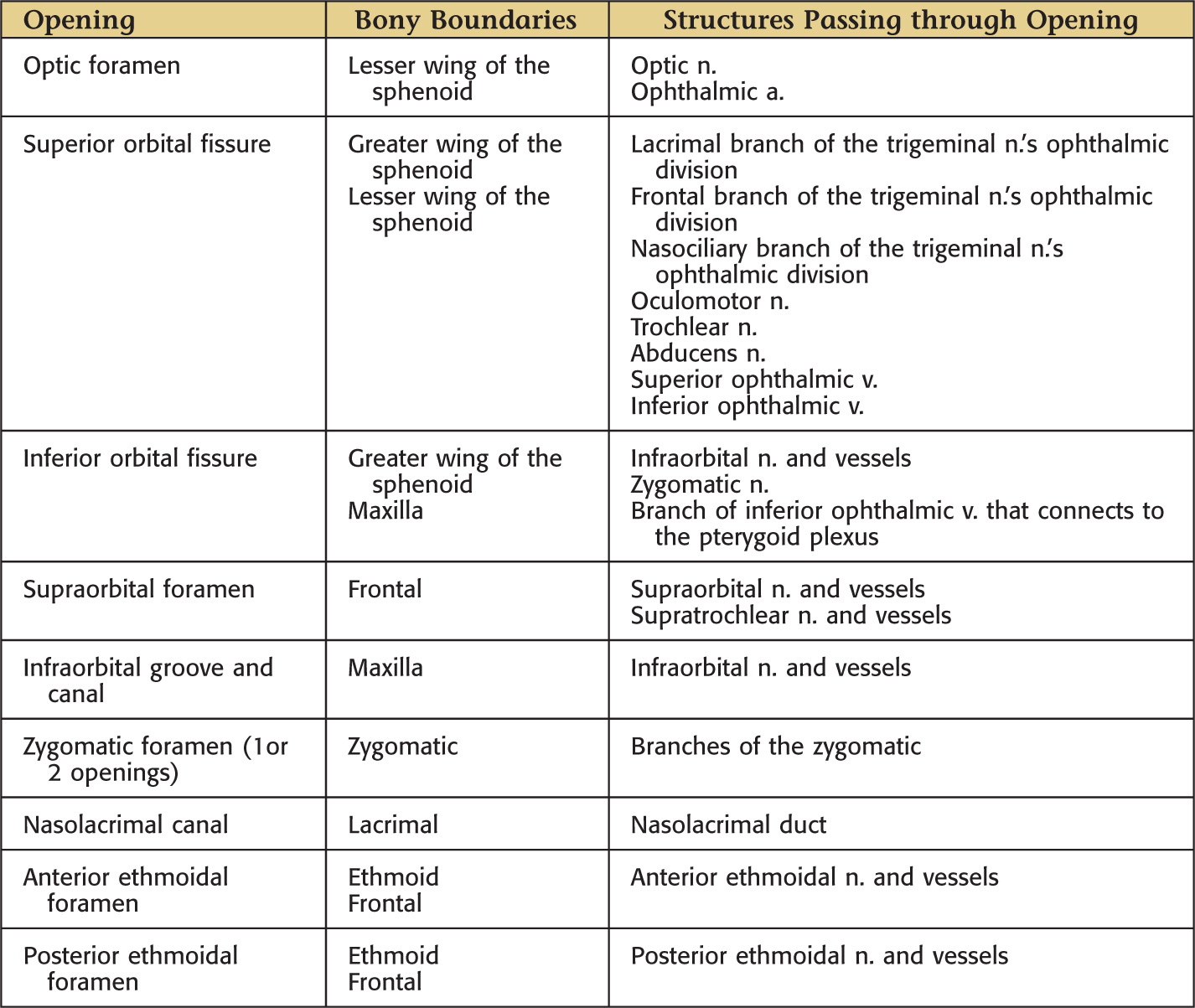
Superior |
|
Inferior |
|
Medial |
|
Lateral |
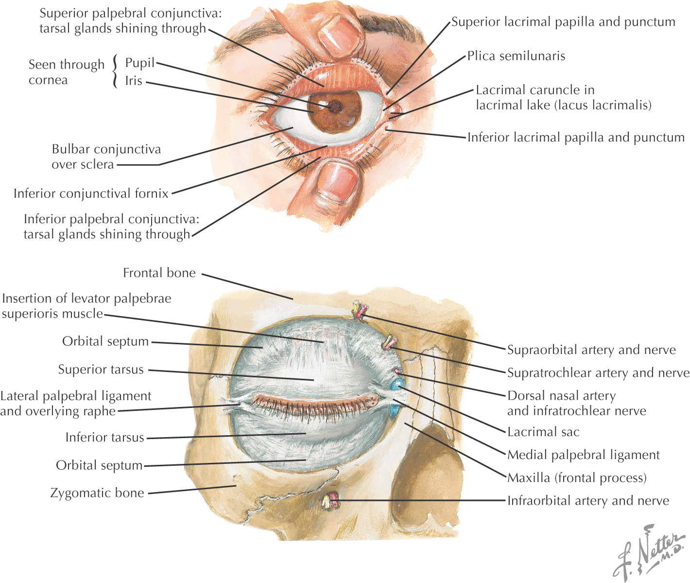
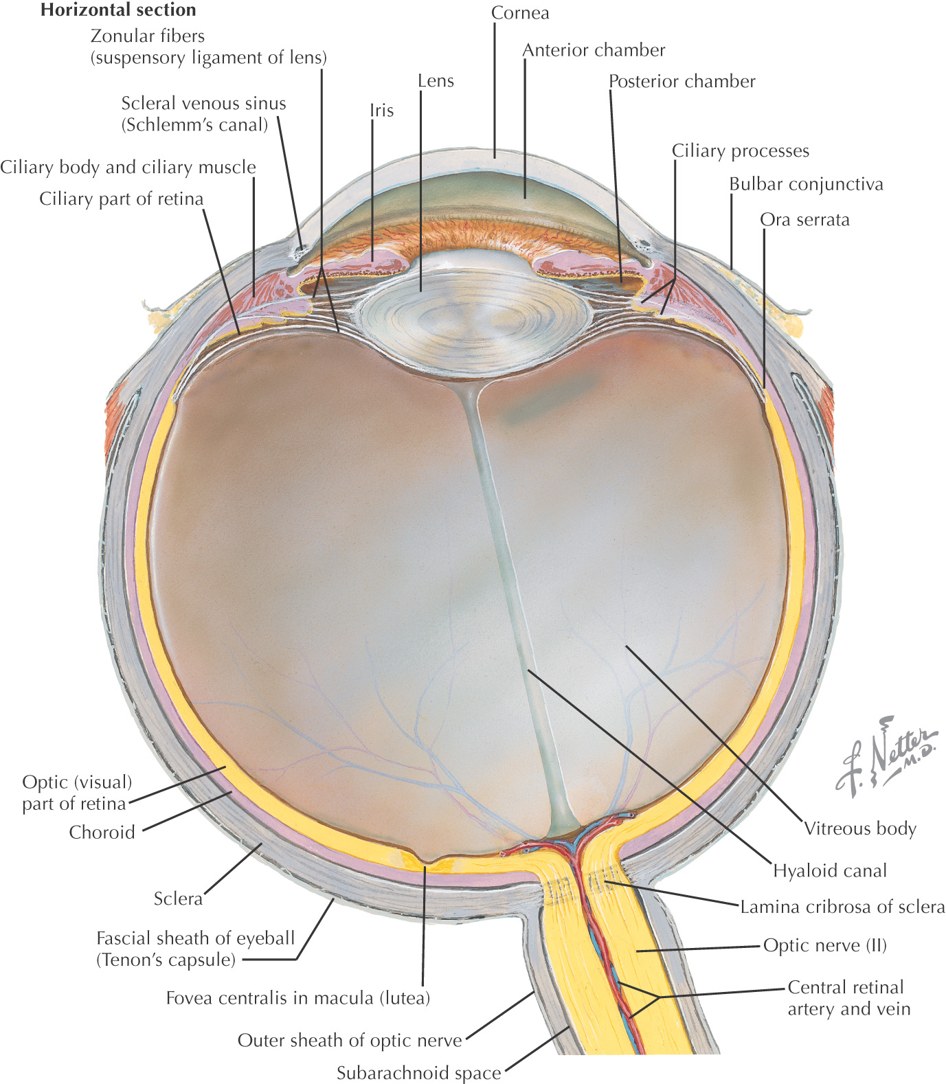
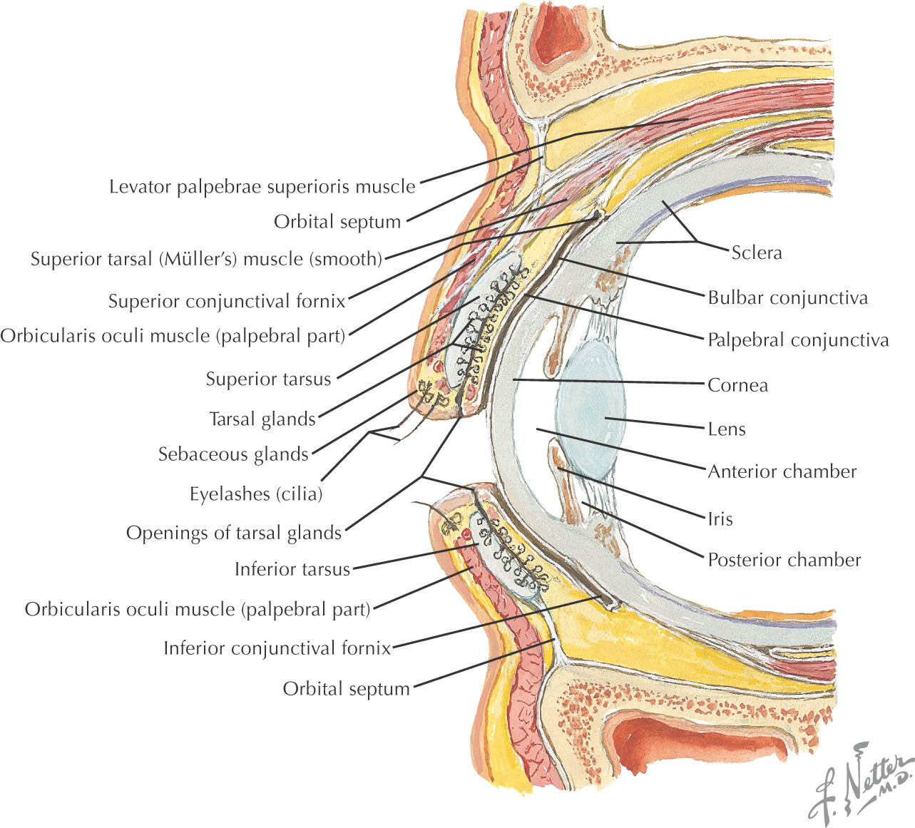
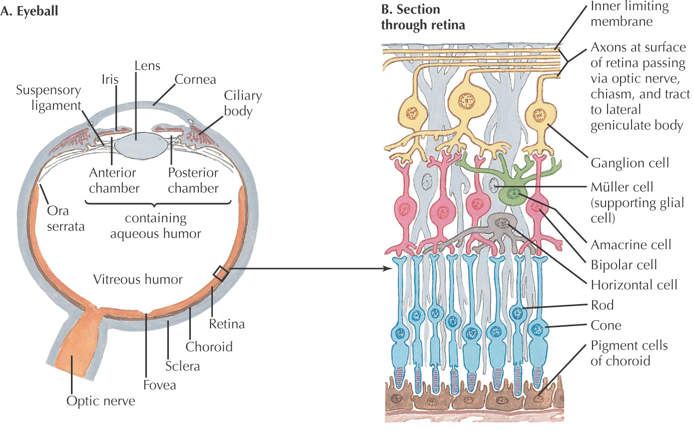
Eye: a spherical globe with a diameter of approximately 2.5 cm that lies in the orbit’s anterior portion
Surrounded by a thin capsule called the fascia bulbi (Tenon’s capsule):
Composed of 3 coats:
Divided into an anterior and a posterior segment:
• Separated into anterior and posterior chambers by the iris
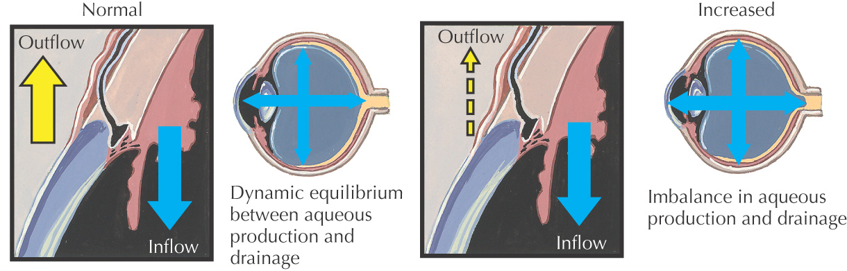
• Contains aqueous humor secreted by the ciliary body and drained through a trabeculated network eventually into the superior ophthalmic vein
• Intraocular pressure is measured in the anterior segment, normally 10 to 20 mm Hg
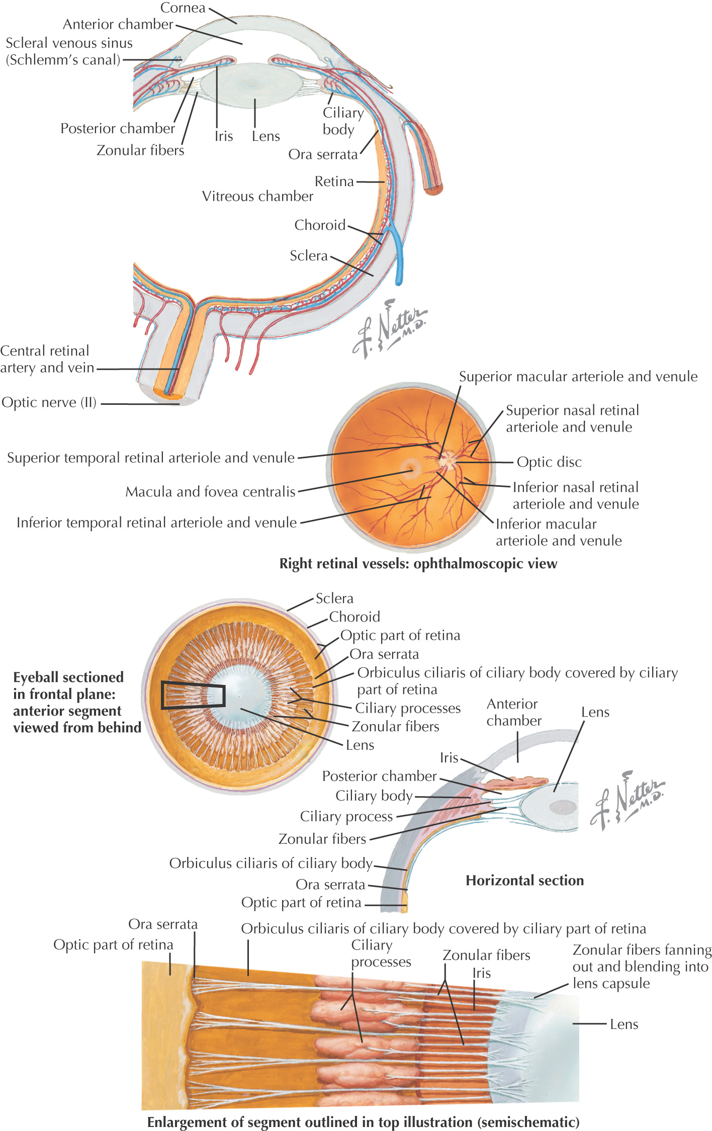
The outermost layer, very fibrous
White along the periphery, except for the anterior portion—the cornea, which is transparent
Composed of choroid layer, ciliary body, and iris
• The pigmented vascular layer between the sclera and the retina
• Extends posteriorly from the region of the optic nerve anteriorly, where it is continuous with the ciliary body near the ora serrata (anterior margin of the retina)
• Located between the choroid and the iris
• Ring-shaped; has a series of transparent fibers that form the suspensory ligament of the lens
• Within it is the ciliary muscle, which changes the shape of the lens
• A thin disclike structure with a central opening—the pupil
• Separates the aqueous humor into the anterior chamber (anterior to the iris) and the posterior chamber (between the iris and the lens)
• Contains the sphincter and dilator pupillae muscles, which change the pupil’s shape in response to light
Located posterior to the iris
A transparent biconcave structure responsible for focusing
Connected to the ciliary body by the suspensory ligaments
The innermost coat of the eye
Thin and highly vascular
Three areas located on the retina’s posterior portion:
Area where the optic nerve enters the retina is called the “blind spot”
Retina’s central artery enters the eye through the optic disc and divides into superior and inferior branches
Lateral to the optic disc
A depressed, yellow-appearing area that contains the fovea centralis in its center
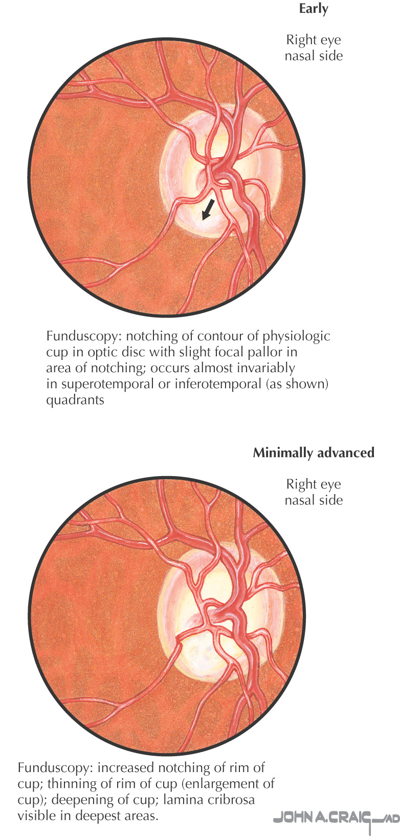
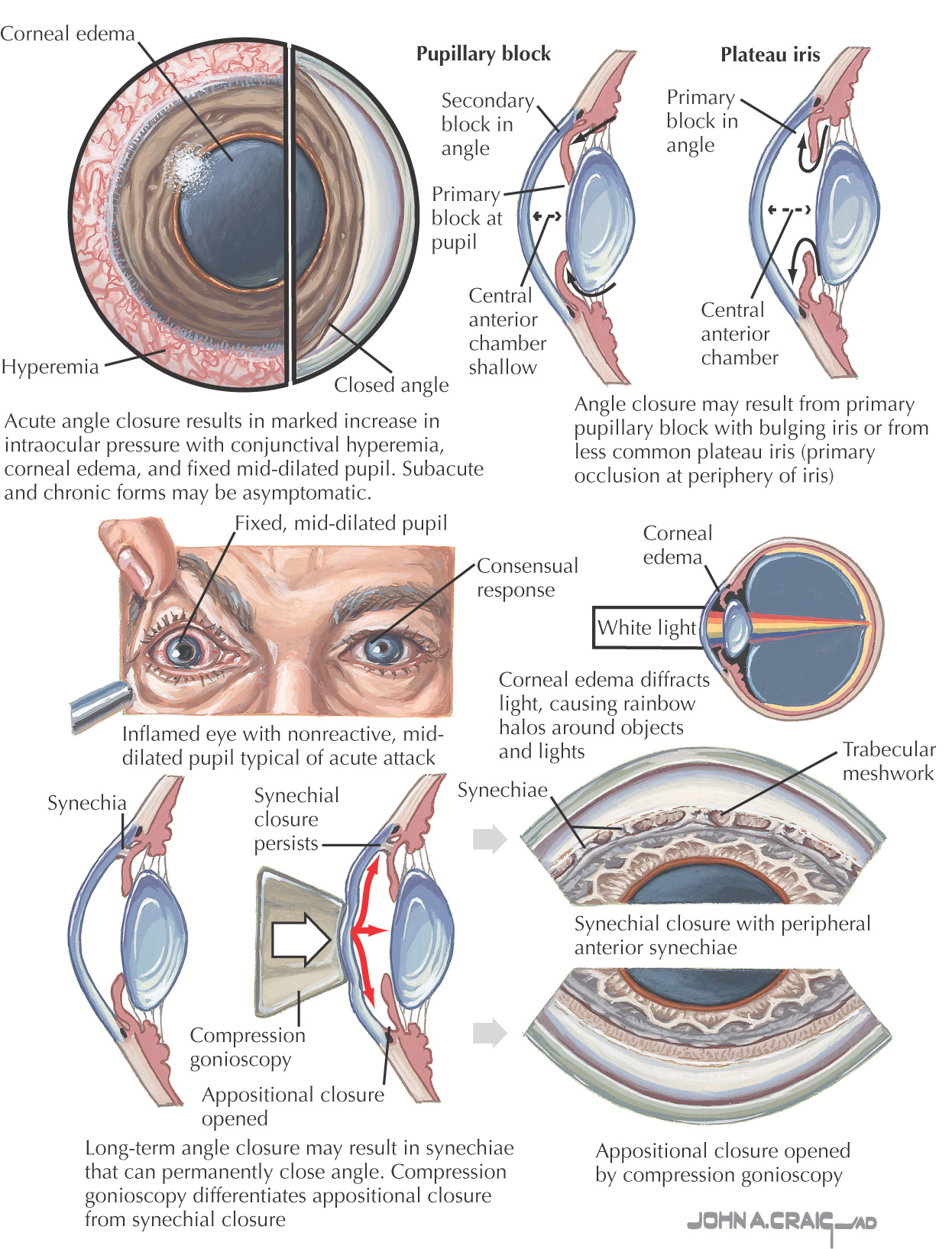
Damage to the optic nerve often due to increased intraocular pressure
The most common form
Gradual and can result in gradual loss of vision
Intraocular pressure elevates due to insufficient drainage within the eye’s canal system located in the angle of the anterior chamber of the anterior segment
Various medications are successful in treating this form
Result of an anatomic blockage of the canal system at the angle of the anterior chamber of the anterior segment
Example: When the iris opens the pupil very wide and blocks the angle, intraocular pressure rises quickly as a result of the possible abrupt blockage
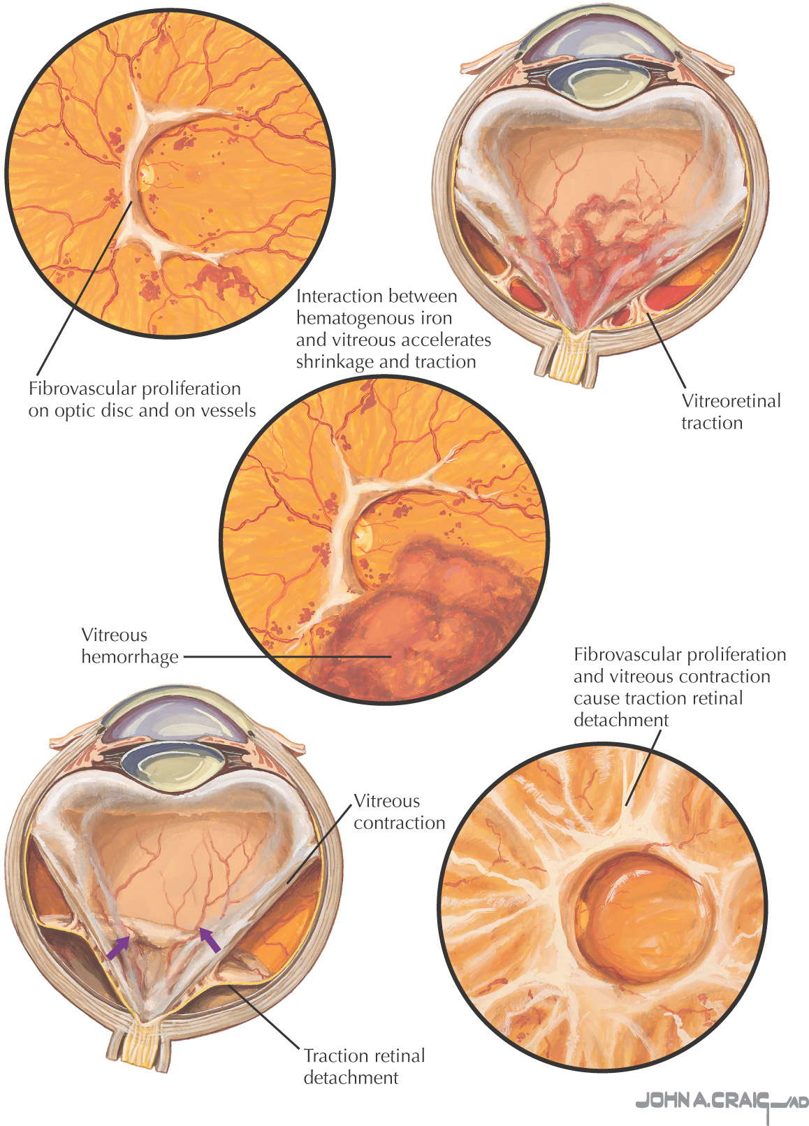
Damage to the retina as a result of damage to the blood vessels in the retina due to diabetes
Can occur in all people with diabetes (types 1 and 2)
As the retinal blood vessels become damaged, they leak fluid into the eye
If the fluid accumulates around the macula lutea (contains the largest amount of cones for acute vision), macula edema occurs in which visual loss is noted
As the permeability of the vessels worsens, lipoprotein is deposited, leading to formation of hard exudates within the retina
As new blood vessels form, they are fragile and bleed, allowing blood to enter the eye, helping to cloud and destroy the retina
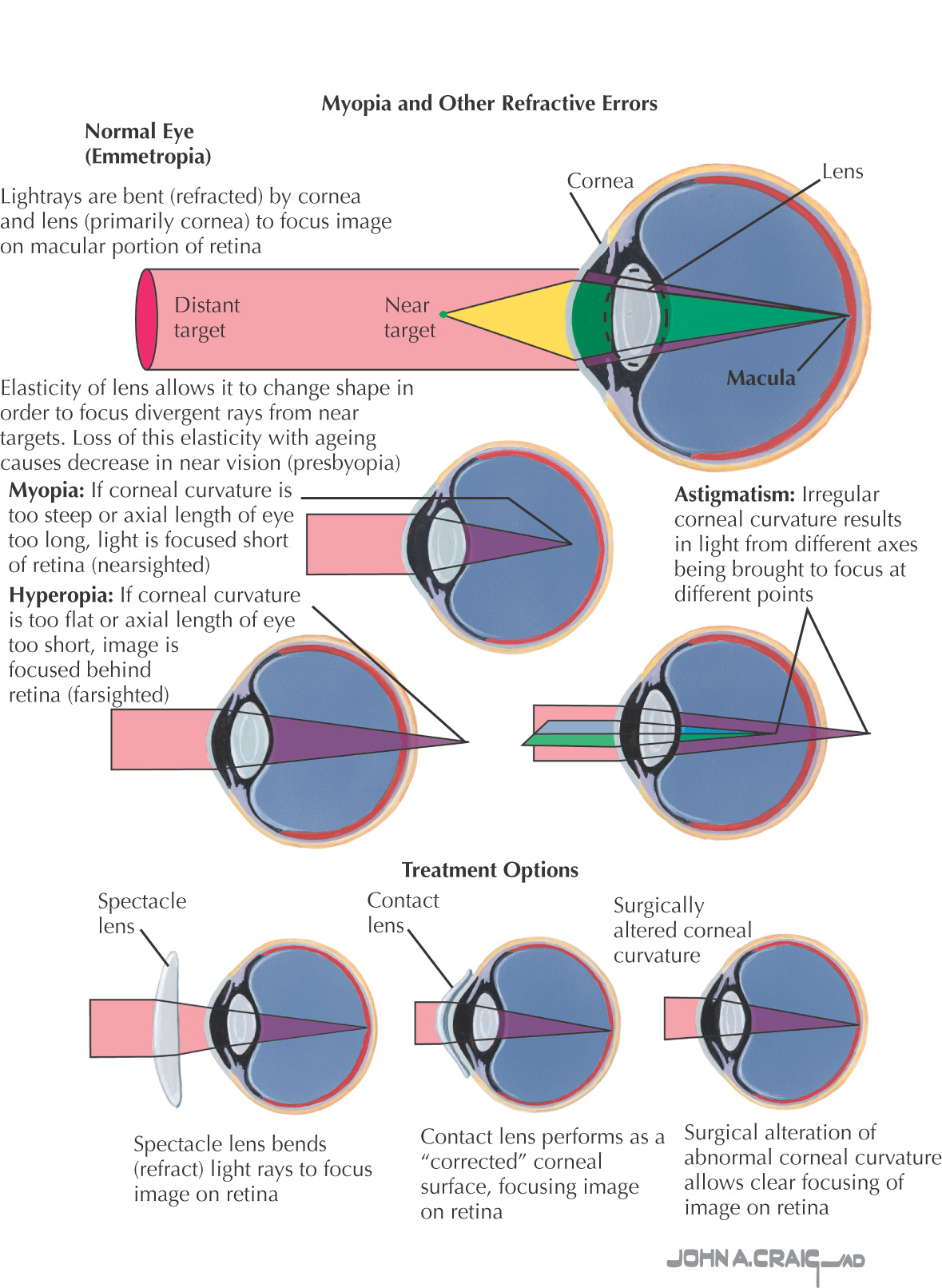
A series of refractive disorders of the eye that cause blurring of the image on the retina
Myopia
• Image is focused anterior to the retina
• Commonly referred to as nearsightedness
Hyperopia
• Image is focused posterior to the retina
• Commonly referred to as farsightedness
Astigmatism
• A nonspherical eye allows the parts of the image to focus at multiple locations, rather than in a single area