Overview and Topographic Anatomy
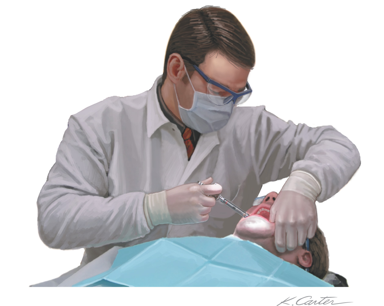
Intraoral injections provide adequate pain control for various dental procedures
Many techniques have been developed
All require detailed understanding of head and neck anatomy to maximize proper administration and minimize complications
Injections should not be performed in areas of infection or inflammation
The application of topical anesthetic to the site of injection will help lessen the pain caused by the insertion of the needle
• Local injections (field blocks)
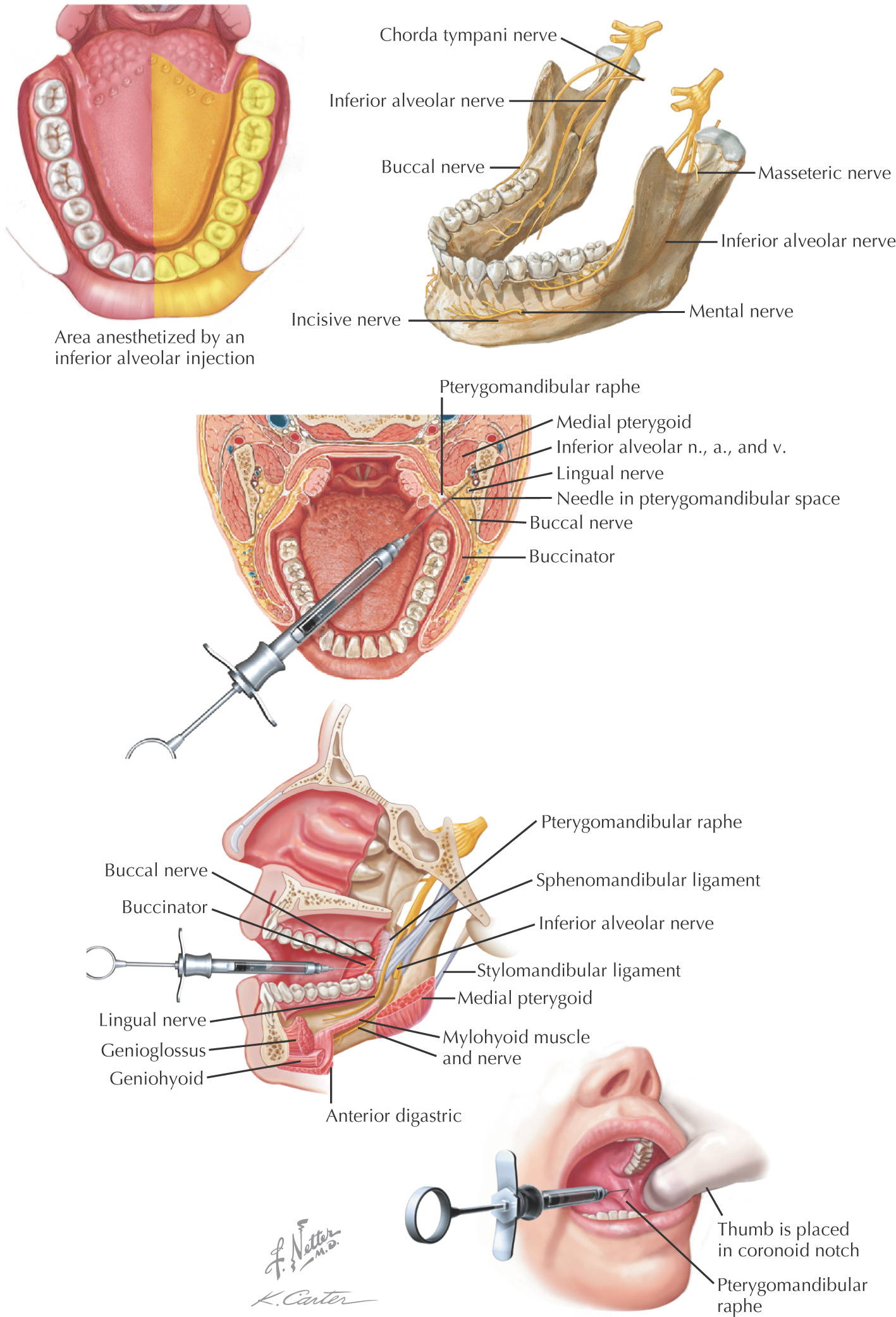
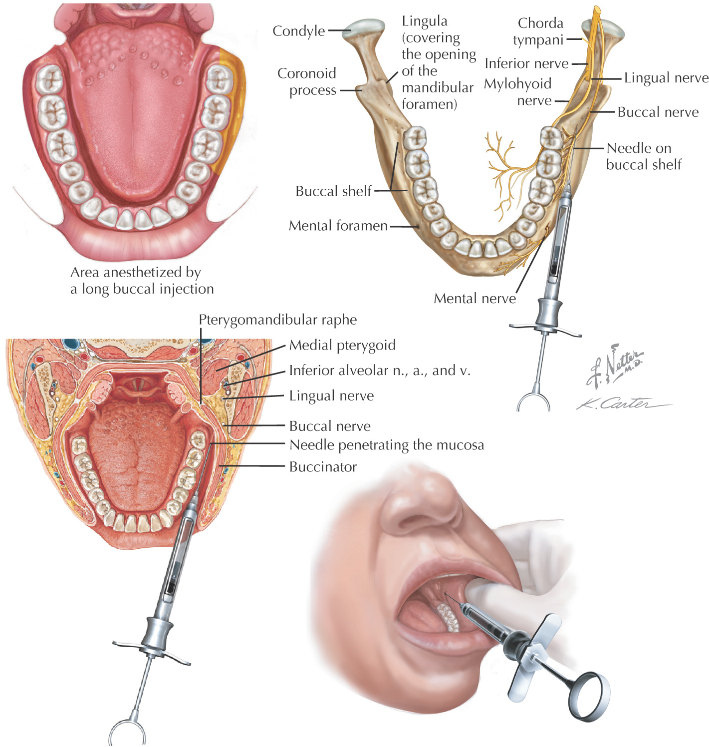
Mandibular:
Maxillary:
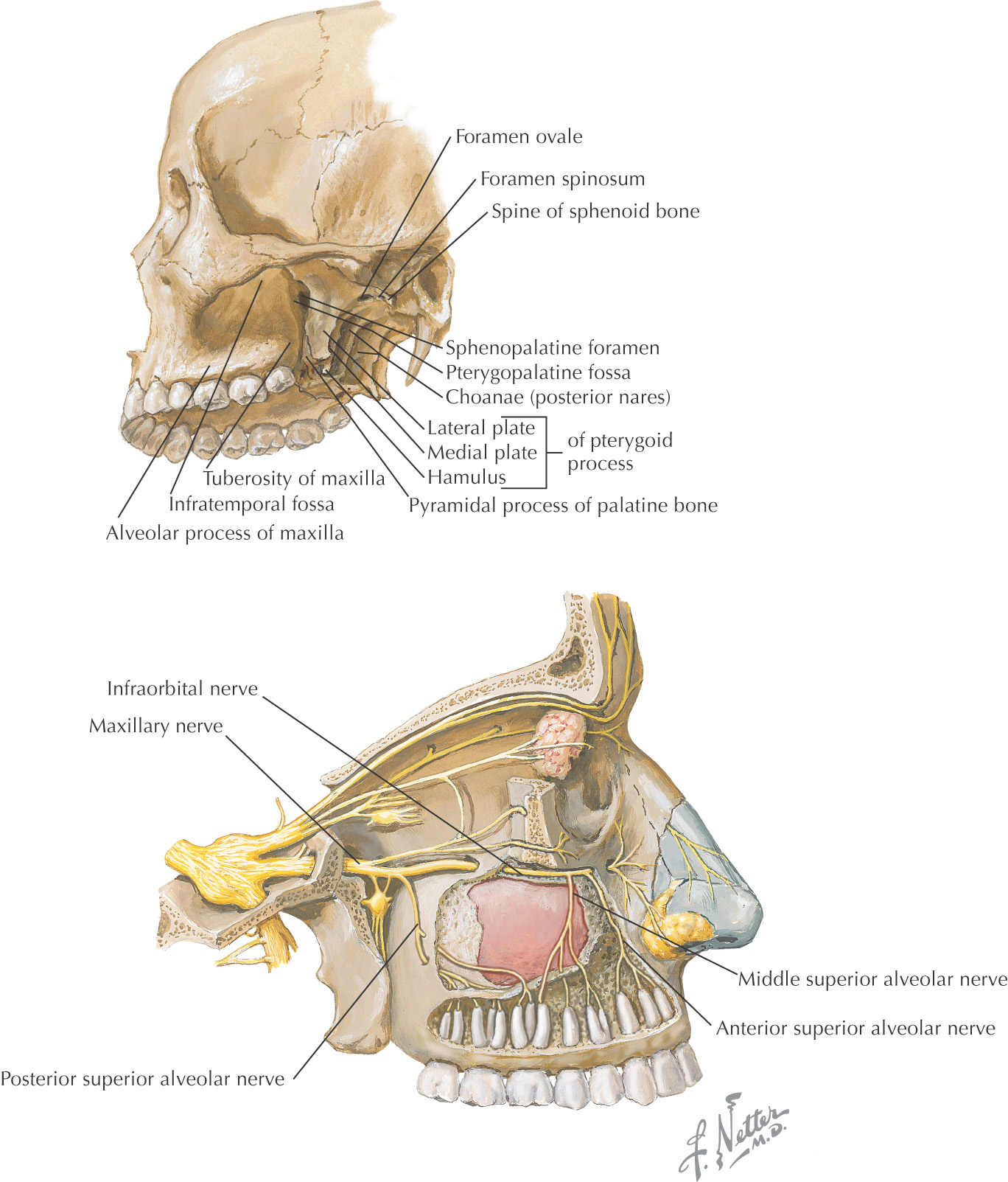
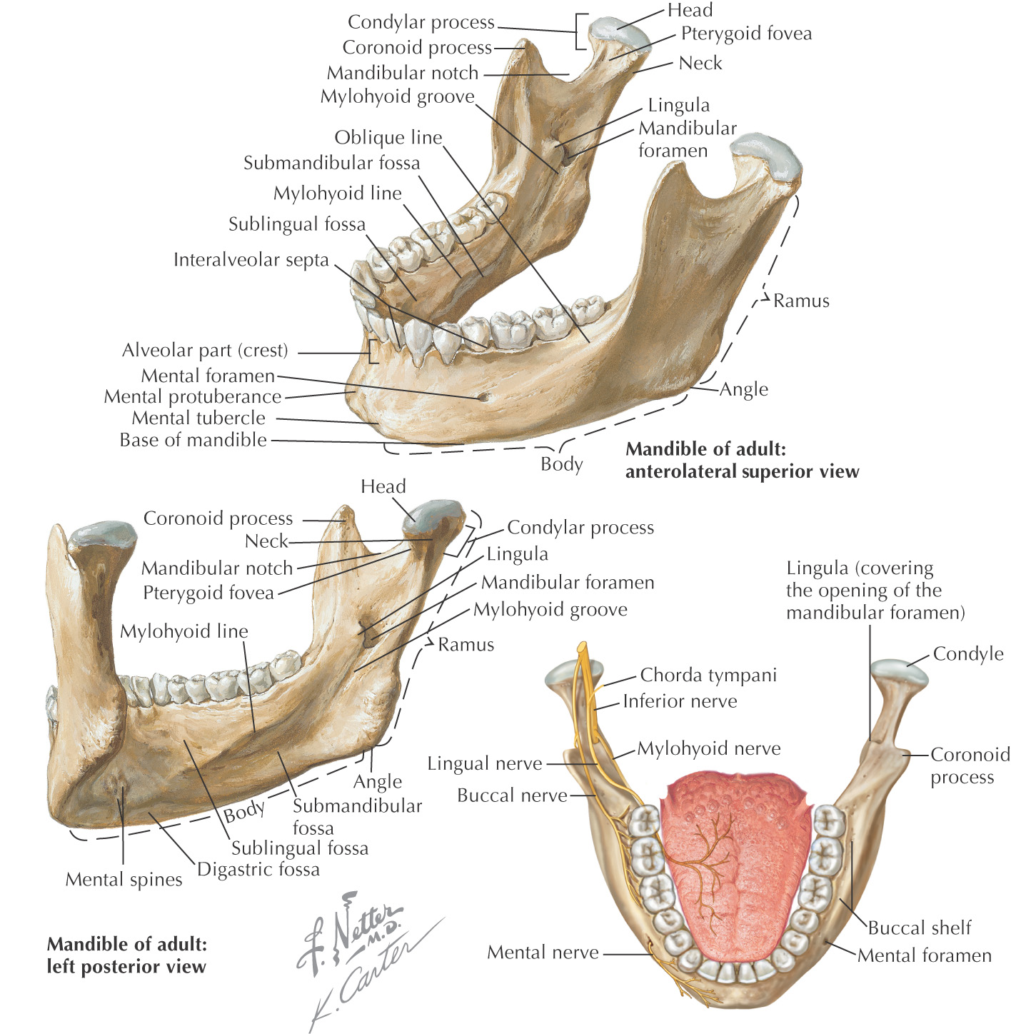
The strongest and largest facial bone
Composed of 2 pieces of thick cortical bone: a lingual plate and a buccal plate
Teeth are contained in the horseshoe-shaped body
Ramus extends superiorly from the angle of the mandible
The coronoid notch is the concavity on the anterior portion of the ramus used to estimate the height of the mandibular foramen, which also is located at the height of the occlusal plane
Associated Nerves
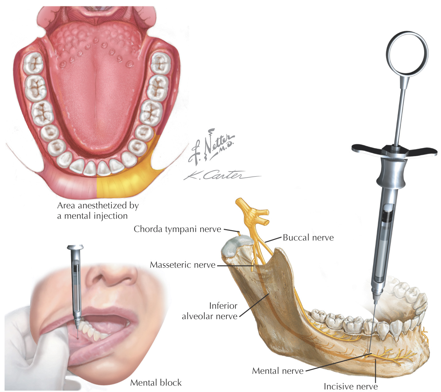
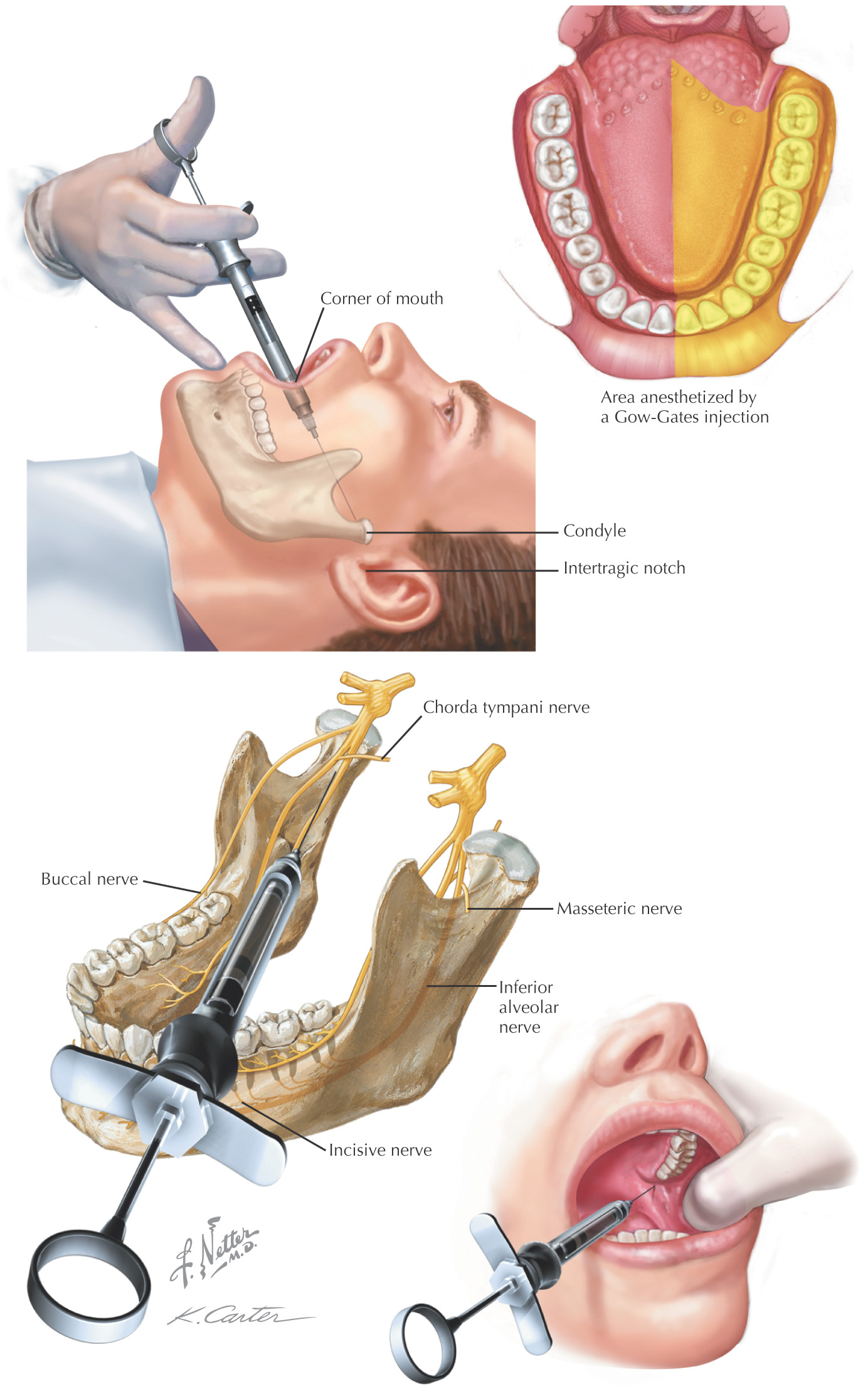
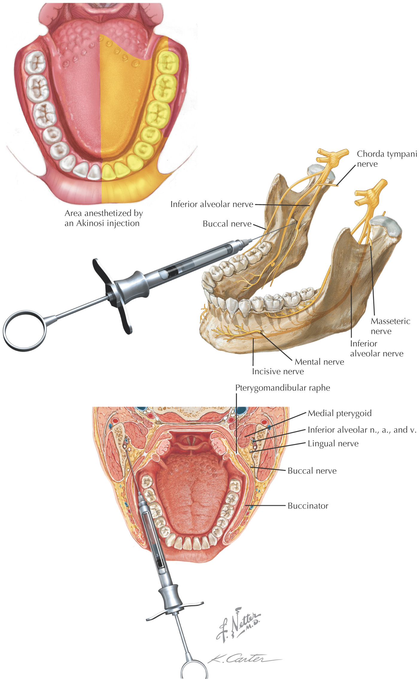
• Inferior alveolar nerve enters the mandible at the mandibular foramen
• Lingual nerve enters the oral cavity passing against the lingual tuberosity
• Buccal nerve lies on the buccal shelf
One of the largest facial bones
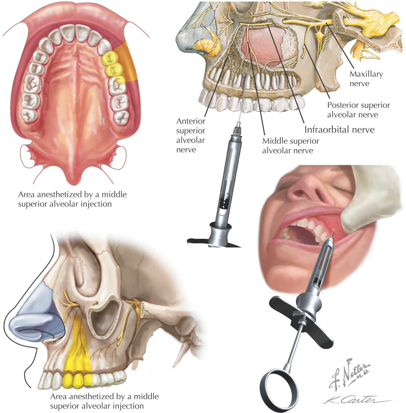
Porous bone, which aids in achieving anesthesia of the maxillary teeth
• Contained in the alveolar bone
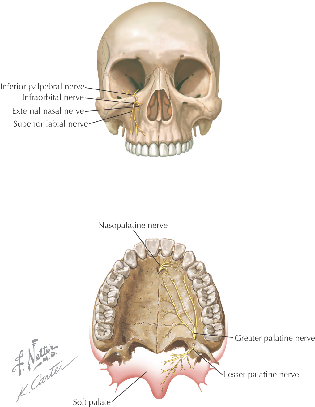
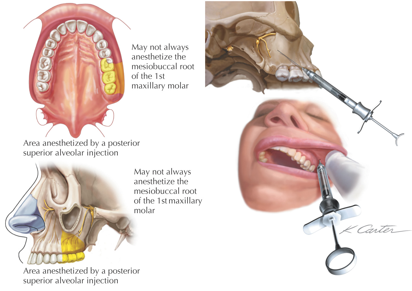
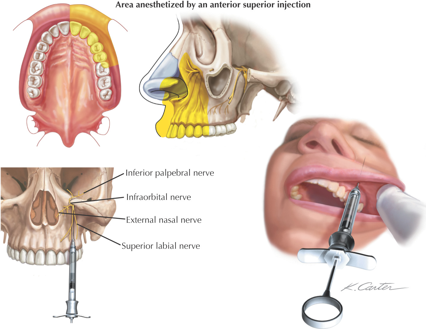
• Maxillary teeth are supplied by the anterior, middle, and posterior superior alveolar nerves (in some patients, the middle superior alveolar nerve may not be present)
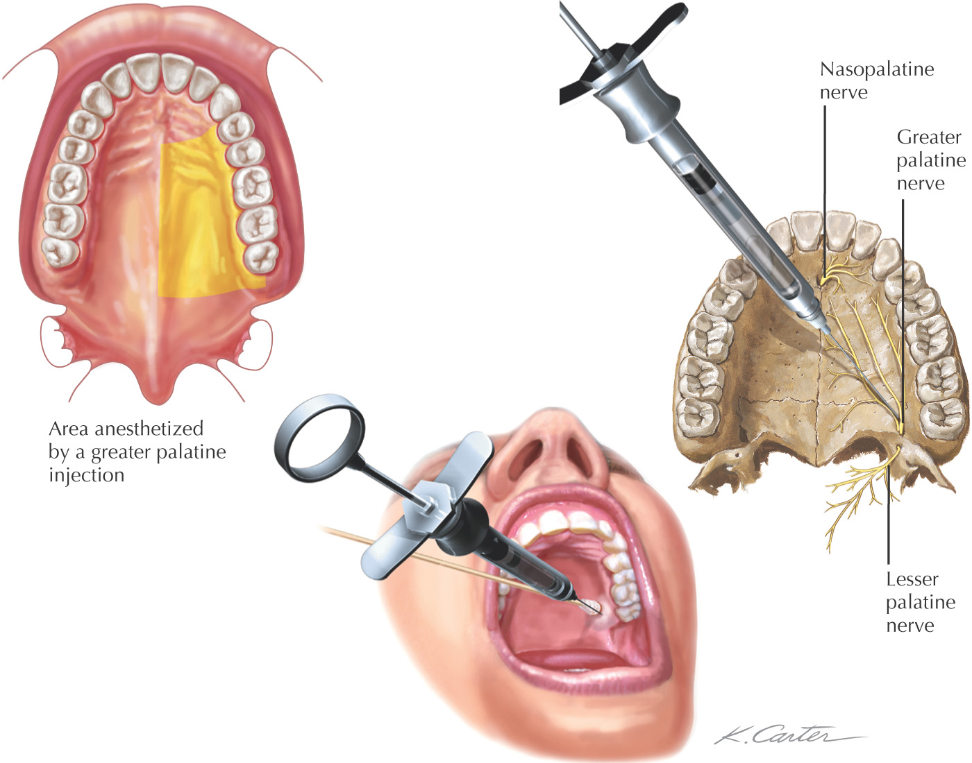
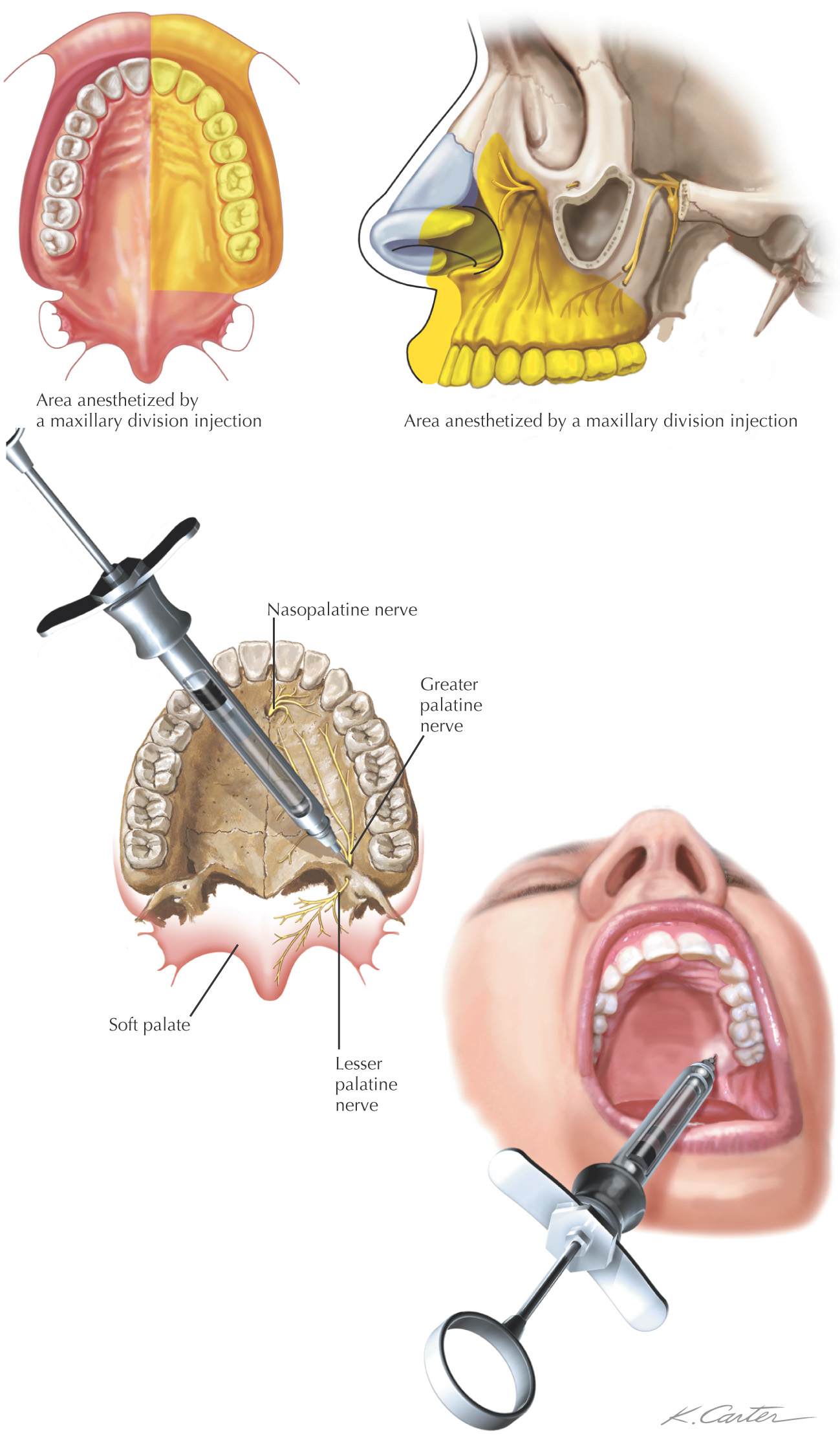
• Composed of the palatal process of the maxilla and the horizontal plate of the palatine
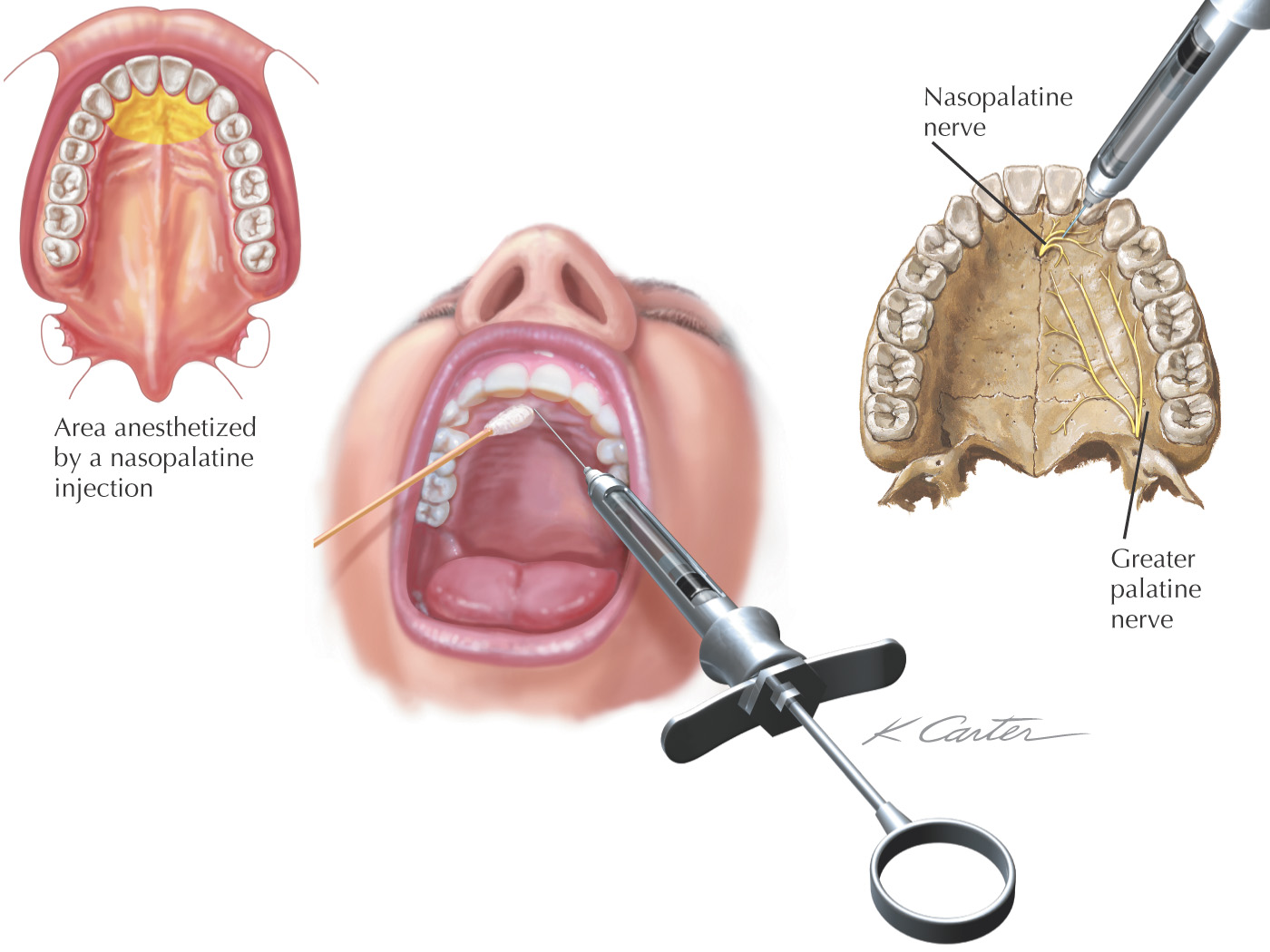
• Supplied by the nasopalatine and greater palatine nerves