56 Paramyxoviruses
A 10-year-old boy presented with cough, conjunctivitis, and coryza plus fever and lymphadenopathy, which progressed to a rash that spread from the hairline down his face and then his body. Within 10 days, the disease appeared to run its course, but a week after the start of the rash, an abrupt onset of headache, vomiting, and confusion progressed to coma, consistent with encephalitis.
1. How does measles replicate?
2. What are the characteristic signs of measles?
Answers
1. Measles binds to sialic acid on glycoproteins and glycolipids, fuses its membrane with the cell’s membrane, delivers its negative-strand genome and RNA-dependent RNA polymerase components into the cytoplasm, where individual mRNAs are generated; a full-length copy (positive-sense template) of the genome is made, and new genomes are transcribed from the template. The proteins of the virus are translated, including glycoproteins, on rough endoplasmic reticulum. The glycoproteins are processed similar to cellular glycoproteins and then inserted into the plasma membrane. The matrix protein associates with these proteins, and the nucleocapsid (genome plus polymerase components) associates with the matrix protein, which promotes the budding of the virion from the membrane, causing it to leave the infected cell. The virus does not have to kill the cell to exit.
2. CCCK: cough, conjunctivitis, coryza, and Koplik spots. Also, photophobia and rash.
3. Measles is transmitted by aerosols.
4. Either the boy never was vaccinated or he did not get a booster to ensure that he has sufficient immune protection.
5. Pneumonia, which can also be a serious complication, accounts for 60% of the deaths caused by measles. Bacterial superinfection is common in patients with pneumonia caused by the measles virus. Encephalitis caused by the virus usually begins 7 to 10 days after the onset of illness, but infection can induce postinfectious encephalitis caused by immunopathologic reactions. Subacute sclerosing panencephalitis is a very late neurologic sequela of measles resulting from a mutant of measles that replicates slowly in neurons until a threshold level induces inflammation.
The Paramyxoviridae include the following genera: Morbillivirus, Paramyxovirus, and Pneumovirus (Table 56-1). Human pathogens within the morbilliviruses include the measles virus; within the paramyxoviruses, the parainfluenza and mumps viruses; and within the pneumoviruses, the respiratory syncytial virus (RSV) and the newly discovered but relatively common metapneumovirus. Their virions have similar morphologies and protein components, and they share the capacity to induce cell-to-cell fusion (syncytia formation and multinucleated giant cells). A new group of highly pathogenic paramyxoviruses, including two zoonosis-causing viruses, Nipah virus and Hendra virus, was identified in 1998 after an outbreak of severe encephalitis in Malaysia and Singapore.
| Genus | Human Pathogen |
|---|---|
| Morbillivirus | Measles virus |
| Paramyxovirus | Parainfluenza viruses 1 to 4 |
| Mumps virus | |
| Pneumovirus | Respiratory syncytial virus |
| Metapneumovirus |
These agents cause some well-known major diseases. Measles virus causes a potentially serious generalized infection characterized by a maculopapular rash (rubeola). Parainfluenza viruses cause upper and lower respiratory tract infections, primarily in children, including pharyngitis, croup, bronchitis, bronchiolitis, and pneumonia. Mumps virus causes a systemic infection whose most prominent clinical manifestation is parotitis. RSV causes mild upper respiratory tract infections in children and adults but can cause life-threatening pneumonia in infants.
Measles and mumps viruses have only one serotype, and protection is provided by an effective live vaccine. In the United States and other developed countries, successful vaccination programs using the live attenuated measles and mumps vaccines have made measles and mumps rare. In particular, these programs have led to the virtual elimination of the serious sequelae of measles.
Structure and Replication
Paramyxoviruses are relatively large viruses with a negative-sense, single-stranded ribonucleic acid (RNA) (5 to 8 × 10⁶ Da) genome in a helical nucleocapsid surrounded by a pleomorphic envelope of approximately 156 to 300 nm (Figure 56-1). They are similar in many respects to orthomyxoviruses but are larger and do not have the segmented genome of the influenza viruses (Box 56-1). Although there are similarities in paramyxovirus genomes, the order of the protein-coding regions differs for each genus. The gene products of the measles virus are listed in Table 56-2.
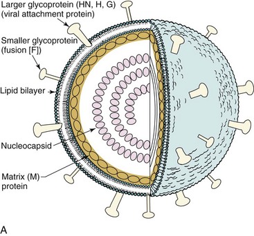
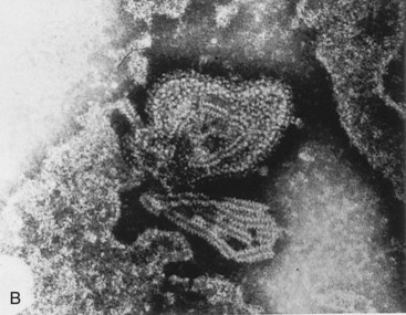
Figure 56-1 A, Model of paramyxovirus. The helical nucleocapsid—consisting of negative-sense, single-stranded RNA and the P protein, nucleoprotein, and large protein—associates with the matrix (M) protein at the envelope membrane surface. The nucleocapsid contains RNA transcriptase activity. The envelope contains the viral attachment glycoprotein (hemagglutinin-neuraminidase [HN], hemagglutinin [H], or G protein [G], depending upon the virus) and the fusion (F) protein. B, Electron micrograph of a disrupted paramyxovirus, showing the helical nucleocapsid.
(A, Modified from Jawetz E, Melnick JL, Adelberg EA: Review of medical microbiology, ed 17, Norwalk, Conn, 1987, Appleton & Lange. B, Courtesy Centers for Disease Control and Prevention, Atlanta.)
Box 56-1
Unique Features of the Paramyxoviridae
Large virion consists of a negative-sense RNA genome in a helical nucleocapsid surrounded by an envelope containing a viral attachment protein (hemagglutinin-neuraminidase [HN] for parainfluenza virus and mumps virus; hemagglutinin [H] for measles virus; or glycoprotein [G] for respiratory syncytial virus [RSV]) and a fusion glycoprotein (F).
The three genera can be distinguished by the activities of the viral attachment protein: HN of parainfluenza virus and mumps virus binds to sialic acid and has hemagglutinin and neuraminidase activity, H of measles virus binds protein receptors and is also a hemagglutinin, but G of RSV binds but is not a hemagglutinin.
Virus replicates in the cytoplasm.
Virions penetrate the cell by fusion with the plasma membrane and exit by budding from the plasma membrane.
Viruses induce cell-to-cell fusion, causing multinucleated giant cells.
Paramyxoviridae are transmitted in respiratory droplets and initiate infection in the respiratory tract.
Cell-mediated immunity causes many of the symptoms but is essential for control of the infection.
Table 56-2 Viral-Encoded Proteins of Measles Virus
| Gene Products* | Virion Location | Function |
|---|---|---|
| Nucleoprotein (NP) | Major internal protein | Protection of viral RNA |
| Polymerase phosphoprotein (P) | Association with nucleoprotein | Part of transcription complex |
| Matrix (M) | Inside virion envelope | Assembly of virions |
| Fusion protein (F) | Transmembranous envelope glycoprotein | Protein promotes fusion of cells, hemolysis, and viral entry |
| Hemagglutinin (H) | Transmembranous envelope glycoprotein | Viral attachment protein |
| Large protein (L) | Association with nucleoprotein | Polymerase |
Modified from Fields BN: Virology, New York, 1985, Raven.
The nucleocapsid consists of the negative-sense, single-stranded RNA associated with the nucleoprotein (NP), polymerase phosphoprotein (P), and large (L) protein. The L protein is the RNA polymerase, the P protein facilitates RNA synthesis, and the NP protein helps maintain genomic structure. The nucleocapsid associates with the matrix (M) protein lining the inside of the virion envelope. The envelope contains two glycoproteins, a fusion (F) protein, which promotes fusion of the viral and host cell membranes, and a viral attachment protein (hemagglutinin-neuraminidase [HN], hemagglutinin [H], or glycoprotein [G] protein) (see Box 56-1). To express membrane-fusing activity, the F protein must be activated by proteolytic cleavage, which produces F1 and F2 glycopeptides held together by a disulfide bond.
Replication of the paramyxoviruses is initiated by the binding of the HN, H, or G protein on the virion envelope to sialic acid on the cell surface glycolipids and glycoproteins. The measles virus can bind to the CD46 (membrane cofactor protein [MCP]) present on most cell types and also CD150 signaling lymphocyte-activation molecule (SLAM), which is expressed on activated T and B cells. CD46 protects the cell from complement by regulating complement activation and is also the receptor for human herpes virus 6 and some strains of adenovirus. SLAM regulates TH1 and TH2 responses, and measles virus may upset this regulation. The F protein promotes fusion of the envelope with the plasma membrane. Paramyxoviruses are also able to induce cell-to-cell fusion, thereby creating multinucleated giant cells (syncytia).
The replication of the genome occurs in a manner similar to that of other negative-strand RNA viruses (i.e., rhabdoviruses). The RNA polymerase is carried into the cell as part of the nucleocapsid. Transcription, protein synthesis, and replication of the genome all occur in the host cell’s cytoplasm. The genome is transcribed into individual messenger RNAs (mRNAs) and a full-length positive-sense RNA template. New genomes associate with the L, N, and NP proteins to form helical nucleocapsids, which associate with the M proteins on viral glycoprotein-modified plasma membranes. The glycoproteins are synthesized and processed like cellular glycoproteins. Mature virions then bud from the host cell plasma membrane and exit without killing the cell. Replication of the paramyxoviruses is represented by the RSV infectious cycle shown in Figure 56-2.
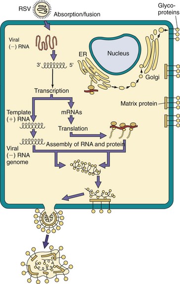
Figure 56-2 Replication of paramyxoviruses. The virus binds to glycolipids or proteins and fuses with the cell surface. Individual messenger RNAs (mRNAs) for each protein and a full-length template are transcribed from the genome. Replication occurs in the cytoplasm. Proteins associate with the genome, and the nucleocapsid associates with matrix and glycoprotein-modified plasma membranes. The virus leaves the cell by budding. (−), Negative sense; (+), positive sense; ER, endoplasmic reticulum; RSV, respiratory syncytial virus.
(Modified from Balows A, et al: Laboratory diagnosis of infectious diseases: principles and practice, New York, 1988, Springer-Verlag.)
Measles Virus
Measles, also known as rubeola, is one of the five classic childhood exanthems, along with rubella, roseola, fifth disease, and chickenpox. Historically, measles was one of the most common and unpleasant viral infections, with serious potential sequelae. Before 1960, more than 90% of the population younger than 20 years had experienced the rash, high fever, cough, conjunctivitis, and coryza of measles. Since the use of the live vaccine began in 1993, fewer than 1000 cases have been reported in the United States. Measles is still one of the most prominent causes of disease (45 million cases per year) and death (1 to 2 million per year) worldwide in unvaccinated populations.
Pathogenesis and Immunity
Measles is known for its propensity to cause cell fusion, leading to the formation of giant cells (Box 56-2). As a result, the virus can pass directly from cell to cell and escape antibody control. Inclusions occur most commonly in the cytoplasm and are composed of incomplete viral particles. Virus production occurs with eventual cell lysis. Persistent infections without lysis can occur in certain cell types (e.g., human brain cells).
Box 56-2
Disease Mechanisms of Measles Virus
Virus infects epithelial cells of respiratory tract.
Virus spreads systemically in lymphocytes and by viremia.
Virus replicates in cells of conjunctivae, respiratory tract, urinary tract, lymphatic system, blood vessels, and central nervous system.
Rash is caused by T-cell response to virus-infected epithelial cells lining capillaries.
Virus causes immunosuppression.
Cell-mediated immunity is essential to control infection.
Sequelae in the central nervous system may result from immunopathogenesis (postinfectious measles encephalitis) or development of defective mutants (subacute sclerosing panencephalitis).
Measles is highly contagious and is transmitted from person to person by respiratory droplets (Figure 56-3). After local replication of virus in epithelial cells of the respiratory tract, the virus infects monocytes and lymphocytes, and the virus is spread through the lymphatic system and by a cell-associated viremia. The wide dissemination of the virus causes infection of the conjunctiva, respiratory tract, urinary tract, small blood vessels, lymphatic system, and the central nervous system. The characteristic maculopapular measles rash is caused by immune T cells targeted to measles-infected endothelial cells lining small blood vessels. Recovery follows the rash in most patients, who then have lifelong immunity to the virus. Death can occur because of pneumonia, diarrhea, or encephalitis. The time course of measles infection is shown in Figure 56-4.
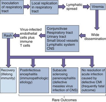
Figure 56-3 Mechanisms of spread of the measles virus within the body and the pathogenesis of measles. CMI, Cell-mediated immunity; CNS, central nervous system.
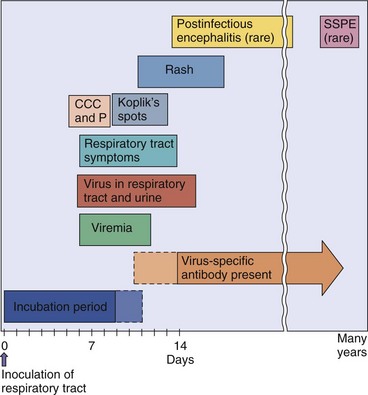
Figure 56-4 Time course of measles virus infection. Characteristic prodrome symptoms are cough, conjunctivitis, coryza, and photophobia (CCC and P), followed by the appearance of Koplik spots and rash. SSPE, Subacute sclerosing panencephalitis.
Measles can cause encephalitis in three ways: (1) direct infection of neurons; (2) a postinfectious encephalitis, which is believed to be immune mediated; and (3) subacute sclerosing panencephalitis (SSPE) caused by a defective variant of measles generated during the acute disease. The SSPE virus acts as a slow virus and causes symptoms and cytopathologic effect in neurons many years after acute disease.
Measles and other paramyxoviruses are excellent inducers of interferon-α and -β, which activate natural killer (NK) cells. Cell-mediated immunity is responsible for most of the symptoms and is essential for the control of measles infection. T-cell–deficient children who are infected with measles have an atypical presentation, consisting of giant cell pneumonia without a rash. Antibody, including maternal antibody and passive immunization, can block the viremic spread of the virus and prevent or lessen disease. Protection from reinfection is lifelong.
During the incubation period, measles causes a decrease in eosinophils and lymphocytes, including B and T cells, and a depression of their response to activation (mitogens). The virus depresses the immune response by (1) directly infecting monocytes and T and B cells and (2) by depressing interleukin 12 (IL-12) production and TH1-type T-cell helper responses. Depression of cell-mediated immune and delayed-type hypersensitivity (DTH) responses increases risk to opportunistic and other infections. This immunosuppression lasts for weeks or months after the disease.
Epidemiology
The development of effective vaccine programs has made measles a rare disease in the United States. In areas without a vaccine program, epidemics tend to occur in 1- to 3-year cycles, when a sufficient number of susceptible people have accumulated. Many of these cases occur in preschool-age children who have not been vaccinated and live in large urban areas. The incidence of infection peaks in the winter and spring. Measles is still common in people living in developing countries, especially in individuals who refuse immunization or have not received a booster in their teenage years. Immunocompromised and malnourished people with measles may not be able to resolve the infection, resulting in death. It is the most significant cause of death in children 1 to 5 years of age in several countries.
Measles, which can be spread in respiratory secretions before and after the onset of characteristic symptoms, is one of the most contagious infections known (Box 56-3). In a household, approximately 85% of exposed susceptible people become infected, and 95% of these people develop clinical disease.
The measles virus has only one serotype, infects only humans, and infection usually manifests as symptoms. These properties facilitated the development of an effective vaccine program. Once vaccination was introduced, the yearly incidence of measles dropped dramatically in the United States, from 300 to 1.3 per 100,000 (U.S. statistics for 1981 to 1988). This change represented a 99.5% reduction in the incidence of infection from the prevaccination years of 1955 to 1962. Incidence of measles must be reported to state and federal health departments.
Despite the effectiveness of vaccination programs, poor compliance and the prevaccinated population (children younger than 2 years) continue to provide susceptible individuals. The virus may surface from within the community or can be imported by immigration from areas of the world lacking an effective vaccine program. Once again, outbreaks of measles are occurring more often in the United States, France, and England. In 2011, most of the cases in the United States were imported from other countries, and most of the patients were unvaccinated. An outbreak of measles in a day-care center (10 infants too young to have been vaccinated and two adults) was traced to an infant from the Philippines.
Clinical Syndromes
Measles is a serious febrile illness (Table 56-3). The incubation period lasts 7 to 13 days, and the prodrome starts with high fever and “CCC and P”—cough, coryza, conjunctivitis, and photophobia. The disease is most infectious during this time.
Table 56-3 Clinical Consequences of Measles Virus Infection
| Disorder | Symptoms |
|---|---|
| Measles | Characteristic maculopapular rash, cough, conjunctivitis, coryza, photophobia, Koplik spots Complications: otitis media, croup, pneumonia, blindness, encephalitis |
| Atypical measles | More intense rash (most prominent in distal areas); possible vesicles, petechiae, purpura, or urticaria |
| Postmeasles encephalitis | Acute onset of headache, confusion, vomiting, possible coma after rash dissipates |
| Subacute sclerosing panencephalitis | Central nervous system manifestations (e.g., personality, behavior, and memory changes; myoclonic jerks; spasticity; and blindness) |
After 2 days of prodromal illness, the typical mucous membrane lesions known as Koplik spots (Figure 56-5) appear. They are seen most commonly on the buccal mucosa across from the molars, but they may appear on other mucous membranes as well, including the conjunctivae and the vagina. The vesicular lesions, which last 24 to 48 hours, are usually small (1 to 2 mm) and are best described as grains of salt surrounded by a red halo. Their appearance with the other disease signs establishes with certainty the diagnosis of measles.
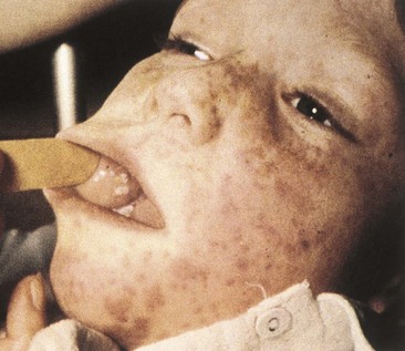
Figure 56-5 Koplik spots in the mouth and exanthem. Koplik spots usually precede the measles rash and may be seen for the first day or two after the rash appears.
(Courtesy Dr JI Pugh, St Albans City Hospital, West Hertfordshire, England; from Emond RTD, Rowland HAK: A color atlas of infectious diseases, ed 3, London, 1995, Mosby.)
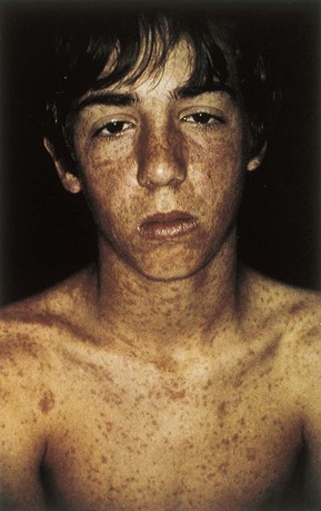
Within 12 to 24 hours of the appearance of Koplik spots, the exanthem of measles starts below the ears and spreads over the body. The rash is maculopapular, usually very extensive, and often the lesions become confluent. The rash, which takes 1 or 2 days to cover the body, fades in the same order in which it appeared. The fever is highest and the patient is sickest on the day the rash appears (Figure 56-6).
Pneumonia, which can also be a serious complication, accounts for 60% of the deaths caused by measles. Similar to the incidence of the other complications associated with measles, the mortality associated with pneumonia is higher in the malnourished and for the extremes of age. Bacterial superinfection is common in patients with pneumonia caused by the measles virus.
One of the most feared complications of measles is encephalitis, which occurs in as few as 0.5% of those infected but carries a fatality rate of 15%. Encephalitis may rarely occur during acute disease but usually begins 7 to 10 days after the onset of illness. This postinfectious encephalitis is caused by immunopathologic reactions, is associated with demyelination of neurons, and occurs more often in older children and adults.
Atypical measles occurred in people who received the older inactivated measles vaccine and were subsequently exposed to the wild-type measles virus. It may also rarely occur in those vaccinated with the attenuated virus vaccine. Prior sensitization with insufficient protection can enhance the immunopathologic response to the challenge by wild measles virus. The illness begins abruptly and is a more intense presentation of measles.
Subacute sclerosing panencephalitis is an extremely serious, very late neurologic sequela of measles that afflicts approximately seven of every one million patients. The incidence of SSPE has decreased markedly because of measles vaccination programs.
This disease occurs when a defective measles virus persists in the brain and acts as a slow virus. The virus can replicate and spread directly from cell to cell but is not released. SSPE is most prevalent in children who were initially infected when younger than 2 years and occurs approximately 7 years after clinical measles. The patient demonstrates changes in personality, behavior, and memory, followed by myoclonic jerks, blindness, and spasticity. Unusually high levels of measles antibodies are found in the blood and cerebrospinal fluid of patients with SSPE.
The immunocompromised and malnourished child is at highest risk for severe outcome of measles (Clinical Case 56-1). Giant cell pneumonia without rash occurs in children lacking T-cell immunity. Whereas the death rate to measles in the United States is only 0.1%, complications, severe bacterial superinfection, and pneumonia in malnourished children result in up to 60% mortality.
Clinical Case 56-1
Measles in the Immunocompromised Child
The lack of cell-mediated immune responses allows measles infection of immunocompromised individuals to progress to serious outcomes. In a case reported by Pullan and associates (Br Med J 1:1562–1565, 1976), within 3 days of exposure to measles, a child on chemotherapy for acute lymphoblastic leukemia (ALL) received pooled immunoglobulin. Despite the IgG therapy, 23 days after exposure, she developed an extensive measles rash, which became hemorrhagic. She had a fever of 39.5° C and bronchopneumonia. Measles was grown from nasopharyngeal secretions, and immunohistochemistry identified giant cells (syncytia) containing measles antigen within the secretions. Her chemotherapy was stopped, and she received several massive doses of immunoglobulin. She started to improve 1 month after the onset of the rash.
In another case, during the 2.5 years that a boy was under treatment for ALL, he suffered severe herpes simplex virus infections around the mouth and herpes zoster on his trunk. During the third year on therapy, he was exposed to measles from his sister and received pooled IgG. After 19 days, he developed mild respiratory symptoms but no rash. After 29 days, he refused to go to school and misbehaved; behavior changes progressed. After 9 weeks, he developed focal motor seizures, increased drowsiness, slurring of speech, and confusion, which progressed to coma and death within 8 days of the onset of seizures. Serology indicated a lack of measles antibody. Autopsy indicated the presence of cytomegalovirus but not measles in the lungs. The brain showed extensive degeneration, but no virus was isolated from the samples. Brain sections indicated large intranuclear and cytoplasmic inclusion bodies with tubular structures that resembled measles nucleocapsids in the cytoplasm. Immunofluorescence with antibody from individuals with subacute sclerosing panencephalitis (SSPE) or antimeasles antibody indicated the presence of measles antigen. These cases illustrate the excessive pathology that measles can cause in the absence of a competent T-cell response. The lack of immune control allowed the progression of the virus to the brain, where it or a variant (SSPE) caused pathology leading to encephalitis.
Laboratory Diagnosis
The clinical manifestations of measles are usually so characteristic that it is rarely necessary to perform laboratory tests to establish the diagnosis. The measles virus is difficult to isolate and grow, although it can be grown in primary human- or monkey-cell cultures. Respiratory tract secretions, urine, blood, and brain tissue are the recommended specimens. It is best to collect respiratory and blood specimens during the prodromal stage and until 1 to 2 days after the appearance of the rash.
Measles antigen can be detected in pharyngeal cells or urinary sediment with immunofluorescence; the measles genome can be identified by reverse transcriptase polymerase chain reaction (RT-PCR) in either of the aforementioned specimens. Characteristic cytopathologic effects, including multinucleated giant cells with cytoplasmic inclusion bodies, can be seen in Giemsa-stained cells taken from the upper respiratory tract and urinary sediment.
Antibody, especially immunoglobulin M (IgM), can be detected when the rash is present. Measles infection can be confirmed by the finding of seroconversion or by a fourfold increase in the titer of measles-specific antibodies between sera obtained during the acute stage and the convalescent stage.
Treatment, Prevention, and Control
As stated previously, a live attenuated measles vaccine, in use in the United States since 1963, has been responsible for a significant reduction in the incidence of measles. The Schwartz or Moraten attenuated strains of the original Edmonston B vaccine are currently being used. Live attenuated vaccine is given to all children at 2 years of age, in combination with mumps and rubella (measles-mumps-rubella [MMR] vaccine) and the varicella vaccines (Box 56-4). Although early childhood immunization is successful in more than 95% of vaccinees, revaccination before grade school or junior high school is required in many states. Misinformation regarding immunization risks caused many parents to refrain from vaccinating their children, putting them at risk of infection and disease. Because of the contagious nature of measles, a decrease in the immunized population to 93% creates a risk of an outbreak of measles.
Box 56-4
Measles-Mumps-Rubella Vaccine
Composition: live attenuated viruses
Measles: Schwartz or Moraten substrains of Edmonston B strain
Vaccination schedule: at age 15 to 24 months and at age 4 to 6 years or before junior high school (12 years of age)
Data from Update on adult immunization. Recommendations of the Immunization Practices Advisory Committee (ACIP), MMWR Recomm Rep 40(RR-12):1–94, 1991.
As noted earlier, a killed measles vaccine introduced in 1963 was not protective; its use was subsequently discontinued because recipients were at risk of the more serious atypical measles presentation on infection. Because measles is strictly a human virus with only one serotype, it is a good candidate for eradication, but this is prevented by difficulties in distributing the vaccine to regions that lack proper refrigeration facilities (e.g., Africa) and distribution networks.
Hospitals in areas experiencing endemic measles may wish to vaccinate or check the immune status of their employees to decrease the risk of nosocomial transmission. Pregnant women, immunocompromised individuals, people with allergies to gelatin or neomycin (components of the vaccine) should not receive the MMR vaccine. Exposed susceptible people who are immunocompromised should be given immune globulin to lessen the risk and severity of clinical illness. This product is most effective if given within 6 days of exposure. High-dose vitamin A treatment reduces the risk of measles mortality and is recommended by the World Health Organization. No specific antiviral treatment is available for measles.
Parainfluenza Viruses
Parainfluenza viruses, which were discovered in the late 1950s, are respiratory viruses that usually cause mild coldlike symptoms but can also cause serious respiratory tract disease. Four serologic types within the parainfluenza genus are human pathogens. Types 1, 2, and 3 are second only to RSV as important causes of severe lower respiratory tract infection in infants and young children. They are especially associated with laryngotracheobronchitis (croup). Type 4 causes only mild upper respiratory tract infection in children and adults.
Pathogenesis and Immunity
Parainfluenza viruses infect epithelial cells of the upper respiratory tract (Box 56-5). The virus replicates more rapidly than measles and mumps viruses and can cause giant cell formation and cell lysis. Unlike measles and mumps viruses, the parainfluenza viruses rarely cause viremia. The viruses generally stay in the upper respiratory tract, causing only coldlike symptoms. In approximately 25% of cases, the virus spreads to the lower respiratory tract, and in 2% to 3%, disease may take the severe form of laryngotracheobronchitis.
Box 56-5
Disease Mechanisms of Parainfluenza Viruses
There are four serotypes of viruses.
Infection is limited to the respiratory tract; upper respiratory tract disease is most common, but significant disease can occur with lower respiratory tract infection.
Parainfluenza viruses do not cause viremia or become systemic.
Diseases include coldlike symptoms, bronchitis (inflammation of bronchial tubes), and croup (laryngotracheobronchitis).
The cell-mediated immune response both causes cell damage and confers protection. IgA responses are protective but short lived. Parainfluenza viruses manipulate cell-mediated immunity to limit development of memory. Multiple serotypes and the short duration of immunity after natural infection make reinfection common, but the reinfection disease is milder, suggesting at least partial immunity.
Epidemiology
Parainfluenza viruses are ubiquitous, and infection is common (Box 56-6). The virus is transmitted by person-to-person contact and respiratory droplets. Primary infections usually occur in infants and children younger than 5 years. Reinfections occur throughout life, indicating short-lived immunity. Infections with parainfluenza viruses 1 and 2, the major causes of croup, tend to occur in the autumn, whereas parainfluenza virus 3 infections occur throughout the year. All of these viruses spread readily within hospitals and can cause outbreaks in nurseries and pediatric wards.
Clinical Syndromes
Parainfluenza viruses 1, 2, and 3 may cause respiratory tract syndromes ranging from a mild coldlike upper respiratory tract infection (coryza, pharyngitis, mild bronchitis, wheezing, and fever) to bronchiolitis and pneumonia. Older children and adults generally experience milder infections than those seen in young children, although pneumonia may occur in the elderly.
A parainfluenza virus infection in infants may be more severe than infections in adults, causing bronchiolitis, pneumonia, and most notably croup (laryngotracheobronchitis). Croup results in subglottal swelling, which may close the airway. Hoarseness, a “seal bark” cough, tachypnea, tachycardia, and suprasternal retraction develop in infected patients after a 2- to 6-day incubation period. Most children recover within 48 hours. The principal differential diagnosis is epiglottitis caused by Haemophilus influenzae.
Laboratory Diagnosis
Parainfluenza virus is isolated from nasal washings and respiratory secretions and grows well in primary monkey kidney cells. Similar to other paramyxoviruses, the virions are labile during transit to the laboratory. The presence of virus-infected cells in aspirates or in cell culture is indicated by the finding of syncytia and is identified with immunofluorescence. Similar to the hemagglutinin of the influenza viruses, the hemagglutinin of the parainfluenza viruses promotes hemadsorption and hemagglutination. The serotype of the virus can be determined through the use of specific antibody to block hemadsorption or hemagglutination (hemagglutination inhibition). Rapid RT-PCR techniques are becoming the method of choice to detect and identify parainfluenza viruses from respiratory secretions.
Treatment, Prevention, and Control
Treatment of croup consists of the administration of nebulized cold or hot steam and careful monitoring of the upper airway. On rare occasions, intubation may become necessary. No specific antiviral agents are available.
Vaccination with killed vaccines is ineffective, possibly because they fail to induce local secretory antibody and appropriate cellular immunity. No live attenuated vaccine is available.
Mumps Virus
Mumps virus is the cause of acute, benign viral parotitis (painful swelling of the salivary glands). Mumps is rarely seen in countries that promote use of the live vaccine, which is administered with the measles and rubella live vaccines.
Mumps virus was isolated in embryonated eggs in 1945 and in cell culture in 1955. The virus is most closely related to parainfluenza virus 2, but there is no cross-immunity with the parainfluenza viruses.
Pathogenesis and Immunity
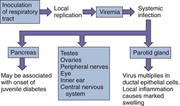
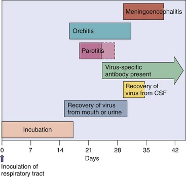
The mumps virus, of which only one serotype is known, causes a lytic infection of cells (Box 56-7). The virus initiates infection in the epithelial cells of the upper respiratory tract and infects the parotid gland, either by way of the Stensen duct or by means of a viremia. The virus is spread by the viremia throughout the body to the testes, ovary, pancreas, thyroid, and other organs. Infection of the central nervous system, especially the meninges, occurs in as many as 50% of those infected (Figure 56-7). Inflammatory responses are mainly responsible for the symptoms. The time course of human infection is shown in Figure 56-8. Immunity is lifelong.
Box 56-7
Disease Mechanisms of Mumps Virus
Virus infects epithelial cells of respiratory tract.
Virus spreads systemically by viremia.
Infection of parotid gland, testes, and central nervous system occurs.
Principal symptom is swelling of parotid glands caused by inflammation.
Cell-mediated immunity is essential for control of infection and responsible for causing some of the symptoms. Antibody is not sufficient because of virus’s ability to spread cell to cell.
Epidemiology
Mumps, like measles, is a very communicable disease with only one serotype, and it infects only humans (Box 56-8). In the absence of vaccination programs, infection occurs in 90% of people by the age of 15 years. The virus spreads by direct person-to-person contact and respiratory droplets. The virus is released in respiratory secretions from patients who are asymptomatic and during the 7-day period before clinical illness, so it is virtually impossible to control the spread of the virus. Living or working in close quarters promotes the spread of the virus, and the incidence of the infection is greatest in the winter and spring.
Clinical Syndromes
Mumps infections are often asymptomatic. Clinical illness manifests as a parotitis that is almost always bilateral and accompanied by fever. Onset is sudden. Oral examination reveals redness and swelling of the ostium of the Stensen (parotid) duct. The swelling of other glands (epididymoorchitis, oophoritis, mastitis, pancreatitis, and thyroiditis) and meningoencephalitis may occur a few days after the onset of the viral infection but can occur in the absence of parotitis. The swelling that results from mumps orchitis may cause sterility. Mumps virus involves the central nervous system in approximately 50% of patients; 10% of those affected may exhibit mild meningitis with 5 per 1000 cases of encephalitis.
Laboratory Diagnosis
Virus can be recovered from saliva, urine, the pharynx, secretions from the Stensen duct, and cerebrospinal fluid. Virus is present in saliva for approximately 5 days after the onset of symptoms and in urine for as long as 2 weeks. Mumps virus grows well in monkey kidney cells, causing the formation of multinucleated giant cells. The hemadsorption of guinea pig erythrocytes also occurs on virus-infected cells, because of the viral hemagglutinin.
A clinical diagnosis can be confirmed by serologic testing. A fourfold increase in the virus-specific antibody level or the detection of mumps-specific IgM antibody indicates recent infection. Enzyme-linked immunosorbent assay, immunofluorescence tests, and hemagglutination inhibition can be used to detect the mumps virus, antigen, or antibody.
Treatment, Prevention, and Control
Vaccines provide the only effective means for preventing the spread of mumps infection. Since the introduction of the live attenuated vaccine (Jeryl Lynn vaccine) in the United States in 1967 and its administration as part of the MMR vaccine, the yearly incidence of the infection has declined from 76 to 2 per 100,000. Antiviral agents are not available.
Respiratory Syncytial Virus
RSV, first isolated from a chimpanzee in 1956, is a member of the Pneumovirus genus. Unlike the other paramyxoviruses, RSV lacks a hemagglutinin and does not bind to sialic acid and therefore does not need or have a neuraminidase. It is the most common cause of fatal acute respiratory tract infection in infants and young children. It infects virtually everyone by 2 years of age, and reinfections occur throughout life, even among elderly persons.
Pathogenesis and Immunity
RSV produces an infection that is localized to the respiratory tract (Box 56-9). As the name suggests, RSV induces syncytia. The pathologic effect of RSV is mainly caused by direct viral invasion of the respiratory epithelium, which is followed by immunologically mediated cell injury. Necrosis of the bronchi and bronchioles leads to the formation of “plugs” of mucus, fibrin, and necrotic material within smaller airways. The narrow airways of young infants are readily obstructed by such plugs. Natural immunity does not prevent reinfection, and vaccination with killed vaccine appears to enhance the severity of subsequent disease.
Box 56-9
Disease Mechanisms of Respiratory Syncytial Virus
Virus causes localized infection of respiratory tract.
Virus does not cause viremia or systemic spread.
Pneumonia results from cytopathologic spread of virus (including syncytia).
Bronchiolitis is most likely mediated by host’s immune response.
Narrow airways of young infants are readily obstructed by virus-induced pathologic effects.
Epidemiology
RSV is very prevalent in young children; almost all children have been infected by 2 years of age (Box 56-10), with global annual infection rates of 64 million and mortality of 160,000. As many as 25% to 33% of these cases involve the lower respiratory tract, and 1% are severe enough to necessitate hospitalization (occurring in as many as 95,000 children in the United States each year).
RSV infections almost always occur in the winter. Unlike influenza, which may occasionally skip a year, RSV epidemics occur every year.
The virus is very contagious, with an incubation period of 4 to 5 days. The introduction of the virus into a nursery, especially into an intensive care nursery, can be devastating. Virtually every infant becomes infected, and the infection is associated with considerable morbidity and occasionally death. The virus is transmitted in aerosols but also on hands and by fomites.
As already noted, RSV infects virtually all children by the age of 4 years, especially in urban centers. Outbreaks may also occur among the elderly population (e.g., in nursing homes). Virus is shed in respiratory secretions for many days, especially by infants.
Clinical Syndromes (Box 56-11)
RSV can cause any respiratory tract illness, from a common cold to pneumonia (Table 56-4). Upper respiratory tract infection with prominent rhinorrhea (runny nose) is most common in older children and adults. A more severe lower respiratory tract illness, bronchiolitis, may occur in infants. Because of inflammation at the level of the bronchiole, there is air trapping and decreased ventilation. Clinically, the patient usually has low-grade fever, tachypnea, tachycardia, and expiratory wheezes over the lungs. Bronchiolitis is usually self-limited, but it can be a frightening disease to observe in an infant. It may be fatal in premature infants, persons with underlying lung disease, and immunocompromised people.
Box 56-11
Clinical Summaries
Measles: An 18-year-old woman had been home for 10 days after a trip to Haiti when she developed a fever, cough, runny nose, mild redness of her eyes; she now has a red, slightly raised rash over her face, trunk, and extremities. There are several 1-mm white lesions inside her mouth. She was never immunized for measles because of an irrelevant “egg allergy.”
Mumps: A 30-year-old man returning from a trip to Russia experienced a 1- to 2-day period of headache and decreased appetite, followed by swelling over both sides of his jaw. The swelling extended from the bottom of the jaw to in front of the ear. Five days after the jaw swelling appeared, the patient began complaining of nausea and lower abdominal and testicular pain.
Croup: A grumpy 2-year-old toddler with little appetite has a sore throat, fever, hoarse voice, and coughs with the sound of a barking seal. A high-pitched noise (stridor) is heard on inhalation. Flaring of the nostrils indicates difficulty breathing.
Table 56-4 Clinical Consequences of Respiratory Syncytial Virus Infection
| Disorder | Age Group Affected |
|---|---|
| Bronchiolitis, pneumonia, or both | Fever, cough, dyspnea, and cyanosis in children younger than 1 year |
| Febrile rhinitis and pharyngitis | Children |
| Common cold | Older children and adults |
Laboratory Diagnosis
RSV is difficult to isolate in cell culture. The presence of the viral genome in infected cells and nasal washings can be detected by RT-PCR techniques, and commercially available immunofluorescence and enzyme immunoassay tests are available for detection of the viral antigen. The finding of seroconversion or a fourfold or greater increase in the antibody titer can confirm the diagnosis for epidemiologic purposes.
Treatment, Prevention, and Control
In otherwise healthy infants, treatment is supportive, consisting of the administration of oxygen, intravenous fluids, and nebulized cold steam. Ribavirin, a guanosine analogue, is approved for the treatment of patients predisposed to a more severe course (e.g., premature or immunocompromised infants). It is administered by inhalation (nebulization).
Passive immunization with anti-RSV immunoglobulin is available for premature infants. Infected children must be isolated. Infection-control measures are required for hospital staff caring for infected children to avoid transmitting the virus to uninfected patients. These measures include hand washing and wearing gowns, goggles, and masks.
No vaccine is currently available for RSV prophylaxis. A previously available vaccine containing inactivated RSV caused recipients to have more severe RSV disease when subsequently exposed to the live virus. This development is thought to be the result of a heightened immunologic response at the time of exposure to the wild virus.
Human Metapneumovirus
Human metapneumovirus is a recently recognized member of the Pneumovirinae subfamily. Use of RT-PCR methods was and remains the means of detecting the pneumoviruses and distinguishing them from other respiratory disease viruses. Its identity was unknown until recently, because it is difficult to grow in cell culture. The virus is ubiquitous, and almost all 5-year-old children have experienced a virus infection and are seropositive.
As with its close cousin RSV, infections by human metapneumovirus may be asymptomatic, cause common cold–type disease, or serious bronchiolitis and pneumonia. Seronegative children, the elderly, and immunocompromised people are at risk for disease. Human metapneumovirus probably causes 15% of common colds in children, especially those which are complicated by otitis media. Signs of disease usually include cough, sore throat, runny nose, and high fever. Approximately 10% of patients with metapneumovirus will experience wheezing, dyspnea, pneumonia, bronchitis, or bronchiolitis. As with other common cold agents, laboratory identification of the virus is not performed routinely but can be performed by RT-PCR. Supportive care is the only therapy available for these infections.
Nipah and Hendra Viruses
A new paramyxovirus, Nipah virus, was isolated from patients after an outbreak of severe encephalitis in Malaysia and Singapore in 1998. Nipah virus is more closely related to the Hendra virus, discovered in 1994 in Australia, than to other paramyxoviruses. Both viruses have broad host ranges, including pigs, man, dogs, horses, cats, and other mammals. For Nipah virus, the reservoir is a fruit bat (flying fox). The virus can be obtained from fruit contaminated by infected bats or amplified in pigs and then spread to humans. The human is an accidental host for these viruses, but the outcome of human infection is severe. Disease signs include flulike symptoms, seizures, and coma. Of the 269 cases occurring in 1999, 108 were fatal. Another epidemic in Bangladesh in 2004 had a higher mortality rate.
An 18-year-old college freshman complained of a cough, runny nose, and conjunctivitis. The physician in the campus health center noticed small white lesions inside the patient’s mouth. The next day, a confluent red rash covered his face and neck.
1. What clinical characteristics of this case were diagnostic for measles?
2. Are any laboratory tests readily available to confirm the diagnosis? If so, what are they?
3. Is there a possible treatment for this patient?
4. When was this patient contagious?
5. Why is this disease not common in the United States?
6. Provide several possible reasons for this person’s susceptibility to measles at 18 years of age.
A 13-month-old child had a runny nose, mild cough, and low-grade fever for several days. The cough got worse and sounded like “barking.” The child made a wheezing sound when agitated. The child appeared well except for the cough. A lateral radiograph of the neck showed a subglottic narrowing.
7. What are the specific and common names for these symptoms?
8. What other agents would cause a similar clinical presentation (differential diagnosis)?
9. Are there readily available laboratory tests to confirm this diagnosis? If so, what are they?
10. Was there a possible treatment for this child?
11. When was this child contagious, and how was the virus transmitted?
1. The three Cs (cough, conjunctivitis, and coryza), rash, and Koplik spots (white lesions in mouth) are characterisitic of measles. Photophobia may also be present.
2. The diagnosis is usually made based on the disease signs. Laboratory tests that may confirm the diagnosis include an RT-PCR analysis of RNA to detect the viral genome or immunofluorescence to detect viral antigens in cells present in respiratory tract secretions, urine or blood.
3. There are no antiviral drugs available for measles but immunoglobulin can limit the severity of the disease.
4. The patient was contagious for approximately 7 days prior to and 3 to 4 days after the onset of disease signs.
5. Incidence of the disease has become rare because of an effective immunization program.
6. The patient had an insufficient immune response (most likely antibody) to prevent the viremic spread of the measles virus and the onset of disease. This could occur if the individual was not immunized or did not receive a booster immunization as a young teenager. In the absence of natural disease, our immune responses (including those established by immunization) do not receive a natural boost and may drop below a threshold of protection.
7. This disease is called laryngotracheobronchitis (croup) and is caused by parainfluenza virus.
8. Haemophilus influenzae can cause an epiglottitis, which would have similar disease signs. RSV, metapneumovirus, and influenza virus may cause croup-like disease.
9. Nasal washings can grow in tissue culture cells and will fuse the cells into multinucleated giant cells (syncytia). RT-PCR can be used to detect and identify the virus in nasal washings.
10. There is no antiviral drug for this disease, but nebulized cold or hot steam can help open the airway.
11. The child is contagious during the symptomatic period. The virus is transmitted by the respiratory route.