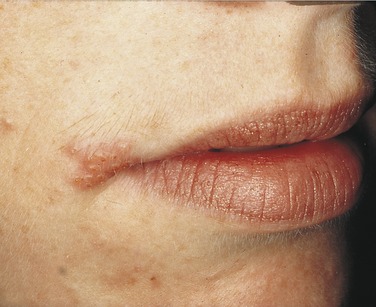
PLATE 1 Herpes labialis 12 hours after onset.
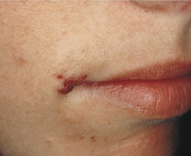
PLATE 2 Herpes labialis 48 hours after onset.
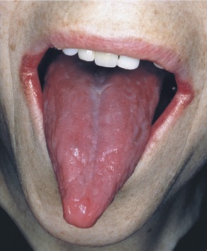
PLATE 3 Pernicious anemia. Note the angular cheilitis and depapillation of the tongue in a patient with pernicious anemia.
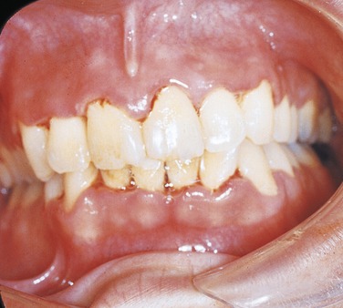
PLATE 4 Necrotizing ulcerative gingivitis.
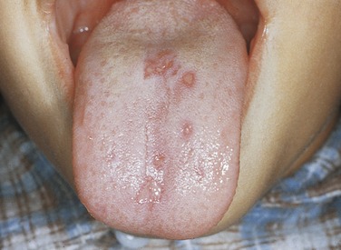
PLATE 5 Primary herpetic gingivostomatitis in a child.
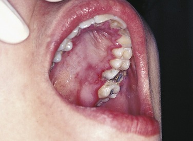
PLATE 6 Primary herpetic gingivostomatitis in an adolescent. Note the painful, swollen gingiva.
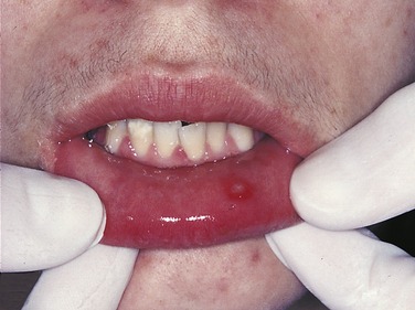
PLATE 7 Primary herpetic gingivostomatitis in an adolescent.
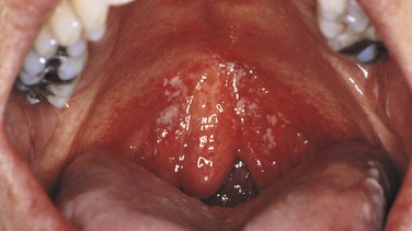
PLATE 8 Candidiasis in a patient with human immunodeficiency virus (HIV) infection. Removable white plaques are present on the mucosa of the soft palate.
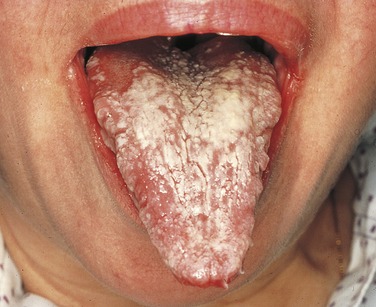
PLATE 9 Chronic hyperplastic candidiasis. The white appearance of the tongue did not wipe off, and it disappeared with antifungal treatment.
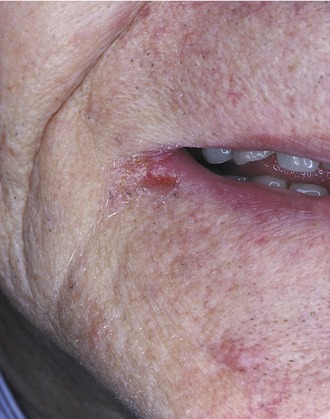
PLATE 10 Angular cheilitis.
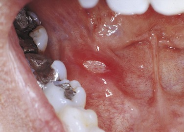
PLATE 11 Example of a minor aphthous ulcer.
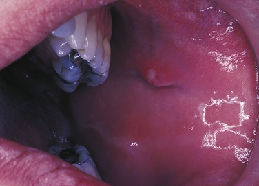
PLATE 12 Minor aphthous ulcer on the buccal mucosa on the papilla of Stensen’s duct.
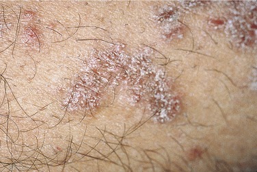
PLATE 13 Skin lesions of lichen planus.
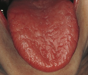
PLATE 14 Sjögren’s syndrome. The patient had severe xerostomia. The filiform papillae are lacking.
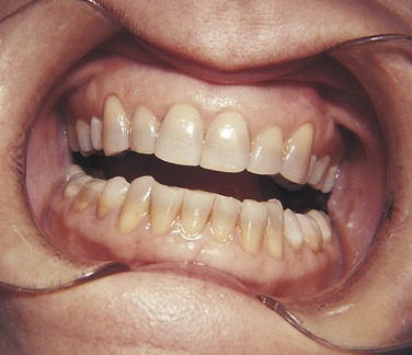
PLATE 15 Discoloration of teeth caused by tetracycline ingestion.
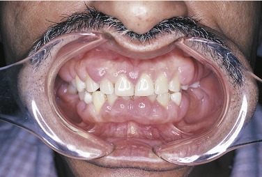
PLATE 16 Fibrous gingival enlargement.
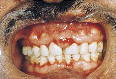
PLATE 17 Inflamed gingival enlargement.