Biochemistry of the skin
Keratins
The important molecules synthesized by the skin include keratin, melanin, collagen and glycosaminoglycans.
Keratins are high-molecular-weight polypeptide chains produced by keratinocytes (Fig. 1). They are the major constituent of the stratum corneum, hair and nails. The stratum corneum comprises 65% keratin (along with 10% soluble protein, 10% amino acid, 10% lipid and 5% cell membrane).
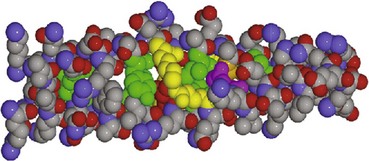
Fig. 1 Molecular structure of alpha-keratin.
The molecule forms a helical coil which, if stretched, unwinds irreversibly to produce the beta form. The covalent bonds linking the cystine molecules provide extra strength.
From J Invest Dermatol 2001: 116; 964–969, with permission of Blackwell Publishing.
Keratin proteins are of varying molecular weight (between 40 and 67 kDa). Different keratins are found at each level of the epidermis, depending on the stage of differentiation. Epidermal keratin contains less cystine and more glycine than the harder hair keratin.
Melanins
Melanin is produced from tyrosine (Fig. 2) in melanocytes and takes two forms:
 eumelanin, which is more common and gives a brown–black colour
phaeomelanin, which is less common and produces a yellow or red colour.
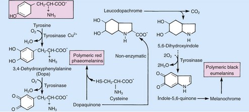
Fig. 2 Biosynthesis of melanin.
Eumelanin is a high-molecular-weight polymer of complex structure formed by oxidative polymerization. The phaeomelanin polymer is synthesized from dopaquinone and cysteine (via cysteinyl dopa).
Most natural melanins are mixtures of eumelanin and phaeomelanin. Melanins act as an energy sink and as free radical scavengers, and absorb the energy of ultraviolet (UV) radiation.
Collagens
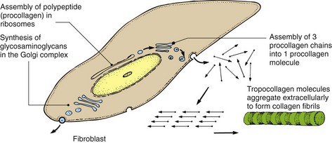
Collagens are synthesized by fibroblasts (Fig. 3) and are the major structural proteins of the dermis, forming 70–80% of its dry weight. The main amino acids in collagens are glycine, proline and hydroxyproline. Collagens are broken down, e.g. in wound healing, by collagenases, of which the matrix metalloproteinases are important. There are over 22 types of collagen; at least five are found in skin:
Glycosaminoglycans (GAGs)
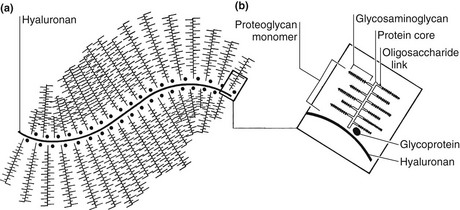
The ‘ground substance’ of skin is largely made up of GAGs, providing viscosity and hydration. In the dermis, chondroitin sulphate is the main GAG, along with dermatan sulphate and hyaluronan.
GAGs often exist as high-molecular-weight polymers with a protein core. These structures are known as proteoglycans (Fig. 4).
Skin surface secretions
The skin surface has a slightly acidic pH (between 6 and 7). Sebum (Table 1), sweat and the horny layer (including intercellular lipid) contribute to the surface conditions, which generally discourage microbial proliferation.
Table 1 Sebum and epidermal lipid composition
| Component | Sebum (%) | Epidermal lipid (%) |
|---|---|---|
| Glyceride/free fatty acid | 58 | 65 |
| Wax esters | 26 | 0 |
| Squalene | 12 | 0 |
| Cholesterol esters | 3 | 15 |
| Cholesterol | 1 | 20 |
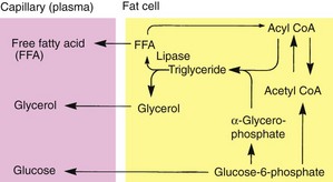
Subcutaneous fat
Triglyceride is synthesized from α-glycerophosphate and acyl coenzyme A (CoA). Triglyceride is broken down by lipase to give free fatty acid (FFA) – an energy source – and glycerol (Fig. 5).
Hormones and the skin
The skin is the site of production of one hormone (vitamin D), but it is often a target organ for other hormones and is frequently affected in endocrine diseases (Table 2).
| Hormone | Site of production | Effects |
|---|---|---|
Vitamin D |
Produced in the dermis from precursors though the action of UV radiation | Important for the absorption of calcium and for calcification |
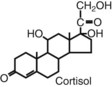
Corticosteroids |
Adrenal cortex | Receptors on several cells in both epidermis and dermis Produce vasoconstriction Reduce mitosis by basal cells Generate anti-inflammatory effects on leucocytes Inhibit phospholipase A |
| Androgens | Adrenal cortex Gonads |
Receptors on hair follicles and sebaceous glands Stimulate terminal hair growth and increased output of sebum |
| Melanocyte-stimulating hormone (MSH) Adrenocorticotrophic hormone (ACTH) |
Pituitary gland | Stimulates melanogenesis |
| Oestrogens | Adrenal cortex Ovaries |
Stimulate melanogenesis |
| Epidermal growth factor (EGF) | Skin (probably produced at several sites in, as well as outside, the skin) | Receptors found on keratinocytes, hair follicles, sebaceous glands and sweat duct cells Stimulates differentiation Alters calcium metabolism |
| Cytokines and eicosanoids | Cell membrane (may be produced by several skin cells, including keratinocytes and lymphocytes) | Effects on immune function, inflammation and cell proliferation |

Web resource
http://www.ncbi.nlm.nih.gov/books/NBK22247/
Biochemistry
Keratins are made up of polypeptide helical coils linked by covalent bonds. They form the horny layer, nails and hair.
Melanin is a complex polymer synthesized from tyrosine. There are eu- and phaeo- types. Melanins absorb free radicals and energy including UV.
Collagens are polypeptide polymers that constitute 75% of the dry weight of the dermis. They are synthesized by fibroblasts.
Glycosaminoglycans make up the ground substance of skin. They provide viscosity and hydration, and can exist as high-molecular-weight polymers.
Vitamin D: cutaneous UV activation produces the active form of vitamin D3 from the inactive 7-dehydrocholesterol via the precursor previtamin D3.
Androgen receptors in hair/sebaceous glands make these structures sensitive to the androgen surge of puberty.