Viral infections – Herpes simplex and herpes zoster
Herpes simplex
Herpes simplex is a very common, acute, self-limiting vesicular eruption due to infection with Herpesvirus hominis.
Aetiopathogenesis and pathology
Herpes simplex virus is highly contagious and is spread by direct contact with infected individuals. The virus penetrates the epidermis or mucous membrane epithelium and replicates within the epithelial cells. After the primary infection, the latent non-replicating virus resides mainly within the dorsal root ganglion, from where it can reactivate, invade the skin and cause recrudescent lesions. There are two types of herpes simplex virus. Type 1 disease is usually facial or non-genital, and type 2 lesions are commonly genital, although this distinction is not absolute. The pathological changes of epidermal cell destruction by the herpes virus result in intraepidermal vesicles and multinucleate giant cells. Infected cells may show intranuclear inclusions.
Clinical presentation
Type 1 primary infection usually occurs in childhood and is often subclinical. Acute gingivostomatitis is a common presentation in those with symptoms. Vesicles on the lips and mucous membranes quickly erode and are painful. Sometimes the cornea is involved. The illness is often accompanied by fever, malaise and local lymphadenopathy, and lasts about 2 weeks.
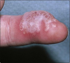
Herpetic whitlow is another presentation (Fig. 1). A painful vesicle or pustule is found on a finger in, for example, a nurse or dentist attending a patient secreting the virus. Similar direct inoculation is sometimes seen in sportsmen such as wrestlers (‘herpes gladiatorum’).
Type 2 primary infection is normally seen after sexual contact in young adults, who develop acute vulvovaginitis or penile or perianal lesions. Culture-positive genital herpes simplex in a pregnant woman at the time of delivery is an indication for caesarean section, as neonatal infection can be fatal.
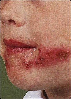
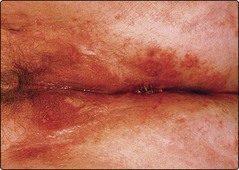
Recurrence is a hallmark of herpes simplex infection; it occurs at a similar site each time, usually on the lips, face (Fig. 2) or genitals (Fig. 3). Rarely, herpes simplex may appear in a zosteriform dermatomal distribution. The outbreak of groups of vesicles is often preceded for a few hours by tingling or burning. Crusts form within 24–48 h, and the infection fades after a week. Attacks may be precipitated by respiratory infection (hence ‘cold’ sore), sunlight or local trauma.
Differential diagnosis
Occasionally, herpes simplex can be confused with impetigo but, in recrudescent disease, the recurrent nature usually indicates the diagnosis. If necessary, the virus can be cultured or detected by an immunofluorescent test.
Complications
Complications are infrequent but can be serious. They include the following:
 Secondary bacterial infection. This is usually due to Staphylococcus aureus.
Eczema herpeticum. Widespread herpes simplex infection is a serious and potentially fatal complication seen in patients with atopic eczema (p. 36) or Darier’s disease (p. 90).
Disseminated herpes simplex. Widespread herpetic vesicles may occur in the newborn or in immunosuppressed patients.
Chronic herpes simplex. Atypical and chronic lesions may be seen in patients with HIV infection.
Herpes encephalitis. This is a serious complication of herpes simplex, not always accompanied by skin lesions.
Carcinoma of the cervix. This is more common in women with serological evidence of infection with type 2 herpes simplex, which may be a predisposing factor.
Erythema multiforme. Herpes simplex infection is the most common cause of recurrent erythema multiforme (p. 82).
Management
Mild herpetic lesions may not require any medication. The treatment of choice for recurrent mild facial or genital herpes simplex is aciclovir (Zovirax) cream (applied five times a day for 5 days), which reduces the length of the attack and the duration of viral shedding, and should preferably be started at the first indication of a recrudescence. More severe episodes warrant oral treatment with aciclovir (200 mg five times a day for 5 days), which shortens the attack. Long-term oral administration is useful in those with frequent recurrent attacks. Intravenous aciclovir may be life-saving in the immunosuppressed and in infants with eczema herpeticum. Genital herpes simplex can also be treated with oral famciclovir or valaciclovir. In those with genital herpes simplex, barrier contraception methods are advisable during intercourse, and intercourse should be avoided during symptomatic episodes.
Herpes zoster
Herpes zoster (shingles) is an acute, self-limiting, vesicular eruption occurring in a dermatomal distribution; it is caused by a recrudescence of Varicella zoster virus.
Aetiopathogenesis and pathology
Herpes zoster nearly always occurs in subjects who have previously had varicella (chickenpox). The virus lies dormant in the sensory root ganglion of the spinal cord but, when reactivated, the virus replicates and migrates along the nerve to the skin, producing pain and ultimately inducing the cutaneous lesions of shingles. A viraemia is frequent, and disseminated involvement may be seen. The pathological changes are identical to those of herpes simplex.
Clinical presentation
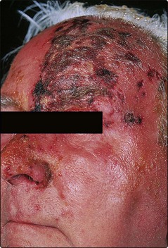
Pain, tenderness or paraesthesia in the dermatome may precede the eruption by 3–5 days. Erythema and grouped vesicles follow, scattered within the dermatomal area (Fig. 4). The vesicles become pustular and then form crusts that separate in 2–3 weeks to leave scarring. Secondary bacterial infection may occur. Herpes zoster is normally unilateral and may involve adjacent dermatomes. The thoracic dermatomes are affected in 50% of cases and, in the elderly, involvement of the ophthalmic division of the trigeminal nerve is particularly common (Fig. 5). Two-thirds of patients with herpes zoster are over 50 years of age, and it is uncommon in children. The lesions shed virus, and contacts with no previous exposure may develop chickenpox.

Some scattering of vesicles outwith the dermatomal distribution is not uncommon, but disseminated or unusually haemorrhagic vesicles raise the possibility of immunosuppression or underlying malignancy. Local lymphadenopathy is usual, as is sensory disturbance of varying degree, including pain, numbness and paraesthesia. Shingles is recurrent in 5% of cases.
Differential diagnosis
The prodromal pain of herpes zoster can mimic cardiac or pleural pain, or an acute abdominal emergency. Once the eruption has appeared, the diagnosis is usually obvious, although herpes simplex may infrequently occur in a dermatomal fashion. Viral culture is sometimes needed.
Complications
Serious complications may occur in herpes zoster. These include the following:
Ophthalmic disease. Corneal ulcers and scarring may result from shingles of the first trigeminal division. Ophthalmological assistance is mandatory.
Motor palsy. Rarely, the viral involvement may spread from the posterior horn of the spinal cord to the anterior horn, and result in a motor disorder. Cranial nerve palsies or paralysis of the diaphragm or other muscle groups may occur.
Disseminated herpes zoster. Immunosuppressed subjects, and patients with Hodgkin’s disease in particular, can develop confluent haemorrhagic involvement, which spreads and may become necrotic or gangrenous. Varicella pneumonia or encephalitis are potentially fatal.
Post-herpetic neuralgia. Neuralgia is infrequent in patients under 40 years but is found in a third of those over 60 years. The pain subsides in the majority within 12 months.
Management
In mild shingles, treatment is symptomatic, with rest, analgesia and bland drying preparations such as calamine lotion. Secondary bacterial infection may require a topical antiseptic or antibiotic. More severe cases may be treated, if seen within 48 h of onset, with oral aciclovir (800 mg five times a day for 7 days) or famciclovir (750 mg once daily for 7 days), which promotes resolution, reduces the viral shedding time and may reduce post-herpetic neuralgia. Immunosuppressed patients often require intravenous aciclovir. Oral prednisolone, given early in the course of herpes zoster for 14 days, reduces the incidence of post-herpetic neuralgia, but must not be used if the patient is immunosuppressed. Post-herpetic neuralgia may respond to topical capsaicin (Axsain).