Advanced dermatological surgery
Some dermatologists specialize in the field of skin surgery. All registrars and residents in dermatology are trained in these techniques. An outline of the subject is given here, including the use of flaps, grafts and Mohs’ surgery, along with mention of lasers and photodynamic therapy, and of some basic cosmetic procedures.
Simple plastic repairs
Simple plastic repairs are carried out by the following:
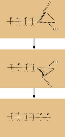
 Dog-ear excision: dog-ears are redundant tissue at the end of an excision line. In sites of high elasticity or where tissue conservation is critical, circular excision of the lesion with appropriate margins is preferable to a predetermined ellipse excision. The redundant skin is lifted like a tent using a skin hook then excised each side (‘dog ears’), and the extended wound is sutured (Fig. 1).
M-plasty: the M-plasty is an excision that reduces the length of an ellipse where space is limited, e.g. on the face. The ‘M’ end of the ellipse is formed by imagining one tip of the ellipse is folded in.
Skin flaps
Side-to-side (‘direct’) closure of a surgical defect is often possible by undermining the edges of the wound using scissors to free the tissue but, when this is not possible, a skin graft or flap is considered. The simplest types of flap are advancement and rotation:
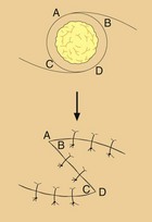
Advancement flap: in this, the skin flap is advanced in one direction over the defect. The flap of skin is created by making excision lines away from the defect to be covered, undermining to free the pedicle, advancing it into the defect and then suturing it in place (Fig. 2).
Rotation flap: a defect may be covered by rotating in skin from one side. One side of the excision wound is extended as an arc that is up to three times the length of the primary defect, depending on the elasticity of the skin at that body site (the scalp and dorsal hand are the least elastic). The pedicle is undermined, rotated in to the defect and sutured in place (Fig. 3).
Skin grafts
When a defect cannot be closed directly or by a flap, healing by secondary intention is often considered. This can produce excellent results on concave aspects of the nose, orbit, ear and temple, but less satisfactory appearances on convex surfaces of the nose, lips, cheeks and chin. If it is essential to cover a defect and other techniques cannot be employed, a skin graft may be used. Skin grafts can give relatively poor cosmetic results if undertaken without careful planning and cause the added complication of creating two wounds. A graft is either full or split thickness:
Full thickness: full-thickness skin is excised completely from the donor site, e.g. behind the ear or upper inner arm, which is then sutured.
Split thickness: in a spilt-thickness graft, the donor site skin is cut through the dermis leaving re-epithialization to occur from the epidermal cells of the hair follicles left behind. This method is usually used by plastic surgeons for covering large defects.
Mohs’ micrographic surgery
Mohs’ surgery describes an approach that maximizes tissue conservation by minimally excising skin cancers. Prior to skin closure, the narrowly excised tumour is examined microscopically. If the cancer is incompletely excised, the surgeon uses a mapping process to make further excisions until the margins are clear. It is indicated mostly for basal cell carcinomas that:
are of the morphoeic type (clinical assessment of margins is unreliable)
have recurred (scarring at site of previous excision can track tumour cells)
have developed in embryonic folds, e.g. nasolabial site
require tissue conservation as a priority (e.g. on the nose, around the eyes).
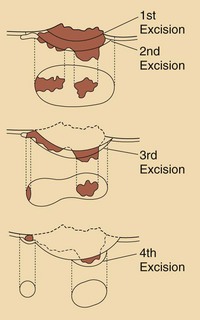
The bulk of the tumour is removed by curettage and then a saucer-like piece of skin is excised. This specimen is marked and flattened, and frozen sections are taken and read immediately by microscope, giving a ‘map’ that shows the extent of the tumour and the areas from which further excision is needed (Fig. 4). The defect is repaired conventionally, sometimes in collaboration with plastic surgeons. The cure rate is 98% for basal cell carcinoma.
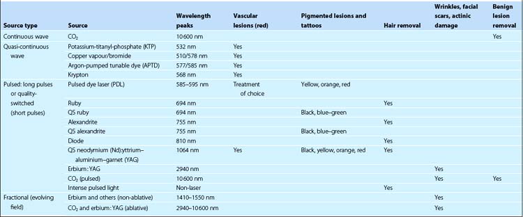
Lasers and intense pulsed light (IPL)
The technology of lasers (light amplification by stimulated emission of radiation) has advanced rapidly, and lasers can be used to treat vascular or pigmented lesions, tumours and tattoos and for hair removal. The variation in absorption of different wavelengths of light means that a range of different lasers is needed (Table 1). Laser therapy is carried out in specialized centres. Treatment is usually painful and several visits are often required. IPL treatment can be used for some conditions such as hair removal, and treatment is similar to laser but is a cheaper alternative. Fractional laser therapy refers to treatment of small zones within the target area patterned like the holes in a watering can rose.
Photodynamic therapy
Photodynamic therapy (PDT), in which the porphyrin precursor 5-amino-laevulinic acid is applied to a lesion that is then irradiated with visible or laser light, is very effective for extensive in situ squamous cell carcinoma, actinic keratoses and superficial basal cell carcinoma.
Cosmetic procedures
Cosmetic procedures are part of the day-to-day practice of the average dermatologist in many countries, although not yet in the UK. Lasers are used extensively for telangiectasia or areas of pigmentation. Other procedures include botulinum toxins for wrinkles, dermabrasion, chemical peels, resurfacing and the use of fillers.
Botulinum toxins: injection of botulinum toxins into facial muscle paralyses the action of the muscle, thus reducing the prominence of frown lines. It is also used for axillary and sometimes palmar hyperhidrosis.
Dermabrasion: the technique of dermabrasion is used for the removal of pitted or depressed scars on the face. It involves abrasive planing in a sedated and prepared patient of the epidermis and superficial dermis using a high-speed rotary brush. Regeneration of the epidermis occurs rapidly due to abundant pilosebaceous structures.
Laser resurfacing: an erbium : YAG laser is used to remove the epidermis with minimal dermal damage, allowing regeneration of epidermis and the elimination of scars or photodamage.
Chemical peels: chemical peel is an alternative to dermabrasion to improve the appearance of photodamaged or wrinkled facial skin. Alpha-hydroxy acids or weak trichloroacetic acid solutions are used.
Fillers: soft tissue defects, e.g. depressed scars or wrinkles, often on the face, may be corrected by the injection of biocompatible materials such as bovine collagen or hyaluronic acid derivatives.
Body sculpturing: removal of subcutaneous body fat by liposuction to produce a slimmer body shape has been widely used. However, newer approaches for lipotransfer and lipolysis by subcision, radiofrequency ablation and pharmacotherapy are available.
Hair transplant: punch biopsies are taken from areas of normal hair density on the scalp. The hair follicles are dissected out one by one and inserted individually into areas of alopecia.
Advanced dermatological surgery
Dog-ear excision removes redundant tissue at the ends of an excision to give a better quality scar.
Skin flaps are used to repair a defect by the mobilization and advancement or rotation of skin.
Skin grafts are used to close a defect, but secondary intention healing may give a better cosmetic result.
Mohs’ surgery describes the microscopically controlled serial excision of difficult-to-treat skin cancers, giving a high cure rate.
Lasers are used to treat vascular or pigmented skin lesions, tattoos, some skin cancers and for hair removal.
Photodynamic therapy is a very effective method of treating widespread in situ squamous cell carcinomas.
Cosmetic procedures are increasingly seen as part of dermatological practice in many countries.