Radiation History
After completion of this chapter, the student will be able to do the following:
• Define the key words associated with radiation history
• Summarize the importance of dental radiographs
• List the uses of dental radiographs
• Summarize the discovery of x-radiation
• Recognize the pioneers in dental x-radiation and their contributions and discoveries
• List the highlights in the history of x-ray equipment and film
• List the highlights in the history of dental radiographic techniques
The dental radiographer cannot appreciate current x-ray technology without looking back to the discovery and history of x-radiation. A thorough knowledge of x-radiation begins with a study of its discovery, the pioneers in dental x-radiation, and the history of dental x-ray equipment, film, and radiographic techniques. In addition, before the dental radiographer can begin to understand x-radiation and its role in dentistry, an introduction to basic dental radiography terms and a discussion of the importance of dental radiographs are necessary. The purpose of this chapter is to introduce basic dental radiography terms, to detail the importance of dental radiographs, and to review the history of x-radiation.
Dentistry and X-Radiation
Before studying the importance of dental radiographs and the discovery and history of x-rays, the student must understand the following basic terms pertaining to dentistry and x-radiation:
Radiation: A form of energy carried by waves or a stream of particles
X-radiation: A high-energy radiation produced by the collision of a beam of electrons with a metal target in an x-ray tube
X-ray: A beam of energy that has the power to penetrate substances and record image shadows on photographic film or digital sensors
Radiology: The science or study of radiation as used in medicine; a branch of medical science that deals with the use of x-rays, radioactive substances, and other forms of radiant energy in the diagnosis and treatment of disease
Radiograph: A two-dimensional representation of a three-dimensional object. In practice, often called an “x-ray”; this is not correct. X-ray (also x ray) is a term that refers to a beam of energy
Dental radiograph: A photographic image produced on an image receptor by the passage of x-rays through teeth and related structures
Radiography: The art and science of making radiographs by the exposure of film to x-rays
Dental radiography: The production of radiographs of the teeth and adjacent structures by the exposure of an image receptor to x-rays
Dental radiographer: Any person who positions, exposes, and processes dental x-ray image receptors
Importance of Dental Radiographs
The dental radiographer must have a working knowledge of the value and uses of dental radiographs. Dental radiographs are a necessary component of comprehensive patient care. In dentistry, radiographs enable the dental professional to identify many conditions that may otherwise go undetected and to see conditions that cannot be identified clinically. An oral examination without dental radiographs limits the dental practitioner to what is seen clinically—the teeth and soft tissue. With the use of dental radiographs, the dental radiographer can obtain a wealth of information about the teeth and supporting bone.
Detection is one of the most important uses of dental radiographs (Box 1-1). Through the use of dental radiographs, the dental radiographer can detect disease. Many dental diseases and conditions produce no clinical signs or symptoms and are typically discovered only through the use of dental radiographs.
Discovery of X-Radiation
Roentgen and the Discovery of X-rays
The history of dental radiography begins with the discovery of the x-ray. Wilhelm Conrad Roentgen (pronounced “ren-ken”), a Bavarian physicist, discovered the x-ray on November 8, 1895 (Figure 1-1). This monumental discovery revolutionized the diagnostic capabilities of the medical and dental professions and, as a result, forever changed the practice of medicine and dentistry.
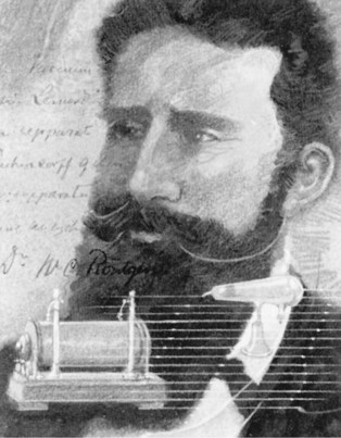
FIGURE 1-1 Roentgen, the father of x-rays, discovered the early potential of an x-ray beam in 1895. (Courtesy: Carestream Health Inc., Rochester, NY.)
Before the discovery of the x-ray, Roentgen had experimented with the production of cathode rays (streams of electrons). He used a vacuum tube, an electrical current, and special screens covered with a material that glowed (fluoresced) when exposed to radiation. He made the following observations about cathode rays:
• The rays appeared as streams of colored light passing from one end of the tube to the other.
While experimenting in a darkened laboratory with a vacuum tube, Roentgen noticed a faint green glow coming from a nearby table. He discovered that the mysterious glow, or “fluorescence,” was coming from screens located several feet away from the tube. Roentgen observed that the distance between the tube and the screens was much greater than the distance cathode rays could travel. He realized that something from the tube was striking the screens and causing the glow. Roentgen concluded that the fluorescence must be the result of some powerful “unknown” ray.
In the following weeks, Roentgen continued experimenting with these unknown rays. He replaced the fluorescent screens with a photographic plate. He demonstrated that shadowed images could be permanently recorded on the photographic plates by placing objects between the tube and the plate. Roentgen proceeded to make the first radiograph of the human body; he placed his wife’s hand on a photographic plate and exposed it to the unknown rays for 15 minutes. When Roentgen developed the photographic plate, the outline of the bones in her hand could be seen (Figure 1-2).
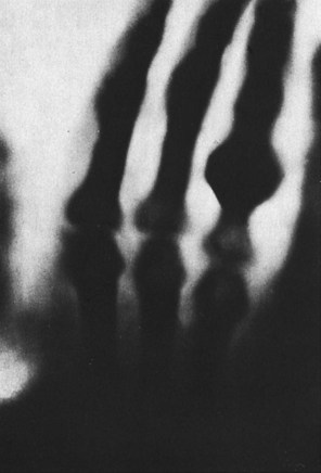
FIGURE 1-2 First radiograph of the human body, showing the hand of Roentgen’s wife. (From Goaz PW, White SC: Oral radiology and principles of interpretation, ed 2, St Louis, 1987, Mosby.)
Roentgen named his discovery x-rays, the “x” referring to the unknown nature and properties of such rays. (The symbol × is used in mathematics to represent the unknown.) He published a total of three scientific papers detailing the discovery, properties, and characteristics of x-rays. During his lifetime, Roentgen was awarded many honors and distinctions, including the first Nobel Prize ever awarded in physics.
Following the publication of Roentgen’s papers, scientists throughout the world duplicated his discovery and produced additional information on x-rays. For many years after his discovery, x-rays were referred to as “roentgen rays,” radiology was referred to as “roentgenology,” and radiographs were known as “roentgenographs.”
Earlier Experimentation
The primitive vacuum tube used by Roentgen in the discovery of x-rays represented the collective findings of many investigators. Before the discovery of x-rays in 1895, a number of European scientists had experimented with fluorescence in sealed glass tubes.
In 1838, a German glassblower named Heinrich Geissler built the first vacuum tube, a sealed glass tube from which most of the air had been evacuated. This original vacuum tube, known as the Geissler tube, was modified by a number of investigators and became known by their respective names (e.g., the Hittorf-Crookes tube, the Lenard tube).
Johann Wilhelm Hittorf, a German physicist, used the vacuum tube to study fluorescence (a glow that results when a fluorescent substance is struck by light, cathode rays, or x-rays). In 1870, he observed that the discharges emitted from the negative electrode of the tube traveled in straight lines, produced heat, and resulted in a greenish fluorescence. He called these discharges cathode rays. In the late 1870s, William Crookes, an English chemist, redesigned the vacuum tube and discovered that cathode rays were streams of charged particles. The tube used in Roentgen’s experiments incorporated the best features of the Hittorf and Crookes designs and was known as the Hittorf-Crookes tube (Figure 1-3).
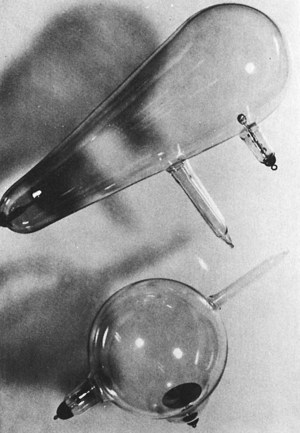
FIGURE 1-3 Hittorf-Crookes tubes used by Roentgen to discover x-rays. (From Goaz PW, White SC: Oral radiology and principles of interpretation, ed 2, St Louis, 1987, Mosby.)
In 1894, Philip Lenard discovered that cathode rays could penetrate a thin window of aluminum foil built into the walls of the glass tubes and cause fluorescent screens to glow. He noticed that when the tube and screens were separated by at least 3.2 inches (8 cm), the screens would not fluoresce. It has been postulated that Lenard might have discovered the x-ray if he had used more sensitive fluorescent screens.
Pioneers in Dental X-Radiation
After the discovery of x-rays in 1895, a number of pioneers helped shape the history of dental radiography. The development of dental radiography can be attributed to the research of hundreds of investigators and practitioners. Many of the early pioneers in dental radiography died from overexposure to radiation. At the time x-rays were discovered, nothing was known about the hidden dangers that resulted from using these penetrating rays.
Shortly after the announcement of the discovery of x-rays in 1895, a German dentist, Otto Walkhoff, made the first dental radiograph. He placed a glass photographic plate wrapped in black paper and rubber in his mouth and submitted himself to 25 minutes of x-ray exposure. In that same year, W.J. Morton, a New York physician, made the first dental radiograph in the United States using a skull. He also lectured on the usefulness of x-rays in dental practice and made the first whole-body radiograph using a 3 × 6 ft sheet of film.
C. Edmund Kells, a New Orleans dentist, is credited with the first practical use of radiographs in dentistry in 1896. Kells exposed the first dental radiograph in the United States using a living person. During his many experiments, Kells exposed his hands to numerous x-rays every day for years. This overexposure to x-radiation caused the development of numerous cancers in his hands. Kells’ dedication to the development of x-rays in dentistry ultimately cost him his fingers, later his hands, and then his arms.
Other pioneers in dental radiography include William H. Rollins, a Boston dentist who developed the first dental x-ray unit. While experimenting with radiation, Rollins suffered a burn to his hand. This initiated an interest in radiation protection and later the publication of the first paper on the dangers associated with radiation. Frank Van Woert, a dentist from New York City, was the first to use film in intraoral radiography. Howard Riley Raper, an Indiana University professor, established the first college course in radiography for dental students.
Table 1-1 lists highlights in the history of dental radiography. The development of dental radiography has moved forward from these early discoveries and continues to improve even today as new technologies become available.
TABLE 1-1
Highlights in the History of Dental Radiography
| Year | Event | Pioneer/Manufacturer |
| 1895 | Discovery of x-rays | W.C. Roentgen |
| 1896 | First dental radiograph | O. Walkhoff |
| 1896 | First dental radiograph in United States (skull) | W.J. Morton |
| 1896 | First dental radiograph in United States (living patient) | C.E. Kells |
| 1901 | First paper on dangers of x-radiation | W.H. Rollins |
| 1904 | Introduction of bisecting technique | W.A. Price |
| 1913 | First dental text | H.R. Raper |
| 1913 | First prewrapped dental films | Eastman Kodak Company |
| 1913 | First x-ray tube | W.D. Coolidge |
| 1920 | First machine-made film packets | Eastman Kodak Company |
| 1923 | First dental x-ray machine | Victor X-Ray Corp, Chicago |
| 1925 | Introduction of bite-wing technique | H.R. Raper |
| 1933 | Concept of rotational panoramics proposed | |
| 1947 | Introduction of long-cone paralleling technique | F.G. Fitzgerald |
| 1948 | Introduction of panoramic radiography | |
| 1955 | Introduction of D-speed film | |
| 1957 | First variable-kilovoltage dental x-ray machine | General Electric |
| 1978 | Introduction of dental xeroradiography | |
| 1981 | Introduction of E-speed film | |
| 1987 | Introduction of intraoral digital radiography | |
| 1998 | Introduction of cone-beam computed tomography (CBCT) for dental use | |
| 1999 | Oral and maxillofacial radiology becomes a specialty in dentistry | |
| 2000 | Introduction of F-speed film |
History of Dental X-Ray Equipment
In 1913, William D. Coolidge, an electrical engineer, developed the first hot-cathode x-ray tube, a high-vacuum tube that contained a tungsten filament. Coolidge’s x-ray tube became the prototype for all modern x-ray tubes and revolutionized the generation of x-rays.
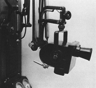
In 1923, a miniature version of the x-ray tube was placed inside the head of an x-ray machine and immersed in oil. This served as the precursor for all modern dental x-ray machines and was manufactured by the Victor X-Ray Corporation of Chicago (Figure 1-4). Later, in 1933, a new machine with improved features was introduced by General Electric. From that time on, the dental x-ray machine changed very little until a variable kilovoltage machine was introduced in 1957. Later, in 1966, a recessed long-beam tubehead was introduced.
History of Dental X-Ray Film
From 1896 to 1913, dental x-ray packets consisted of glass photographic plates or film cut into small pieces and hand-wrapped in black paper and rubber. The hand wrapping of intraoral dental x-ray packets was a time-consuming procedure. In 1913, the Eastman Kodak Company manufactured the first prewrapped intraoral films and consequently increased the acceptance and use of x-rays in dentistry. The first machine-made periapical film packets became available in 1920.
The films currently used in dental radiography are greatly improved compared with the films of the past. At present, fast film requires a very short exposure time, less than 2% than the initial exposure times used in 1920, which, in turn, reduces the patient’s exposure to radiation.
History of Dental Radiographic Techniques
The intraoral techniques used in dentistry include the bisecting technique, the paralleling technique, and the bite-wing technique. The dental practitioners who developed these radiographic techniques include Weston Price, a Cleveland dentist, who introduced the bisecting technique in 1904, and Howard Riley Raper, who redefined the original bisecting technique and introduced the bite-wing technique in 1925. Raper also wrote one of the first dental radiography textbooks in 1913.
The paralleling technique was first introduced by C. Edmund Kells in 1896 and then later, in 1920, used by Franklin W. McCormack in practical dental radiography. F. Gordon Fitzgerald, the “father of modern dental radiography,” revived interest in the paralleling technique with the introduction of the long-cone paralleling technique in 1947.
The extraoral technique used most often in dentistry is panoramic radiography. In 1933, Hisatugu Numata of Japan was the first to expose a panoramic radiograph; however, the film was placed lingually to the teeth. Yrjo Paatero of Finland is considered to be the “father of panoramic radiography.” He experimented with a slit beam of radiography, intensifying screens, and rotational techniques.
Summary
• An x-ray is a beam of energy that has the power to penetrate substances and record image shadows on photographic film.
• A radiograph is a two-dimensional representation of a three-dimensional object.
• Radiography is the art and science of making radiographs by the exposure of image receptors to x-rays.
• A dental radiographer is any person who positions, exposes, and processes dental x-ray image receptors.
• Disease detection is one of the most important uses for dental radiographs.
• Wilhelm Conrad Roentgen discovered the x-ray in 1895.
• Following the discovery of the x-ray, numerous investigators contributed to advancements in dental radiography.
Frommer, HH, Savage-Stabulas, JJ, Ionizing radiation and basic principles of x-ray generation. Radiology for the dental professional, ed 9, St Louis, Mosby, 2011.
Haring, JI, Lind, LJ. The importance of dental radiographs and interpretation. In: Radiographic interpretation for the dental hygienist. Philadelphia: Saunders; 1993.
Johnson, ON, Thomson, EM, History of dental radiography. Essentials of dental radiography for dental assistants and hygienists, ed 8, Upper Saddle River, NJ, Pearson Education, Inc, 2007.
Langlais, RP. Exercises in oral radiology and interpretation, ed 4. St Louis: Saunders; 2004.
Langland, OE, Langlais, RP. Early pioneers of oral and maxillofacial radiology. Oral Surg Oral Med Oral Pathol. 1995;80(5):496.
Langland, OE, Langlais, RP, Preece, JW, Production of x-rays. Principles of dental imaging, ed 2, Baltimore, MD, Lippincott Williams and Wilkins, 2002.
Miles, DA, Van Dis, ML, Williamson, GF, Jensen, CW, X-ray properties and the generation of x-rays. Radiographic imaging for the dental team, ed 4, St Louis, Saunders, 2009.
White, SC, Pharoah, MJ, Radiation physics. Oral radiology: principles and interpretation, ed 6, St Louis, Mosby, 2009.
White, SC, Pharoah, MJ, Radiation safety and protection. Oral radiology: principles and interpretation, ed 6, St Louis, Mosby, 2009.
Matching
For questions 1 to 9, match each term (a to i) with its corresponding definition.
____1. A photographic image produced on film by the passage of x-rays through teeth and related structures.
____2. A beam of energy that has the power to penetrate substances and record image shadows on photographic film.
____3. A form of energy carried by waves or a stream of particles.
____4. Any person who positions, exposes, and processes x-ray image receptors.
____5. The production of radiographs by the exposure of image receptors to x-rays.
____6. A high-energy radiation produced by the collision of a beam of electrons with a metal target in an x-ray tube.
____7. The science or study of radiation as used in medicine.
____8. The production of radiographs of the teeth and adjacent structures by the exposure of image receptors to x-rays.
____9. A two-dimensional representation of a three-dimensional object.
For questions 10 to 19, match the dental pioneers with their contributions (a to j).
a. Used paralleling technique in practical dental radiography
d. Introduced bisecting technique
e. Exposed first dental radiograph
f. Wrote first paper on the danger of x-radiation
g. Exposed first dental radiograph in United States (skull)
h. Introduced long-cone paralleling technique
i. Wrote first dental text; introduced bite-wing technique
j. Exposed first dental radiograph in United States (living patient)