Chapter 10 The nervous system
The nervous system forms mainly from the ectoderm layer, at the beginning of the third week (see Chapter 1), the process known as neurulation. The neural plate forms as a thickening which is widest at its cranial end (Fig. 10.1A). Laterally, the plate edges thicken to form the neural folds (Fig. 10.1B). This is by a process of induction by the underlying notochord and somites. As these neural folds develop they turn towards each other forming the neural groove (Fig. 10.1C), and ultimately fusing as the neural tube. The neural tube structure begins in the cervical region and ends caudally. At the cranial end of the tube the brain develops, whereas the remainder of the tube gives rise to the spinal cord. At each end of the tube are the anterior and posterior neuropores which close in the middle and end of the fourth week respectively (Fig. 10.1D).
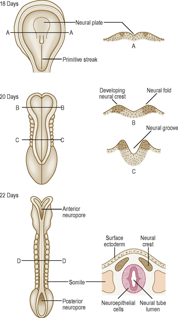
Fig. 10.1 Folding of the neural plate to form the neural tube between days 18 and 22. By day 22 the neural tube is detached from the overlying surface ectoderm. Transverse sections through the embryo (A, B, C and D) are shown in corresponding diagrams on the right.
The lumen of the neural tube is lined by neuroepithelial cells (derived from the neuroectoderm). These cells give rise to the neurons of the grey matter and their processes (dendrites and axons). Some of the axons become invested by myelin and extend into the white matter of the spinal cord. The wall of the neural tube thickens as a consequence of mitosis of the neuroepithelial cells, which become neuroblasts. These cells differentiate into mature neurons. The glial cells also differentiate from the neuroepithelial cells once the differentiation of neuroblasts has ended. Whilst the astrocytes and oligodendrocytes arise from the neuroectoderm the microglial cells are bone marrow derived, arising from mesoderm.
In addition to the neuroectoderm cells from which the neurons develop, neural crest cells develop on the edges along the length of the neural folds (Fig. 10.1). These cells detach themselves from the edges of the folds lying beneath the surface ectoderm, and migrate laterally to form a variety of structures. The principal derivatives of neural crest cells are:
Development of the spinal cord (Fig. 10.2A, B)
Most of the length of the neural tube gives rise to the spinal cord. As the wall of the tube thickens it comes to consist of three zones. The innermost is the neuroepithelial (ventricular) layer and, with the mantle layer, it forms the rest of the wall as well as the lining of the central canal of ependymal cells (Fig. 10.2A). The neuroepithelial cells in the mantle layer differentiate to become neuroblasts which will eventually be the neurons of the grey matter. The outermost layer of the developing spinal cord becomes the marginal zone and contains the axons entering and leaving the mantle zone (Fig. 10.2A). After myelination, this layer looks whitish and constitutes the white matter of the spinal cord. There is further development of the mantle zone through differentiation of the neuroblasts forming thickenings in the dorsal and ventral regions of the cord: the alar and basal plates (Fig. 10.2B). The alar plate becomes the sensory region (or dorsal horn) of the grey matter and the basal plate becomes the motor region (ventral horn). The lumen of the neural tube in the region of the spinal cord becomes diamond shaped. The pointed dorsal aspect of the tube is the roof plate, whilst the floor plate lies at the opposite pole. Dividing the alar and basal plates is the sulcus limitans, a groove. The neurons in the ventral horn give rise to axons that enter the ventral roots. The sensory bipolar neurons in the dorsal root ganglia give rise to axons that enter at the dorsal roots synapsing with perikarya (neuronal cell bodies) in the dorsal horns.
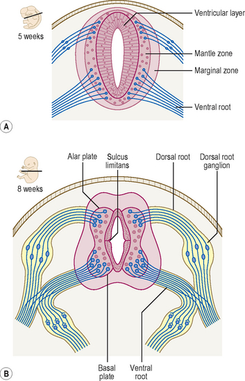
Until the third month of development the spinal cord extends along the entire length of the vertebral column. Thus, spinal nerves exit at the intervertebral foramen opposite their appropriate segmental level. Subsequently, the vertebral column grows longer than the spinal cord. The spinal nerves, however, still exit at their appropriate intervertebral foramina, thus lengthening accordingly, and forming the cauda equina. The spinal cord ends at about the level of the second lumbar vertebra in the adult.
Development of the brain
At the cranial end of the tube three dilations develop during the fourth week, the primary brain vesicles (the prosencephalon, the mesencephalon and the rhombencephalon; Fig. 10.3). They are also known respectively as the forebrain, midbrain and hindbrain. Mainly because of the limited space in which the cranial end of the neural tube is forming there is insufficient space for the lengthening tube. It thus has to bend, and does so in two places: the cervical and cephalic flexures. The former lies between the rhombencephalon and spinal cord, whereas the latter lies in the region of the mesencephalon (Fig. 10.4B).
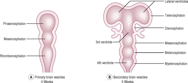
Fig. 10.3 Development of the brain showing primary brain vesicles at 4 weeks (A) and secondary vesicles at 5 weeks (B).
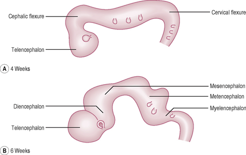
The three primary vesicles develop into five secondary vesicles by the fifth week. The prosencephalon becomes the telencephalon and the diencephalon. The telencephalon has bilateral portions which become the two cerebral hemispheres. The diencephalon becomes the thalamus and hypothalamus. The rhombencephalon comprises two regions, separated by the pontine flexure: the metencephalon and the myelencephalon (Fig. 10.4B). The metencephalon becomes the pons and cerebellum, and the myelencephalon gives rise to the medulla.
The lumen of the neural tube becomes the ventricular system in the region of the brain and brainstem, and the central canal in the spinal cord. The part of the ventricular system lying within the rhombencephalon becomes the fourth ventricle, and that in the diencephalon becomes the third ventricle (Fig. 10.3B). The part of the neural tube lumen between the lateral ventricles and the third ventricle is the interventricular foramen and between the third and the fourth ventricle is the cerebral aqueduct, which is continuous with the central canal of the spinal cord.
 Clinical box
Neural tube defects
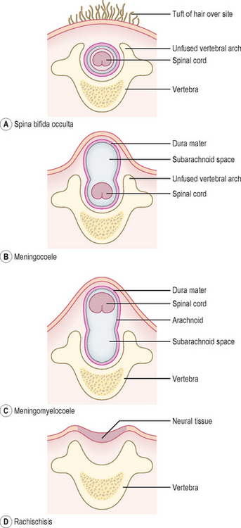
Abnormal closure of the neural tube may occur, especially between the third and fourth weeks, and results in a range of anomalies known as spina bifida. This relates to the usual finding of a divided vertebral arch, which is present in all cases of spina bifida. Other changes may involve the underlying neural tube tissue. The clinical problems with spina bifida include problems with lower limb movements, and control of bowel and bladder function. Spina bifida occulta is the form where there is a divided vertebral arch, but no other abnormality (Fig. 10.5A). Often in such cases the site of the abnormality is marked by a tuft of hairy skin. In the more serious case of spina bifida cystica, neural tissues and their coverings protrude through the vertebral arches and skin, forming cyst-like arrangements. There are two types: meningocoele, where the neural tube lies in its normal position, with a cyst formed by the protruding subarachnoid space (Fig. 10.5B), or meningomyelocoele, in which the neural tube lies ectopically within the cystic space (Fig. 10.5C). Spina bifida cystica is often associated with hydrocephalus. More rarely, the neural folds do not round up but remain as folds continuous with the surface ectoderm, with no lumen for the neural tube. In this type of spina bifida, the neural tube tissue is exposed and folded, and is known as rachischisis (Fig. 10.5D).
Cerebral hemispheres
The cerebral hemispheres expand in all directions during the first few months and overgrow other parts of the brain. The neurons in the floor of the hemispheres aggregate and form the corpus striatum, which will form the caudate and lentiform nuclei of the basal ganglia. Initially, the surface of the hemispheres remain smooth and as growth continues, the convolutions (gyri) appear, separated by sulci and fissures. A number of fibre bundles known as commissures and which connect the two hemispheres develop between weeks 7 and 10. The most prominent commissure is the corpus callosum connecting large areas of the hemispheres. The cerebral cortex differentiates by continuous mitotic activity in the ventricular layer, just outside the lateral ventricle. The neuroblasts migrate superficially to the outer surface of the hemispheres to form the layers of the cortex.
Development of the meninges
The meninges around the cephalic end of the neural tube develop from the neural crest cells, whereas those surrounding the future spinal cord arise from the mesenchyme of the paraxial mesoderm. The dural sac that surrounds the spinal cord ends at the level of the second sacral vertebra.
Formation of the pituitary gland
The pituitary gland develops from two sources: a downgrowth from the floor of the diencephalon (or infundibulum), and an upgrowth from the stomodaeum (the ectodermal portion of the oral cavity) known as Rathke’s pouch (Fig. 10.6). At about the third week Rathke’s pouch appears as an evagination growing towards the infundibulum. By 8 weeks the pouch loses its connection with the oral cavity, and lies immediately adjacent to the infundibulum (Fig. 10.6). Thus the pituitary gland has two components: a posterior part derived from the diencephalon with which it is in direct communication, and an anterior part derived from the oral cavity. These two parts are known as the neurohypophysis and the adenohypophysis respectively.
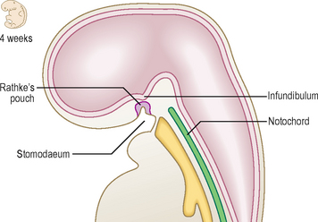
Fig. 10.6 Sagittal section of the cranial part of the embryo at 4 weeks showing development of the pituitary gland.
Anomalies of pituitary gland formation
Rarely, a small amount of pituitary tissue from Rathke’s pouch develops in the posterior wall of the pharynx, a pharyngeal hypophysis. Remnants of Rathke’s pouch may give rise to tumours (craniopharyngiomas), which are usually benign, though sometimes functional.
Anomalies of cranial development
Anencephaly results from the failure of the cephalic end of the neural tube to close (which then degenerates), and is characterized by failure of parts of the brain to develop normally.
Hydrocephalus is an abnormal accumulation of cerebrospinal fluid in the ventricular system and results from a blockage in the normal drainage pathway for the fluid. This anomaly rarely results in the enlarged head typifying hydrocephalus in the past because of surgical intervention to clear the blockage. The brain and skull tissues may be considerably thinned by this increase in cerebrospinal fluid, though mental impairment is not normally seen.
If the neural tube fails to develop normally in size then the cranial cavity is accordingly smaller. This condition is known as microcephaly, and results in mental impairment in many cases.
 The nervous system develops from ectoderm at the start of the third week, initially as neural folds which then close to form the neural tube in the process known as neurulation. The neural crest detaches from the folds, and disseminates widely to contribute to a range of structures throughout the body. The spinal cord develops initially as three layers: the neuroepithelial layer, the mantle layer and the marginal zone. The neuroepithelial layer becomes the ependymal layer, the mantle layer forms the neuroblasts that will become the neurons of the grey matter and the white matter contains the axons. Further development of the mantle zone gives rise to the alar and basal plates, the sensory and motor regions of the spinal cord. The cranial end of the neural tube gives rise to three primary brain vesicles: the prosencephalon, the mesencephalon and the rhombencephalon. They become the five secondary vesicles: the telencephalon, the diencephalon, the mesencephalon, the metencephalon and the myelencephalon respectively.