Chapter 19 Mycobacteria and legionellae
Mycobacteria
According to the World Health Organization (WHO), nearly 2 billion people, one-third of the world’s population, have disease caused by mycobacteria, particularly tuberculosis. Mycobacteria are widespread both in the environment and in animals and cause two major human diseases – tuberculosis and leprosy. They are aerobic, acid-fast bacilli (not stained by the Gram stain because of the high lipid component of the cell wall). The major medically important pathogens are:
Mycobacterium tuberculosis
Habitat and transmission
Found in infected humans, mainly in the lungs; in the body, it resides primarily in the cells of the reticuloendothelial system; transmission is by coughing (droplet spread).
Characteristics
Acid- and alcohol-fast, slender, beaded bacilli; non-sporing. As the organisms do not take up the Gram stain because of the long-chain fatty acids (mycolic acid) in the cell wall, a special stain (the Ziehl–Neelsen stain) is required to visualize them. However, fluorescent microscopy, with auramine stain, is now used commonly for this purpose.
Culture and identification
This species does not grow on ordinary media and requires Löwenstein–Jensen medium for growth (constituents: whole egg, asparagine, glycerol, malachite green). Slow-growing (2–3 weeks; sometimes up to 6 weeks) at 37°C. They grow as ‘rough, tough and buff’ colonies – rough due to dry, irregular growth; tough due to difficulty in lifting the colony from the surface; and buff due to the pale yellow colour (Fig. 19.1).

Fig. 19.1 Growth of Mycobacterium tuberculosis on Löwenstein–Jensen medium: the bottle on the left is uninoculated; the bottle on the right shows ‘rough, tough and buff’ colonies of the organism.
In general, identification of mycobacteria is based on their rate of growth, optimum temperature requirements and pigment production in the presence or absence of light; biochemical tests are also helpful. These slow procedures are being supplanted by more efficient nucleic acid probe techniques.
Pathogenicity
This organism is the agent of tuberculosis, a chronic, granulomatous, slowly progressive infection, usually of the lungs; eventually, many other organs and tissues may be affected. A pandemic disease, tuberculosis is especially common in the developing world owing to HIV infection (15–20% of individuals with HIV disease may have tuberculosis). The oral cavity is affected secondary to primary disease elsewhere (see Chapter 35). The hallmark of the disease is granuloma formation and caseation mediated by cellular immunity. No exotoxins or endotoxins.
Antibiotic sensitivity and control
Long-term therapy (6–9 months) with antituberculous drugs (isoniazid, rifampicin, pyrazinamide, ethambutol). As drug resistance is growing and a persistent problem, combination therapy should always be given. Tubercle bacilli resistant to a number of antituberculous drugs (multidrug-resistant tuberculosis (MDR-TB)) is a growing problem. Hence, regimentation of drug delivery is a cornerstone of managing the disease, which is achieved by a global programme termed directly observed therapy (DOT).
Prevention is by bacille Calmette–Guérin (BCG) vaccination containing live attenuated organisms, in childhood. Pasteurization of milk and general improvement of living standards have played a valuable role in prevention.
Mycobacterium bovis
This organism infects cattle. Humans become infected by ingesting M. bovis-contaminated milk. Infection is rarely seen in the West owing to eradication of the disease in cattle. The organism specifically causes the childhood disease scrofuloderma, characterized by enlarged, caseous cervical lymph nodes. M. bovis is similar in many respects to M. tuberculosis; in the laboratory, it can be distinguished from the latter by its poor growth on Löwenstein–Jensen medium and ready infection of rabbits.
Mycobacterium leprae
Habitat and transmission
Humans are the only known hosts of M. leprae, which resides mainly in the skin and nerves. Prolonged contact is thought to be the mode of transmission.
Characteristics
Aerobic, acid-fast bacilli (they are not alcohol-fast, i.e. decolourized by alcohol); no known toxins.
Culture and identification
Cannot be cultured in vitro but grows on the footpads of mice or armadillos, yielding chronic granulomas at the inoculation site.
Pathogenicity
The leprosy bacillus causes a slow, progressive, chronic disease that mainly affects the skin and the nerves; the lesions are predominantly seen in the cooler parts of the body. Two forms of leprosy are recognized (Table 19.1).
Table 19.1 Comparison of the different types of leprosy
| Tuberculoid | Lepromatous | |
|---|---|---|
| Cell-mediated immunity | ++ | – or ± |
| Antibody response | − | ++ |
| Widespread lesions | − | + |
| Numbers of Mycobacterium leprae in lesions | ± | ++ |
++, predominant; +, common; ±, uncommon; –, absent.
Lepromatous leprosy
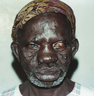
The cell-mediated immune response is depressed or absent; M. leprae bacilli are usually seen in large numbers in the lesions and in blood; commonly involves mucosae, especially the nose (Fig. 19.2); leads to much disfigurement.
Tuberculoid leprosy
Associated with an intense cell-mediated immune response to the organisms; principally involves the nerves, with resultant anaesthesia and paraesthesia. Hence, damage to extremities is caused, with resultant loss of fingers and toes (see Chapter 35 for oral manifestations).
Mycobacteria other than tuberculosis (MOTT)
MOTT is a collective name given to a group of mycobacteria of low human pathogenicity. These species include M. avium, M. intracellulare, M. kansasii, M. marinum, M. fortuitum and others.
Culture and identification
Grow on Löwenstein–Jensen medium but differ from ‘pathogenic’ mycobacteria in the colour of pigment produced and temperature requirements. Some species produce pigments in the dark (scotochromogens), others after exposure to light (photochromogens), and still others are non-chromogenic.
Pathogenicity and antibiotic sensitivity
In the main, MOTT cause pulmonary infection, often with M. tuberculosis; infections are especially seen in compromised individuals (e.g. in HIV disease). These mycobacteria are thought to be passengers in the disease process. They are usually sensitive to the normal antituberculous drugs.
M. marinum, associated with the keeping of tropical fish, causes skin ulcers.
Legionella
There are currently some 39 recognized species belonging to the genus Legionella, but Legionella pneumophila, the species first described, is the most important human pathogen. They cause atypical pneumonia, both in community dwellers and hospitalized patients.
Legionella pneumophila
Habitat and transmission
Ubiquitous organism found in soil and water, including air-conditioning units, domestic and hospital water supplies, and sometimes in dental unit water systems. Spread is known to occur by contaminated aerosols.
Culture and identification
Does not grow on ordinary media; grows slowly (3 weeks) in a special medium (cysteine-charcoal-yeast extract agar) under 5% carbon dioxide. Identification is by direct immunofluorescence.
Pathogenicity
The portal of entry is the respiratory tract and infection results in legionnaires’ disease, a severe form of pneumonia. Older men who smoke and drink alcohol in excess are typically affected. Other risk factors are cancer and immunosuppression. The clinical picture is variable, ranging from mild influenza-like illness to severe pneumonia with mental confusion, diarrhoea, haematuria and proteinuria. A less severe form of pneumonia (Pontiac fever) may be produced by some legionellae. Although there has been some concern on the legionella in stagnant dental unit water lines, and the possibility of legionellosis in dental patients, there is no firm evidence for such speculation.
Antibiotic sensitivity and control
Erythromycin is the drug of choice and may be combined with rifampicin or ciprofloxacin.
It is impossible to eradicate the organism from water supplies as it is ubiquitous, but protective measures include increasing chlorine concentration and the temperature of hospital water supplies; aerosolization of water should be minimized.
Key facts
Bagg J. Tuberculosis: A re-emerging problem for health care workers. British Dental Journal. 1996;180:376-381.
Fallen R.J. Legionellaceae. In Collee J.G., Fraser A.G., Marmion B.P., Simmons A., editors: Mackie and McCartney’s practical medical microbiology, 14th ed., Edinburgh: Churchill Livingstone, 1996.
Greenwood D., Slack R., Peutherer J., editors. Medical microbiology, 16th ed., Edinburgh: Churchill Livingstone, 2003. Chs 19 and 34
Review questions (answers on p. 353)
Please indicate which answers are true, and which are false.