CHAPTER 7 Aging
Introduction
The age of a horse can be an important consideration when forecasting its useful working life, when purchasing the animal, for insurance policies and for the prognosis of diseases. Furthermore, as long as no indelible identification methods for horses are imposed, age estimation contributes to the identification of an animal.
Why does the horse, of all animals, have teeth that lend themselves to age determination? Most domestic animals (cattle, carnivores, etc.) have brachydont incisor teeth, i.e., low-crowned teeth that erupt fully prior to maturity and that are strong enough to survive for the life of the animal. Equine teeth, which are subjected to much higher levels of dietary abrasive forces, are hypsodont, which means that they erupt continuously over most of the horse’s life. It is important in this context to make a distinction between tooth growth and tooth eruption. Tooth growth implies the lengthening of the tooth in its apical part due to the deposition of new layers of dentin and cement. Tooth eruption is the progressive protrusion of the tooth out of its alveolus. It is now generally assumed that tooth eruption is caused by a continuous remodeling of the periodontal ligament fibers and not by root lengthening as was claimed before. The deposition of bone at the bottom of the alveolus should be the result rather than the cause of tooth eruption.1
Equine incisors erupt lifelong, whereas their intrinsic growth ceases at the age of about 17.2 As the total length of horse incisors remains unchanged from the age of six until the age of 17, the continuous loss of occlusal dental tissue is compensated by an equal amount of newly formed dental tissue at the apical end of the tooth. During this period of time, tooth root growth makes up for occlusal wear, which is estimated to occur at a rate of 2.5 mm a year.3 In horses aged over 17 years, occlusal wear is no longer compensated by apical tooth growth, and the total incisival length diminishes progressively. In horses of this age category, continuous tooth eruption is the only mechanism to provide maintenance of occlusal contact between upper and lower incisors.
Because of the marked wear on the surface of hypsodont equine teeth, occlusal exposure of dentin and cement is inevitable after the protecting enamel is worn off. This leads to the presence of alternate layers of the three calcified tissues at the occlusal surface, whose continuously changing configuration allows a macroscopic dental age estimation.4–15
The most appropriate teeth for aging horses are the (lower) incisors. The premolars and molars can be used with considerable accuracy to determine the horse’s age, but their distal position has limited their use.16 Recent work has shown that cheek teeth morphology data can be used to predict age in horses that possess all permanent dentition.17 Radiographic assessment of cheek teeth root morphology can also help in determining age, especially in the young horse.8 Because contact between upper and lower canines is seldom made, canines do not wear down in a regular way and have no age-related occlusal surface.
When estimating a horse’s age by its incisors, the eruption dates and the changes in appearance of the occlusal surfaces are the main criteria. Neither is wholly dependable but the first is the more reliable, although limited in application to younger animals. The second may be used throughout the life span but becomes increasingly inaccurate with age.18 Incisival characteristics that are frequently used for dental aging in horses are summarized below.
Eruption
In this context, gingival emergence is used as a reference point for eruption.
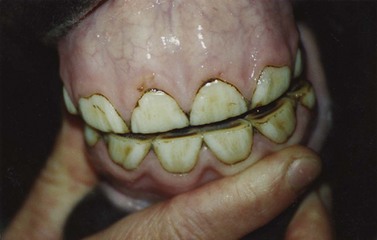
Eruption of the deciduous incisors (Fig. 7.1)
The deciduous incisors are smaller than the permanent ones. The surface of their crown is white and presents several small longitudinal ridges and grooves. The occlusal tables of deciduous incisors are oval in the mesiodistal direction.
Eruption of the permanent incisors (Figs 7.2 & 7.3)
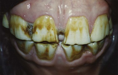
Permanent incisor teeth are larger and more rectangular than the deciduous incisors. Their crown surface is largely covered with cement and has a yellowish aspect. The upper incisors generally present two distinct longitudinal grooves on their labial surface; the lower incisors have only one clearly visible groove.
Changes of the occlusal surface (Fig. 7.4)
Appearance of the dental star
The dental star is a yellow-brown structure on the occlusal surface situated between the labial edge of the incisor and the infundibular cavity or ‘cup.’ It consists of secondary and tertiary dentin that occludes the pulpal chamber when it risks being exposed by wear. In young animals the dental star appears as a linear stripe because the occlusal end of the original pulp cavity is not conical but elongated in a mesiodistal direction. With age dental stars become oval and then round and move towards the center of the occlusal table. These progressive patterns reflect cross-sections through the stuffed pulp cavity at various levels.
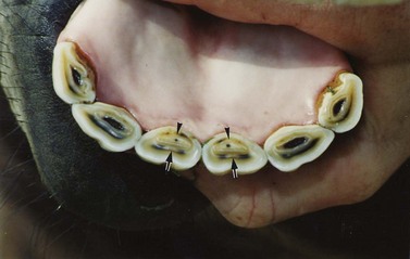
Fig. 7.4 Standardbred horse, 8 years old. Dark-colored dental stars are present in all lower incisors. The characteristic white spot in the center of the dental star appears in the centrals (arrows). Cups have disappeared from the central incisors. The remaining marks are oval (arrowheads). Deep cups are still present on the middle and the corner incisors.
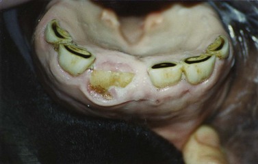
Disappearance of the cups
The infundibulum is an enamel infolding in the occlusal surface of the equine incisor. The superficial half of the infundibulum is empty or filled with food particles. This part is called the ‘cup.’ The bottom of the infundibulum is filled with cement. When wear has brought this infundibular cement layer into the occlusal surface, the cup is filled in or has disappeared. The exposed cement core and the surrounding enamel ring are called the ‘mark.’
Disappearance of the marks
The shape of the mark generally corresponds to the contour of the occlusal table of the incisor. In young horses marks are oval in the mesiodistal direction. When wear progresses, marks become smaller and rounder and move caudally (lingually) on the occlusal surface. With age, the cement of the infundibular bottom wears away and eventually the remaining enamel spot disappears from the occlusal surface.
Changes in shape of the incisors
Changes in shape of the occlusal surfaces (Fig. 7.5)
Due to extensive wear, the sequential shapes of the occlusal tables represent the cross-sections of the incisor teeth at various levels. The sequence ranges from oval in the mesiodistal direction, to trapezoid and triangular, and finally to oval in the labiolingual direction.
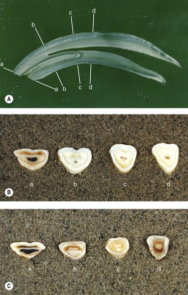
Fig. 7.5 (A) Longitudinal section of the lower central incisor of a Standardbred horse (4 years old). (B) Lower central incisor of a 5-year-old Standardbred horse. Cross-sections at various levels as indicated in (A). In the sections c and d, the pulpal cavity is open. (C) Occlusal tables of the lower central incisor of Standardbreds aged: a, 5 years; b, 8 years; c, 14 years; d, 20 years, respectively. In the occlusal tables c and d the pulpal cavity is occluded by secondary dentin.
Direction of upper and lower incisors (Fig. 7.6)
When incisors are viewed in profile, the angle between the upper and lower incisors changes with age. In young horses, the upper and lower incisors are positioned in a straight line (angle ±180°) with each other. With age, the occlusal portions of the crowns wear off, and we look at different cross-sections of the crown shape in profile. The angle between upper and lower incisors becomes, therefore, increasingly acute. The lower incisors are the first to obtain an oblique position followed at a later date by the upper incisor teeth.
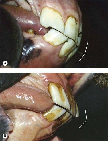
Fig. 7.6 (A) Belgian draft horse, 6 years old. The upper and lower incisors are positioned in a straight line with each other. The crown of the upper corner (103) is wider than it is tall. Notice the presence of a hook on the upper corner (arrow). (B) Standardbred horse, 16 years old. The angle between upper and lower incisors is more acute. The crown of the upper corner (103) is taller than it is wide. The upper corner presents a Galvayne’s groove over the entire length of its labial surface.
Length versus width of the upper corner incisor (Fig. 7.6)
The shape of the upper corner incisor has been used recently to categorize a horse’s age into three groups from 5–20 years of age.8 Between 5 and 9 years of age the crown of this tooth is generally wider than it is tall. At ages 9–10, the upper corner appears square in most horses and then progresses to taller than it is wide as age increases.
The hook on the upper corner incisor (Fig. 7.6A)
The caudal edge of the upper corner sometimes exceeds the occlusal surface of the lower corner, especially when the lower incisors have acquired their oblique position. If the caudolateral portion of the upper corner is no longer in contact with its lower counterpart, it wears more slowly, forming a hook in the occlusal surface. Later, when the upper incisors obtain their oblique position and the caudal edge of the upper corner is in contact with its lower counterpart again, this notch can disappear.
The Galvayne’s groove (Fig. 7.6B)
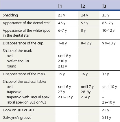
The Galvayne’s groove is a shallow, longitudinal groove on the labial surface of the upper corner and is filled with dark stained cement. In the unworn tooth the groove starts halfway from occlusal surface to apex and continues three-fourths of the distance to the apex. It is buried within the alveolus when the tooth first comes into wear.19 With age, and due to the prolonged eruption of the tooth, the Galvayne’s groove first appears at the gumline. As the tooth continues to erupt, it extends down the labial surface to reach the occlusal edge, then starts to disappear at the gumline and finally disappears completely. The appearance of the groove and its usefulness in aging horses were mentioned for the first time in the early 1880s by an American horsetamer called Sample. Later, his theory was adopted by Sidney Galvayne, an Australian horseman.20 It was in his first work, Horse dentition: showing how to tell exactly the age of a horse up to thirty years (published prior to March 1886) that Galvayne described the groove, which now bears his name, on the vestibular surface of the permanent upper corner incisor.21 The presence and length of the Galvayne’s groove as an accurate guide to the age of the older horse became known throughout the English-speaking world. However, it was not until World War I that several investigations were undertaken to validate his theory.20 Contrary to Galvayne’s statements, these investigations showed that the groove may be absent in more than 50 % of the horses between the ages of 10 and 30 years.
Dental star morphology
The appearance of the dental star is, next to eruption times, one of the more reliable dental features, and the correlation between dental star morphology and age is stronger than for any other feature.22
Horses at pasture have obvious darkly colored dental stars, whereas individuals without access to pasture or grass fodder usually have pale yellowish dental stars (Fig. 7.7). This suggests that the coloration of the dental star is caused by an impregnation of grass pigments. Two small experiments support this theory:23
1. When equine incisors are sectioned longitudinally one can observe that the brown color of the dental star extends only a few millimeters beneath the occlusal surface and that the color intensity fades towards the pulpal chamber. This indicates that the color originates from the ‘outside world’ rather than from the pulp as suggested in older literature reports.3,24
2. When incisors with pale dental stars are stored in a mush of crushed grasses, dental stars become darkly colored after a few days; when they are stored in a buffered (pH 6.8) solution of various diphenols (caffeic acid, 3,4-dihydroxybenzoic acid and 3,4-dihydrophenylalanine (10 mmol/l) together with thyrosinase, the dental stars obtain a deep brown color after 72 hours (Fig. 7.8).
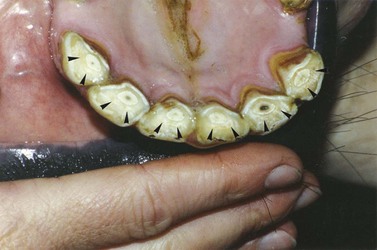
Fig. 7.7 Standardbred horse, 12 years old, deprived of fresh grass. Dental stars are yellowish (arrowheads). It is difficult to distinguish the white spot in the center of the stars.
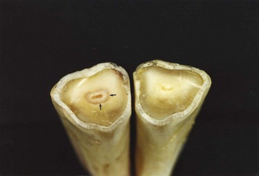
Fig. 7.8 Lower central incisors of a 20-year-old Standardbred without access to pasture. The right tooth (401) was stored in a buffered Ringer’s lactate solution; and the left one (301) was immersed in a buffered solution of 3,4-dihydroxybenzoic acid (10 mmol/l) for 48 hours. In 301, the periphery of the dental star has obtained a brown color (arrows).
This suggests that food pigments are responsible for the dark color of the dental star.
Dental stars also present a topical coloration pattern. In young horses, the dental stars have a uniform color, whereas in older individuals they are composed of a darker periphery that surrounds an uncolored central zone, the so-called ‘white spot’ (Fig. 7.9). The reason why absorption of food pigments occurs only in the peripheral rim of the dental star and not in the white spot nor in the surrounding primary dentin can be found by examining the diameter, extent and orientation of the dentinal tubules.
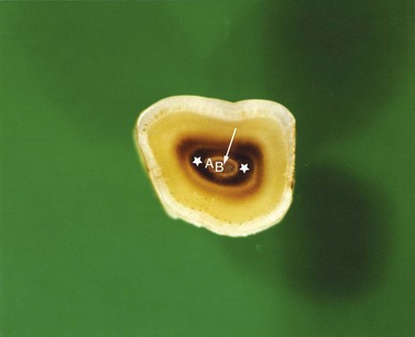
Fig. 7.9 Occlusal surface of the left lower central incisor (301) of a 15-year-old draft horse. The dental star consists of a dark peripheral rim (asterisk) and a central white spot (arrow). The white spot is composed of secondary dentin (A) and a core of tertiary dentin (B).
Dentinal tubules are formed as the odontoblasts retreat centripetally and leave behind a cytoplasmatic process around which the dentin matrix is deposited and mineralized. The tubules can therefore be regarded as hollow cylinders traversing the dentin. Each tubule starts peripherally at the interface between the primary dentin and the enamel, and extends centripetally toward the pulpal border. The first dentin produced by the odontoblasts is located peripherally in the tooth, i.e., underneath the enamel, and is called primary dentin. It surrounds the younger and more centrally located secondary dentin, whereas tertiary dentin is only formed in the restricted areas between the tip of the pulp chamber and the occlusal surface.
The only obvious feature characterizing the transition between primary and secondary dentin of equine teeth is the presence of peritubular dentin (Fig. 7.10), which is hypercalcified tissue, deposited as a collar inside the tubular walls of primary dentin. The term peritubular dentin is anatomically incorrect because this dentin forms within the dentinal tubule (not around it) and narrows the tubular lumen. It is, therefore, sometimes (more accurately) referred to as intratubular dentin.25 Apart from the presence of peritubular dentin, the structure of dentinal tubules is identical in primary and secondary dentin. Peritubular dentin deposit is thickest at the outer end of the primary dentinal tubules and disappears at the transition between primary and secondary dentin.23 The presence of peritubular dentin gives the tubules a tapered shape with the wider lumen at the pulpal side and the narrower luminal diameter near the enamel.
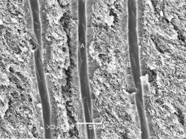
Fig. 7.10 SEM image of longitudinally fractured dentinal tubules. A, tubular lumen; B, peritubular dentin; C, intertubular dentin (×3500).
The dental star consists of a central core of tertiary dentin and a much broader ring of secondary dentin, in neither of which peritubular dentin is deposited. Tertiary dentin, situated in the very center of the dental star, is formed between the tip of the pulp chamber and the occlusal surface and protects the pulp from exposure to attrition. Penetration of food pigments in this zone of the dental star is prevented by the small number of tubules, which are, for the most part, discontinuous with those of the surrounding secondary dentin, their irregular arrangement, and their small diameter (Fig. 7.11). This explains the colorless aspect of the central core of tertiary dentin inside the dental star.
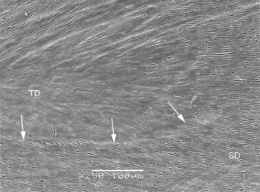
Fig. 7.11 SEM image of the occlusal surface at the center of the dental star. The boundaries of the tertiary dentin are indicated by arrows. TD, tertiary dentin; SD, secondary dentin (×250).
The secondary dentin around the core of tertiary dentin consists of a pale inner zone and a brown peripheral zone. Both zones contain regularly arranged dentinal tubules that are continuous with those of the surrounding primary dentin and are completely devoid of peritubular dentin. The high numerical tubular density, the regular tubular arrangement, and the large tubular diameters of the secondary dentin are suggestive of an easy and uniform penetration of food pigments in this area. The only difference between the pale inner zone and the dark peripheral zone of secondary dentin is the spatial arrangement of the dentinal tubules. In the periphery of the dental star, tubules end perpendicularly into the occlusal surface (Fig. 7.12). This orientation allows an optimal inflow of food pigments, which is far superior to the dye penetration in the more central, uncolored secondary dentin, where tubules lie nearly parallel to the occlusal surface (Fig. 7.13). Penetration of food pigments in the latter zone is nearly negligible because due to the horizontal position of the tubules, the maximal penetration depth of food pigments in this zone cannot exceed the tubular diameter, which is 3 µm. Even when the horizontally exposed tubules are filled with food pigments, this 3 µm-thick mass of colored dentin is worn off in less than 1 day by the severe occlusal attrition, which amounts to 2500 µm a year. Food pigments can, therefore, not be accumulated in the inner zone of the secondary dentin of the dental star. This contrasts with the more peripheral zone of secondary dentin, where food pigments can permeate a longer distance in the perpendicularly debouching tubules. The pigments can accumulate within these tubules and thus cause the dark coloration of the dental star periphery. This mechanism is fully compatible with the aforementioned preliminary experiments, showing that secondary dentin acquires its dark color within 72 hours after immersion in a pigmented solution.23
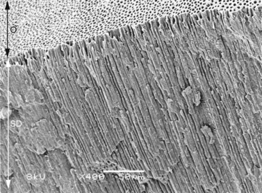
Fig. 7.12 SEM image of the etched occlusal surface (O) and the subjacent secondary dentin (SD) at the dark peripheral rim of the dental star of a longitudinally fractured incisor. Dentinal tubules end perpendicular to the occlusal surface (×400).
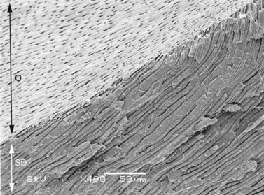
Fig. 7.13 SEM image of the etched occlusal surface (O) and the subjacent secondary dentin (SD) at the pale inner zone of secondary dentin in the dental star. The orientation of the tubules is almost parallel to the occlusal surface (×400).
The microstructure and spatial arrangement of the dentinal tubules can also explain the color differences between primary and secondary dentin. Primary dentin has a smaller number of tubules per area unit than secondary dentin and thus offers fewer pathways for food pigments to penetrate into the occlusal surface. In primary dentin, exposed onto the occlusal surface peripheral to the dental star, tubules debouch obliquely. For the reasons described above, penetration of food pigments into these tubular lumina is considerably less than in the dark peripheral rim of the dental star, where tubules end perpendicularly. Furthermore, primary dentin contains high levels of peritubular dentin and has an almost translucent appearance, which is similar to the complexion of enamel, as both tissue types are highly mineralized.26 In contrast, the less mineralized secondary dentin has a dull opaque appearance. Additionally, because tubules in secondary dentin are devoid of peritubular dentin, they have wider lumina in which plant pigments can penetrate more easily and give the dentinal tissue a dark brown color.
Dental aging in different horse breeds
Many standard textbooks dealing with aging of horses suggest that the above-mentioned characteristics give an accurate indication of a horse’s true age. However, some reports are inconsistent in their guidelines and show large discrepancies in the dental features described at specific ages. A possible explanation for the non-uniformity of existing guidelines is the lack of evidence that any system was used to validate an author’s recommendations for aging.27,28 A study performed by Richardson et al29 casts serious doubts on the belief that the age of a horse can be determined accurately from an examination of its teeth. In this study a large group of horses with documented evidence of birth were examined, and age was estimated both by experienced clinicians and by a computer model. There was little difference between the accuracy of the computer model and the clinical observers, but neither method was accurate when compared with the actual age. In older horses, there was much greater variability between the dental age and the actual age, which means that the accuracy of dental aging declines markedly with age. Most standard texts do not provide exact data concerning breed, sex, and nutrition of the examined horses. However, anatomical, physiological, environmental, and behavioral differences between individuals ensure differences in rate of equine dental wear.30 The concealment of these data may explain the discrepancies between different reports. Inaccuracies in the dental aging system of horses may also result from differences between breed and type of horse involved. Eisenmenger and Zetner stated that the teeth of Thoroughbreds erupt earlier than those of Lipizzaners and coldblood horses. Teeth of ponies may also have rates of eruption and wear that differ from the teeth of horses.3 As for donkeys, both ancient literature data31 and recent investigations32 have suggested that the degree of dental attrition in donkeys is slower than in horses.
The nature of diet can also play a part in the abrasion of horse incisors. Dental wear is caused not only by grinding of opposing crowns against one another, but also by contact with abrasive particles in food, such as silicate phytoliths which form part of the skeleton of grasses. Other plant-borne abrasives include cellulose and lignin.30 In order to preclude the influence of the quality of nutrition on the rate of dental wear, it is necessary that horses that are examined for breed variability are raised and kept under similar environmental and nutritional conditions.
Based on the suggestions that the degree of attritional dental wear is correlated with the breed of horse, four unrelated horse breeds have been subjected to a comparative study.33–35 All horses examined here were raised in Western Europe, were given access to daily pasture, and were fed concentrates and hay. None of the horses was a crib-biter nor suffered from other vices with a possible influence on dental wear. The incisor teeth had not been rasped in any individual. It is evident that in practice one has to be vigilant for these considerations. Factors that are difficult to control and that could not be taken into consideration are the individual chewing habits and the amount of food intake.
A critical evaluation of the dental aging technique revealed that the rate of attritional dental wear is different in different horse breeds. Indentation hardness tests, performed with a Knoop diamond indenter, showed slight breed differences in the hardness of equine enamel and dentin. These different microhardness values seem to contribute to the differences in the rate of attritional dental wear.36
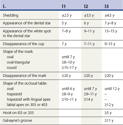
The following text describes the appearance of lower incisor teeth at various ages as generally seen in the Standardbred horse, the Belgian draft horse, the Arabian horse, and the mini-Shetland pony population of Western Europe. It must be emphasized that this text is not a truism. When determining a horse’s age, one must register all dental features together and take account of clinical factors that may have influenced the aspect of the horse’s teeth. The following descriptions will therefore be accurate in many cases, but may be incorrect for any individual.
Eruption of the deciduous incisors
The central incisors generally erupt during the first week of life. The middle incisors emerge through the gums at 4–6 weeks, and the corners erupt between the sixth and the ninth month of life. In the mini-Shetland pony, eruption of the middle and the corner incisors is retarded. The middle incisor starts erupting at the age of 4 months, whereas the corner incisor breaks through the gums between 12 and 18 months of age.
Eruption of the permanent incisors
The upper and lower permanent incisors erupt almost simultaneously. In some horses shedding begins with the maxillary, in others with the mandibular incisor teeth. Arabian horses shed their central, middle, and corner incisors at 2.5, 3.5 and 4.5 years of age, respectively. In Standardbreds and in Belgian draft horses, shedding generally occurs later, namely at nearly 3, nearly 4, and nearly 5 years of age (Fig. 7.14). In mini-Shetland ponies, eruption of the permanent incisors is still further delayed by 2 or 3 months. In male horses, the canines erupt at about 4.5–5 years of age. Generally, these teeth are absent or rudimentary in mares.
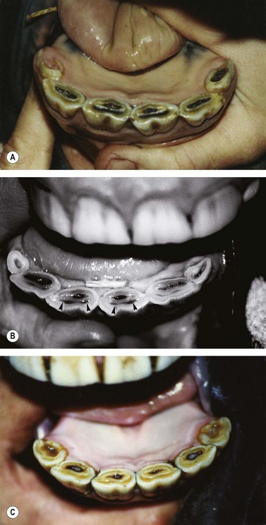
Fig. 7.14 (A) Standardbred horse, 5 years old. The permanent central and middle incisors are in place, the corner incisor is emerging through the gums. The dental star is present on the centrals, absent on the middles and the corners. All lower incisors have deep cups. (B) Belgian draft horse, 4 years and 8 months. The permanent central and middle incisors are in place. The corner incisors are still deciduous. Dental stars are present on the centrals (arrowheads) and appear also on the middles. Cups are present in the central and the middle incisors. (C) Arabian horse, 5 years old. All lower incisors are permanent, the corners are not yet fully in wear. There are no obvious dental stars. Deep cups are present on all lower incisors.
Appearance of the dental star
Dental stars appear sequentially in the central, the middle and the corner incisors. In Standardbreds and in Arabian horses they appear on the centrals at 5 years, on the middles at 6 years, and on the corners at 7–8 years. In Belgian draft horses and mini-Shetland ponies, stars appear somewhat earlier, namely on the centrals at 4.5 years, on the middles at 5.5 years and on the corners at 6.5–7 years (Fig. 7.14). With age, the characteristic white spot becomes visible in the center of the dental star (Figs 7.15–7.17). In Standardbreds and in Arabian horses this white spot appears on the central incisors from the age of 7–8 years onwards, and on the middle incisors from the age of 9–11 years onwards. In Belgian draft horses and in mini-Shetland ponies the white spot becomes visible on the centrals at the age of 6–7 years and on the middles at the age of 8. In all breeds, the appearance of the white spot in the dental star of the corner incisors is variable and occurs between 9 and 15 years.
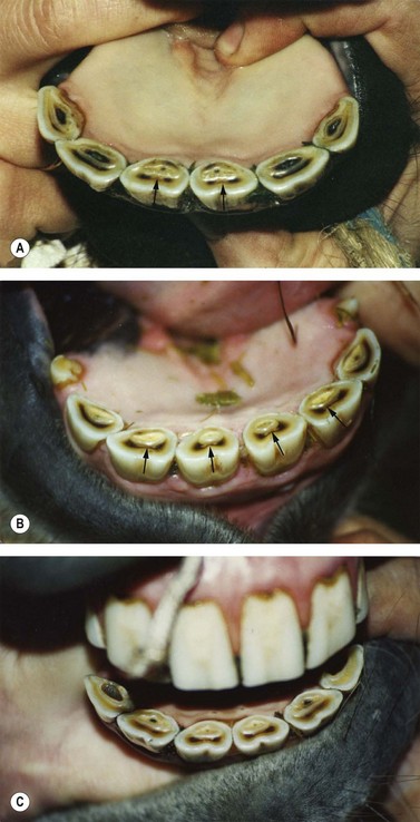
Fig. 7.15 (A) Standardbred horse, 8 years old. Dental stars are present on all incisors. In the central incisor, the white spot in the dental star becomes apparent (arrows). Cups are filled-in on the centrals. On the middles and the corners, cups are still present. The occlusal tables of the central incisors are becoming trapezoid, those of the middles and the corners are still oval. (B) Belgian draft horse, 8 years and 6 months. Dental stars are present on all incisors. In the central and the middle incisors, the white spot in the dental star becomes apparent (arrows). Cups are filled-in on all lower incisors. The remaining marks are oval. The occlusal tables of the centrals and the middles are becoming trapezoid. (C) Arabian horse, 8 years and 6 months. Dental stars are present on the central and the middle incisors. The white spot in the dental star is appearing in the central incisor. Cups on the centrals and the middles have nearly disappeared; the remaining marks are oval (middles) to triangular (centrals). Deep cups are still present on the corner incisors. The occlusal tables of the centrals become trapezoid.
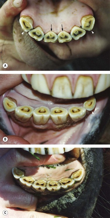
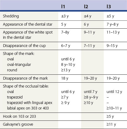
Fig. 7.16 (A) Standardbred horse, 12 years old. Dental stars, consisting of a white spot and a dark periphery, are present on all lower incisors. Cups have disappeared, and the marks are small oval to rounded. The occlusal tables of the central and the middle incisors are trapezoid. On the central incisor, the lingual apex is visible (arrows). The corner incisors have an apex on the labial side (arrowheads). (B) Belgian draft horse, 12 years old. Dental stars, consisting of a white spot and a dark periphery, are present on all lower incisors. Marks are rounded, and on the central incisors they have almost disappeared. The occlusal tables of the centrals and the middles are trapezoid. On the corner incisor, the labial apex is obvious (arrowheads). (C) Arabian horse, 12 years old. Dental stars are present on all lower incisors. On the central and the middle incisors, the white spot in the dental star is visible. Cups have disappeared. The remaining marks are oval and still clearly visible. The occlusal tables of the centrals and the middles are trapezoid. The corner incisor presents a labial apex.
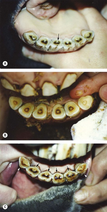
Fig. 7.17 (A) Standardbred horse, 18 years old. Marks are small and rounded. On the central incisors, they have almost disappeared (arrow). The occlusal tables of the centrals and the middles are trapezoid, those of the corner incisors are triangular with an apex to the labial side. (B) Belgian draft horse, 18 years old. Marks have disappeared from all lower incisors. The occlusal surfaces are triangular; those of the central and the middle incisors have a lingual apex, and those of the corners have a labial apex. (C) Arabian horse, 18 years old. Round marks are still clearly visible on all lower incisors. The occlusal surfaces of the centrals and the middles are trapezoid with a lingual apex (arrows); those of the corner incisors are oval with a labial apex (arrowheads).
Disappearance of the cups
The disappearance of the cups is an unreliable feature for age determination because it does not occur between narrow age limits. In all breeds, cups on the central incisors disappear at the age of 6–7 years, whereas cups on the middle incisors are filled in variably between 7 and 11 years and those on the corners between 9 and 15 years. The variations in the age at which the cups disappear may be due to a difference in the depth of the cup. The accumulation of cement in the infundibulum is variable, i.e., superabundant in some individuals and almost non-existent in others.
Changes in shape of the marks
On the central incisors, big oval marks are visible until the age of 6–7 years. These marks become oval to triangular from the age of 7–8 years onwards in Belgian draft horses, from the age of 8–10 years onwards in Standardbreds and Arabian horses, and from the age of 10 years onwards in mini-Shetlands. Round marks on the central incisors are visible at 9–10 years in Belgian draft horses, at 13–14 years in Standardbreds and mini-Shetlands, and at 15–17 years in Arabian horses (Fig. 7.17).
Disappearance of the marks (Fig. 7.17)
From all age-related dental characteristics, the disappearance of the marks is the one with the highest interbreed variability. In draft horses, marks on the central incisors disappear from the age of 12–15 years, and those on the middles and the corners from the age 14–15 years onwards. In mini-Shetland ponies, marks on the central, the middle, and the corner incisors disappear at the age of 15, 16, and 17 years, respectively. In Standardbred horses, marks disappear some years later. On the centrals, they vanish in 18-year-old horses while disappearing on the middle and the corner incisors in 19- to 20-year-olds. In Arabian horses, marks on the lower incisors may persist for a very long time. They start disappearing at the age of 20 but exhibit considerable individual variations.
Changes in shape of the occlusal surfaces
Changes in shape of the occlusal surfaces of the lower incisors are useful but inaccurate indicators of age. The changes are difficult to judge objectively because successive shapes shade off into one another and are not easily distinguishable. The sequential shapes of the tables of the central and the middle incisors are oval, trapezoid, triangular with the apex pointing to the lingual side, and biangular. A survey of the most important changes is given in Tables 7.1–7.4. It is striking that the shape of the lower corner incisor does not conform to the sequential changes described above. The lower corners remain oval for a long time and gradually develop an apex at the labial side. In Belgian draft horses and mini-Shetland ponies, a labial apex appears at the age of 9, and in Standardbreds at the age of 11. In Arabian horses, the apex is a constant characteristic in individuals aged over 12 years (Figs 7.16 & 7.17).
Direction of upper and lower incisors
The arch formed by the incisors of the opposing jaws as they meet, when viewed in profile, changes as the teeth advance from their alveoli and undergo attrition (Fig. 7.6). In young horses, the upper and lower incisors are positioned in a straight line (±180°). From the age of approximately 10 years onwards, the angle between upper and lower incisors becomes more acute. Because exact measurements of the age-related incisival angle are not available, the evaluation of the angle provides only a rough estimate of an animal’s age. The same applies for the curvature of the dental arch formed by the lower incisive tables. In young horses this arch is a semicircle, whereas in older individuals it forms a straight line. In view of the gradual character of this change in direction, however, it is impossible to determine the exact age at which it occurs.
The hook on the upper corner
The hook on the caudal edge of the upper corner incisor has long been considered as the typical characteristic for a 7- or 13-year-old horse. However, hooks on 103 and 203 are seen in a minority of horses and occur at practically any age over 5 years. Only 13 % of all 7-year-olds and 8 % of all 13-year-olds that were examined for this study presented a hook on one or both upper corners. On the other hand, hooks were also seen in 14 % of the 5- and 6-year-old horses, in 22 % of the horses aged between 8 and 12, and in 13 % of all horses aged over 13 years. As the presence of hooks on 103 and 203 cannot be related to any specific age category, it is considered irrelevant for the estimation of age in horses.
Conclusion
Teeth provide a practical available tool for estimating age in horses. Aging an individual horse from its dentition, however, is a complex process and all above-mentioned features should be carefully examined. It must be emphasized that dental aging in horses can only provide an approximate guess rather than an exact evaluation. In older horses, most of the so-called characteristic features can only be judged subjectively. It is obvious that the accuracy of the dental age determination declines markedly with age.
An important factor that can interfere with an accurate dental age determination in horses is the breed-dependence of the attritional dental wear. A comparison of the dental criteria in different breeds revealed that, in general, the incisor teeth of draft horses and mini-Shetland ponies are more liable to attrition, whereas the incisors of Arabian horses wear more slowly than those of Standardbred horses.
A variety of other factors such as nature and quality of food, environmental conditions, heredity, injury, and disease can also influence dental wear. It is, therefore, important that equine clinicians do not claim levels of accuracy that are unjustifiable. As it is impossible to assign specific ages to each dental feature, accuracy of age estimation in certain individuals can be very low.
Therefore, it is advisable to make written records at the time of examination to show the dental features upon which the age estimate was made. In some countries, there have been legal guidelines established to distance veterinarians from trying to state the age of a horse solely from dental findings. In case of insurance policies or legal questions, the veterinarian should indicate explicitly that he is providing an ‘estimate of age.’ It is also advisable that the incisor tables are photographed. When necessary, the pictures can be submitted to others for a second opinion and can be stored with appropriate identification for further use as well.8
1 Ten Cate AR. Physiologic tooth movement, eruption and shedding. In: Ten Cate AR, ed. Oral histology, development, structure and function. 5th edn. St Louis: CV Mosby; 1998:289–314.
2 Muylle S, Simoens P, Lauwers H. Age-related morphometry of equine incisors. Journal of Veterinary Medicine A. 1999;46:633–643.
3 Eisenmenger E, Zetner K. Veterinary dentistry, Lea & Febiger, Philadelphia, 1985:2–26.
4 Kertesz P. In search of Mr Bishop. Veterinary Record. 1993;133:608.
5 Zipperlen W. Over de ouderdomskennis van het paard of de tandleer. In: Geïllustreerd veeartsenijkundig handboek. Utrecht: B. Dekema; 1871:171.
6 Dupont M. L’âge du cheval. Paris: Librairie J B Baillière; 1901.
7 Frateur JL. De Ouderdomsbepaling van het Paard door het Gebit. Brussel: E Marette; 1922.
8 American Association of Equine Practitioners. Official Guide for Determining the Age of the Horse, 6th edn. Iowa: Fort Dodge; 2002.
9 Barone R. Dents. In Anatomie comparée des mammifères domestiques. Tome 3, 3rd edn, Paris: Vigot; 1997:91.
10 Dyce KM, Wensing CJ. Anatomie van het paard. Utrecht: Scheltema-Holkema; 1980.
11 Willems A. Ouderdomsbepaling van het Paard, 5th edn. Oud-Heverlee: Van de Sompele; 1980.
12 Habermehl KH. Wie sicher ist die Altersbestimmung beim Pferd? Berliner und Münchener Tierärztliche Wochenschrift. 1981;94:167.
13 McMullan WC. Dental criteria for estimating age in the horse. Equine Practice. 1983;5:10. 36
14 Walmsley JP. Some observations on the value of ageing 5–7-year-old horses by examination of their incisor teeth. Equine Veterinary Education. 1993;5:295.
15 Sack WO. Rooney’s Guide to the dissection of the horse, 6th edn. Ithaca: Veterinary Textbooks; 1994.
16 Navin JN. The age. In: Navin’s Veterinary practice. Indianapolis: John B. Hann; 1882:431–446.
17 Carmalt JL, Allen AL. Morphology of the occlusal surfaces of premolar and molar teeth as an indicator of age in the horse. J Vet Dent. 2008;25(3):182–188.
18 Dyce KM, Sack WO, Wensing CJ. Textbook of veterinary anatomy, 2nd edn. Philadelphia: W B Saunders; 1996.
19 St Clair LE. Teeth. In Sisson and Grossman’s The anatomy of the domestic animals, 5th edn, Philadelphia: W B Saunders; 1975:460.
20 McCarthy PH. Galvayne: the mystery surrounding the man and the eponym. Anatomia Histologia Embryologia. 1987;16:330.
21 Galvayne S. Horse dentition: showing how to tell exactly the age of a horse up to thirty years. Glasgow: Thomas Murray; 1886.
22 CD Equus – Vetstream Ltd, Three Hills Farm, Bartlow, Cambridge CB1 6EN, UK
23 Muylle S, Simoens P, Lauwers H. A study of the ultrastructure and staining characteristics of the dental star of equine incisors. Equine Veterinary Journal. 2002;34:230–234.
24 Joest E, Becker E. Zähne. In Handbuch der speziellen pathologischen Anatomie der Haustiere, 3rd edn, Berlin: Verlag Paul Parey; 1970:83–315.
25 Torneck CD. Dentin-pulp complex. In: Ten Cate AR, ed. Oral histology, development, structure and function. 5th edn. St Louis: CV Mosby; 1998:150–196.
26 Kilic S, Dixon PM, Kempson SA. A light microscopic and ultrastructural examination of calcified dental tissues of horses: III Dentine. Equine Veterinary Journal. 1997;29:206–212.
27 Richardson JD, Cripps PJ, Hillyer MH, et al. An evaluation of the accuracy of ageing horses by their dentition: a matter of experience? Veterinary. Record. 1995;137:88.
28 Richardson JD, Lane JG, Waldron KR. Is dentition an accurate indication of the age of a horse? Veterinary Record. 1994;135:31.
29 Richardson JD, Cripps PJ, Lane JG. An evaluation of the accuracy of ageing horses by their dentition: can a computer model be accurate? Veterinary Record. 1995;137:139.
30 Hillson S. Teeth. Cambridge: Cambridge University Press; 1986.
31 Marcq J, Lahaye J. Extérieur du cheval. Gembloux: J Duculot; 1943.
32 Misk NA. Radiographic studies on the development of incisors and canine teeth in donkeys. Equine Practice. 1997;19:23.
33 Muylle S, Simoens P, Lauwers H. Ageing horses by an examination of their incisor teeth: an (im)possible task? Veterinary Record. 1996;138:295–301.
34 Muylle S, Simoens P, Lauwers H, van Loon G. Ageing draft and trotter horses by their dentition. Veterinary Record. 1997;141:17–20.
35 Muylle S, Simoens P, Lauwers H, van Loon G. Ageing Arab horses by their dentition. Veterinary Record. 1998;142:659.
36 Muylle S, Simoens P, Verbeeck R, et al. Dental wear related to the microhardness of enamel and dentine. Veterinary Record. 1998;144:558–561.