Chapter 22 Gait Analysis for the Quantification of Lameness
Evaluation of lameness in horses is a skill first learned by training and then sharpened with experience over time. The standard of practice is an initial subjective evaluation of the horse while moving to detect and then localize the source of pain causing lameness to the affected limb or limbs. It is a difficult endeavor in horses with mild lameness, when only subtle changes in movement from normal are present. There is substantial disagreement, even between experienced clinicians, in the identification and localization of the lame limb(s) in horses with mild lameness.1-3 There is also disagreement between experts in the amount of improvement in lameness after nerve blocks,4 and equine veterinarians, because they are human, have been shown to be biased. The amount of disagreement between veterinarians on the results of subjective evaluation of lameness in horses is what would be expected for any difficult diagnostic test.
A contributing source of disagreement is the limited sensitivity of the human eye, which has an estimated time resolution of about 10 to 15 samples per second.5,6 Events in stride of a horse trotting at 4 meters per second, which is about 1.5 strides per second, occur at a frequency of twice the stride rate, or about 3 times per second.7 To prevent significant errors in detection of signal amplitude, the sampling frequency should be, at a minimum, 5 times the frequency of the event being detected.8 Therefore the natural capability of the human eye is below or just at the minimum required to detect important asymmetry in motion events used to judge lameness in horses. All the objective methods of lameness evaluation discussed in this chapter sample data at frequencies higher than that of the naked eye, and thus are theoretically capable of higher accuracy. An objective, precise, and accurate method of lameness detection and quantification in horses is certainly justifiable.
The purpose of this chapter is to introduce and summarize the gait analysis methodology currently being studied or used. Emphasis is given to those that could be used by private practitioners. To be effective, any such system has to be easy to use, and data collection and analysis must be quick. It must be affordable and capable of providing the veterinarian with adequate return on investment. It may be most useful to veterinarians as an aid in detecting and evaluating horses with subtle lameness and lameness in several limbs. Natural stride-to-stride variability must be overcome, so that small differences in subtle lameness can be detected. For example, to detect subtle differences after diagnostic analgesia or after treatment, any such system must be capable of collecting data from multiple contiguous strides. Lameness over ground may be different from lameness on a treadmill; therefore any system must be capable of collecting data when the horse is moving over ground. Also, ideally, any system should be capable of detecting and evaluating lameness in all limbs simultaneously.
There are two general approaches to using gait analysis to detect and measure lameness in horses: kinetics and kinematics. A kinetic technique measures ground reaction forces. A kinematic technique measures motion of the body. There are advantages and disadvantages for each general approach.
Kinetics
Kinetics can rightly be considered a more direct method (compared with kinematics) for detecting and measuring lameness in horses. If a horse has pain during the weight-bearing portion of the stride because of lameness, it bears less weight on that limb, resulting in lower peak vertical ground reaction forces (GRFs) on that limb. Certain lameness conditions may decrease horizontal or transverse GRFs, but the effect on the vertical GRFs is usually most prominent.
Kinetic methods to measure GRFs include a stationary force plate, pressure-measuring pads, instrumented horse shoes, and force-measuring treadmills. A stationary force plate is the most commonly used and cited method, but each method is briefly discussed. A stationary force plate, because of its widespread use in lameness research centers around the world, is discussed in more detail.
Pressure-measuring pads are force or pressure distribution measurement systems consisting of force sensor elements installed between matting surfaces. A horse is led over the pressure-sensitive pad to collect data from single or, if the pad is long enough, a few consecutive strides. Pressure-sensitive pads can be cut and customized to fit the bottom of a horse’s foot and then placed between the bottom of the foot and a shoe in an attempt to collect data from multiple contiguous strides.9,10 This technology is ideal for mapping the force or pressure profile of a surface, but peak force or pressure can also be quantified. With further development and marketing this technology may become useful for routine evaluation of shoeing techniques designed to alter force distribution within the foot. Currently there are only a few equine practices in the United States using this technology clinically to evaluate lame horses.
Instrumented horse shoes are custom-built systems designed to either directly, with force transducers, or indirectly, with strain gauges, measure vertical GRFs to the hoof during weight bearing.11-14 Their main advantage is the ability to collect data from multiple contiguous strides in an overground, field-like setting. The main difficulties and disadvantages of instrumented horse shoes include the complexity, size, and weight of the instrumentation, which affect normal hoof and limb movement and gait. Although there are several recent reports of force-measuring horse shoes used successfully in research investigations, currently there are no commercially available systems for a practicing veterinarian to use. Strain gauges glued to the hoof wall, with the resulting strain to the hoof during weight bearing estimating GRF, can act as a surrogate for an instrumented horse shoe. Disadvantages of any hoof-mounted system for measuring GRFs to detect lameness include the need to instrument all four feet simultaneously and, more importantly, the extreme dependence of results on surface characteristics. Because of these disadvantages and the technical and design expertise required for development, it is unlikely that such a device will quickly become adopted for use during routine lameness evaluations.
A force-measuring equine treadmill is a unique piece of equipment consisting of piezoelectric load-sensitive sensors in a treadmill platform (Figure 22-1).15,16 The only working system in regular use is at the University of Zurich in Switzerland. The force-measuring equine treadmill is exceptional for detecting small changes in lameness between treatments because of its ability to collect data from multiple, contiguous strides. It is also exceptional for determining severity and location of compensatory lameness because it is capable of measuring vertical GRFs in all four limbs simultaneously.17 It is a one-of-a-kind, custom-built, installed piece of equipment, and as such is unlikely to be adopted for use as a lameness diagnostic aid in any but the most sophisticated equine private practices or research centers.
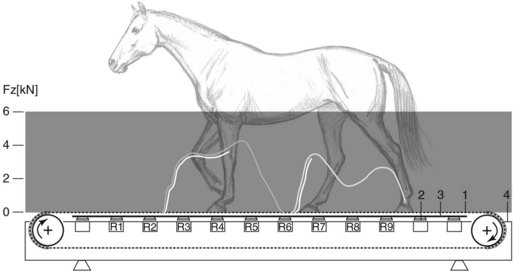
Fig. 22-1 Treadmill-integrated force-measuring system capable of determining the vertical ground reaction forces and the hoof positions during stance phase of all limbs simultaneously. Fz, vertical ground reaction force in kilo-Newtons. 1, Traction belt; 2, shock absorber; 3, treadmill platform; 4, treadmill frame. R1 through R9, piezoelectric force sensors of the right side of the treadmill.
(Drawing courtesy Dr. Michael Weishaupt, University of Zurich, Switzerland.)
A stationary force plate is the most widely used and available kinetic technique for objective evaluation of lameness in horses.18,19 Under controlled conditions of constant speed the coefficient of variation of vertical GRF between strides in both lame and sound horses is remarkably low.20-22 Thus a lesser number of stride repetitions are required to achieve repeatable results, and small differences between treatments can be detected. A stationary force plate is also sensitive to subtle lameness detection, with some studies suggesting that force plate detection of lameness (decreased vertical GRF) is more sensitive than the human eye in subjective evaluation of lameness.23,24 A stationary force plate, unlike the aforementioned other kinetic techniques, is also capable of measuring horizontal GRFs, which may be helpful in further differentiating type of lameness, that is, acceleratory or deceleratory. There are several force plates worldwide that are used, at least occasionally, for evaluation of lame horses. Despite the high accuracy and precision of a stationary force plate, most experts agree that five or six strikes on the force plate are needed for acceptable results.10,20-22 Because a horse does not always strike the force plate on every attempt, the process is time consuming. Also, the size of commercially available force plates is too small, such that data from only one hoof (occasionally two hooves) are collected at one time. Collection of data from contiguous strides and simultaneous measurement of all four limbs to detect and evaluate compensatory or secondary lameness is not possible. Therefore a stationary force plate has not been readily accepted as a tool for routine, clinical evaluation of lameness.
Kinematics
Using kinematics is an indirect method of detecting and quantifying lameness. Pain of lameness causes the horse to bear less weight on the affected limb. The decreased vertical GRF during weight bearing perturbs, in some way, the normal, expected motion of the torso, head, neck, and limbs, usually by increasing asymmetry of movement between the right and left strides. However, motion of the torso, head, neck, and limbs is perturbed by causes other than lameness as well; for example, conscious movement of the head in a curious or anxious horse. Thus in lame horses motion perturbation is more variable than change in GRF. Therefore kinematic techniques must be able to collect numerous (more than is required for a stationary force plate), contiguous strides to attain the sensitivity required to detect and evaluate mild to moderate lameness. However, because most veterinarians observe changes in motion in a horse’s gait during their subjective evaluation, kinematic techniques generate results that are generally more intuitive and well understood. Important findings in kinematic studies can be easily applied by a practicing veterinarian in a standard lameness evaluation.
Until only very recently the most commonly used kinematic technique consisted of filming the horse in motion with cameras and then analyzing the collected motion with software on a remote station. The horse is not instrumented except for possibly attaching lightweight reflective markers for easier identification of the body parts of interest. There are many different commercially available, camera-based systems for kinematic evaluation of lameness in horses. These systems use different types of cameras with different capabilities and various, custom-designed software packages with varying capabilities for data analysis. They all depend on unobstructed, line-of-sight light transmission.
I have used camera-based kinematic gait analysis to study and evaluate lameness in horses since the early 1990s and have come to the following conclusions.1,25-38 Camera-based techniques are reliable, accurate, and sensitive for detecting and evaluating lameness if two conditions are met: (1) multiple, contiguous strides must be collected to overcome the natural stride-by-stride variability, so that small differences can be detected; and (2) the size of the field of view compared with the size of the subject must be controlled and kept as small as possible for constant and precise spatial resolution. These limitations can be relaxed somewhat for lameness in the moderate to severe range but are absolutely vital for detection and evaluation of mild lameness. Essentially these limitations confine the use of this technique for evaluation of lameness to a treadmill. Adequate data can be acquired with the horse moving over ground in front of the camera, but to capture the movement from enough strides with reasonable spatial resolution, multiple passes are required. Because of these limitations it is unlikely that camera-based kinematic gait analysis technique can be used in clinical practice. However, it is a useful investigative tool, and there are many camera-based kinematic studies of lameness from which practical information for an equine practitioner can be extracted.1,25-80
The long list of motion parameters that may have some association with lameness posed a problem in early studies attempting to develop objective kinematic techniques. There was always the question of what parameters were best at differentiating a sound from a lame horse? Many investigators used a shotgun approach, measuring many different variables, on small numbers of horses, calculated from a relatively small number of contiguous strides. Under these conditions, moderately severe lameness was necessary to find significant differences between sound and lame horses or between lameness grades. Results were frequently conflicting, with studies using different models of induced lameness finding different motion parameters as the most sensitive for lameness detection and quantification. Most kinematic studies of lameness involve evaluations of horses trotting on a treadmill, where assumptions about torso acceleration and deceleration or the biomechanics of limb impact and push-off are not the same as in the natural overground state. Despite these limitations, there are numerous motion parameters that have been shown to be useful for detecting and quantifying lameness in horses.
In horses with unilateral lameness, fetlock extension and distal interphalangeal joint flexion are less during weight bearing of the lame limb compared with the sound contralateral limb.* This is true for both forelimb and hindlimb lameness. These kinematic measures are considered to be two of the most sensitive indicators of weight-bearing lameness. Fetlock extension during the lame limb stance phase was 8 degrees less than in the nonlame limb stance phase in a sole pressure model of induced grade 1 (of 5) lameness.76 However, whether 8 degrees of difference in fetlock extension can be detected by the unaided human eye in a horse at a trot is a matter of conjecture. Carpal extension during stance is reduced, but only in horses with moderate to severe lameness.27,56,76 Proximal limb joints become more flexed during weight bearing of the lame limb, resulting in an overall limb shortening during stance,47,76 a compensatory mechanism to reduce peak vertical GRF. Because of the competing opposite mechanisms of increased proximal joint flexion and decreased fetlock extension during lame limb stance, overall limb shortening during lame limb stance is probably not a very sensitive indicator of lameness.
In weight-bearing lameness of mild to moderate intensity, stance duration of the lame limb is increased compared with the sound limb.16,17,25-27,81 This may seem counterintuitive but is an effective method to reduce peak vertical GRF by spreading the entire vertical GRF impulse over a longer time. The difference between lame and sound limbs is small at the trot even when lameness is of moderate intensity.27,43,82 It is unlikely to be seen by an unaided human eye but can be measured by expanding the time domain with slow motion review of high sampling-rate video. Length of forelimb retraction, or the extent to which the horse will keep the forelimb on the ground between full weight bearing at midstance and end of breakover, has also been reported to change with forelimb lameness.27,48,76 In a sole pressure-induced lameness model, forelimb retraction was shortened in the lame forelimb at the walk.27 Sound forelimb retraction has also been reported to be reduced in forelimb lameness.76 Results reported for the association of forelimb retraction with lameness are not clear enough for forelimb retraction to be considered a sensitive indicator of forelimb lameness.
The length and shape of the limb flight arc during the swing phase of the stride are commonly perceived to be associated with both forelimb and hindlimb lameness. Forelimb protraction, or the extent to which the horse swings the limb forward before impact, has been reported to increase or decrease with lameness, depending on the location of pain within the limb.27,57,76 Heel pressure-induced forelimb lameness, similar to navicular disease, causes decreased forelimb protraction, and toe pressure-induced lameness, like laminitis, causes increased forelimb protraction.27 Hindlimb protraction, by contrast, seems to be a sensitive indicator of most causes of weight-bearing hindlimb lameness.28,48 It can also be easily appreciated when looking from the side of the horse as it passes by the evaluator. Comparing the space between the retracting forelimb and protracting hindlimb during each half of the stride during the walk and especially the trot is easy to do and can be a highly productive exercise for detecting difficult hindlimb lameness in the horse.
The association of other swing phase parameters with lameness and their sensitivity for detection of lameness are less clear. Stride length of the lame limb is usually less than the sound limb, but significant differences are not seen until lameness severity is of moderate intensity.41,43 Step length, or the distance between placement of opposite limbs, is less between placement of the lame and then sound limbs than between placement of the sound and then lame limb.76 Height of foot flight arc may be increased or decreased in a lame forelimb compared with the sound forelimb, and the shape may be different, depending on the cause of lameness.1,21,48,60 Although it is commonly perceived that horses with hindlimb lameness drag the toe on the affected side because of low hoof flight arc, this is not always the case. In a hindlimb, height of hoof flight arc is determined by two competing factors, with the strongest determining the overall effect. Decreased propulsion during push-off of the lame hindlimb causes the hind torso to rise less. To bring the affected limb forward during the swing phase of the stride without dragging it on the ground, the proximal limb joints flex more. The comparative extents of the decreased torso rise and increased limb flexion determine the height of the hoof flight arc. Anecdotally, limb abduction or adduction during the swing phase of the stride is thought to be helpful for determination of forelimb and hindlimb lameness in horses, and the direction of horizontal swing has been associated with lameness in specific locations; for example, forelimb abduction (swing out) for carpal lameness, hindlimb adduction (swinging in) for distal tarsal lameness, and hindlimb abduction for stifle lameness. The specificity of forelimb or hindlimb abduction to detect and locate lameness has not been studied adequately. In one study of amphotericin-induced carpal lameness, carpal abduction during swing was not increased in the lame limb.43
There is substantial evidence from numerous studies indicating that the most sensitive kinematic measures for detecting lameness in horses are the motion parameters associated with asymmetry of vertical torso movement; more specifically, vertical displacement, velocity, and acceleration of the head and neck for forelimb lameness and of the pelvis for hindlimb lameness.* In sound horses, asymmetry of trunk and proximal limb movement is less than that of the distal limb.51 In horses with weight-bearing lameness, asymmetry of trunk and proximal limb movement is greater than that of the distal limb.60 Thus if using asymmetry between movement of the right and left sides of the body as the metric for detecting and quantifying lameness, one should concentrate on movement of the torso and put less credence in movement of the limbs. In a directed attempt to determine the best indicators of forelimb lameness, vertical movement of the head provided the best classification between sound and lame limbs than any movement in the forelimbs.31 As stated by one prominent equine veterinarian with experience using kinematics to determine lameness in horses, “Load redistribution in lameness is possible only by altered vertical movements of the trunk, head, and neck.”76
Asymmetric vertical torso movement in forelimb lameness is best expressed in movement of the head and neck.* Asymmetric vertical movement of the head amplifies asymmetric vertical movement of the body’s center of mass because of the long moment arm of the neck.34,47,76,82,83 In contrast, asymmetric vertical movement of the withers was shown to be less sensitive for detecting forelimb lameness.43,84 Asymmetric vertical movement of the head was shown in many studies to be one of the most sensitive kinematic indicators of forelimb lameness.† This, of course, is what many equine practitioners see when they evaluate horses for forelimb lameness, and it is routinely known as the “head bob.” “Down on sound” and “up on bad” are both used to describe the vertical asymmetric motion of the head in horses with forelimb lameness. Simply put, the head is supposed to move down more and to a lower height when the sound forelimb is in stance, and it is supposed to move up more and to a higher height after push-off of the lame limb. It is not as simple as this because both of these statements are not mutually exclusive. In addition, whether one is interpreting total vertical head excursion or the end maximum or minimum heights of the head during the strides of right and left forelimbs, neither is correct in every horse. Minimum head position was proposed to be most diagnostic of impact forelimb lameness, the most common type of forelimb lameness in the horse, with the head moving down to its lowest position during the stance phase of the sound limb.37 Additionally, maximum head position after push-off of the lame limb when the pain of lameness occurs in the second half of stance (lameness likely of lower incidence in the forelimb) is higher than after push-off of the sound limb.37 This description of head movement associated with forelimb lameness has not been sufficiently proven experimentally and is solely based on hypothetical mathematical modeling of the head and torso acting as free bodies moving in opposite directions during the trot. If this description is true, then a careful study of vertical head motion may give the equine practitioner valuable information for localizing lameness within the affected forelimb.
†References 7, 31, 37, 41, 43, 47, 50, 63, 76.
The best indicators of asymmetric hind torso movement for detection of hindlimb lameness in the horse are (1) differential vertical movements of the tubera coxae, the “pelvic rotation” or “hip hike” technique,28,39,84 and (2) asymmetric vertical movement of the entire pelvis between left and right hindlimb stance phases, paralleling the “head bob” technique for detection of forelimb lameness.* The first method is easier to see in most horses with hindlimb lameness but has been criticized as misleading in the occasional horse with asymmetric pelvic anatomy and insensitive to rapid alleviation of lameness after nerve blocks. The second method, because the overall vertical movement is less, is more difficult to appreciate in horses with mild hindlimb lameness. However, because whole pelvic fall better mimics the association of vertical GRFs, it is probably more sensitive and accurate. The “pelvic rotation” or “hip hike” method centers on the fact that vertical displacement of the lame-side hemipelvis is greater than of the sound-side hemipelvis. The pelvis appears to rotate toward the side of lameness. Easily visible markers fixed to the right and left tubera coxae may help to detect this asymmetric movement. The best position for the evaluator using this method is behind the horse moving away from the evaluator. The vertical displacement method detects the whole pelvis moving down to a lower height during the stance phase of the sound hindlimb or the pelvis moving up to a greater height after push-off of the sound limb. An easily visible marker fixed to the most dorsal aspect of the pelvis between the tubera sacrale may help to detect this asymmetric movement. Using this method the evaluator can be either behind the horse as it moves away or beside the horse as it passes by. Horses with an impact type of lameness, that is, greatest pain in the deceleratory phase of stance, should primarily display asymmetric downward movement in the pelvis. Horses with an impulsive type of lameness, that is, greatest pain in the acceleratory phase of stance, should primarily display asymmetric upward movement of the pelvis.37
Body-mounted inertial sensors are the best for an objective kinematic system of lameness evaluation in horses.30,32,84,85 Body movement can be detected from multiple contiguous strides and wirelessly transmitted to a remote hand-held computer in a field situation. To be clinically useful, a body-mounted inertial sensor system must satisfy several criteria, including (1) the inertial sensors must be small and light enough so that normal movement is not affected; (2) data from sensors must be sampled at a rate greater than the sampling rate of the human eye using digital transmission of sufficient bit size for accurate signal representation; (3) range of data transmission should be far enough to allow measurement of movement in the environment normally used by the veterinarian; (4) data collection and analysis should be quick; and (5) the reported measures must be valid for quantification of lameness and easy to understand.
Size of the actual sensing element is not the limiting factor in the end size and weight of the entire apparatus attached to the horse’s body. Micromachined accelerometers and gyroscopes are very small. Apparatus size is dictated by the size of commercially available radio components and batteries needed for adequate transmission range and power usage. True, vertical movement, the measure of most importance for lameness detection, requires multiple, simultaneously collected sensing elements (accelerometers, gyroscopes, and magnetometers). The power requirement for transmission, and coincidentally the size and weight or “footprint” of the sensor, increases with increasing number of sensors sampled. Higher sampling (e.g., 200 Hz over 50 Hz), increased bit size (e.g., 12-bit transmission over 8-bit), and range of transmission (e.g., 100 m vs. 30 m) also increase demands for power usage, which translates into bigger and less ideal body-mounted sensors. Using data-logging equipment attached to the horse to circumvent this size-transmission tradeoff is not convenient for a veterinarian. A veterinarian does not want to instrument the horse, collect data, stop the horse, take equipment off the horse, or download data after data collection, and then do data analysis. The veterinarian wants results quickly while performing the normal lameness evaluation, without watching computer screens or worrying about whether data are being collected properly. The end result or completed analysis should be accomplished, if not in real time, in as little time as possible. The results should be understandable to practicing veterinarians, some of whom may not have much experience in computers, mathematics, statistics, or biomechanics and who may not have the luxury of spare time to gain such.
At the time of the writing of this chapter there are a few private companies and university laboratories around the world investigating and developing a body-mounted inertial sensor-based kinematic evaluation system for use in horses (Figure 22-2). The remainder of this chapter briefly describes some of these systems.
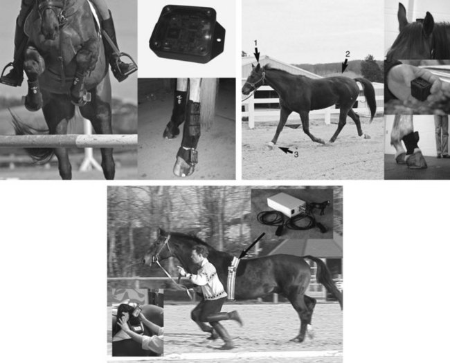
Fig. 22-2 Currently available inertial sensor systems for field detection and evaluation of lameness in horses. Top left: EquuSense system from EquuSys. Clockwise from left: sensor nodes on forelimbs during jump; bare sensor node; closeup of sensor node on left forelimb. Top right: Lameness Locator from Equinosis. Clockwise from left: position of 1, head accelerometer; 2, pelvic accelerometer; and 3, right forelimb pastern gyroscope during live field data collection; head accelerometer attached with 3M (St. Paul, Minnesota, United States) Dual Lock tape to felt head bumper; bare sensor; right forelimb pastern sensor in pastern wrap pouch. Bottom: Equimetrix system from Centaure-Métrix. Live horse data collection. Insert top right: sensors and data logger attached to girth strap. Insert bottom left: placing data logger in saddle pad.
(Top photos courtesy Michael Davies, EquuSys. Bottom photos courtesy Dr. Eric Barrey, Centaure-Métrix.)
EquuSys Inc. (Sudbury, Massachusetts, United States) is marketing EquuSense Equine sensor systems for analysis of equine performance, including lameness. EquuSense Equine sensor systems are composed of sensors with multiple sensing units (e.g., accelerometers, gyroscopes, and magnetometers) and telemetry capability (WiFi, GPS, and Bluetooth) that are attached to the horse, and accompanying hardware (a laptop computer) and software. Each sensor provides objective and accurate information on its position, velocity, acceleration, orientation, and rotation relative to the horse or to a global reference frame. The software can track from 8 to 18 sensors, depending on the product platform selected, at up to 2000 frames/s in real time. The sensors weigh less than 100 g and can be attached to the horse with specially designed boots and pouches. The data are analyzed to give the user a graphical output of sensor trajectories. This system is a more practical, field-ready replacement for the more familiar camera and marker kinematic systems. The user determines what trajectory variables should be measured and how measured values are analyzed.
Lameness Locator is a wireless, body sensor-based lameness evaluation system developed by equine veterinarians and engineers at the University of Missouri in collaboration with engineers at the Hiroshima Institute of Technology in Japan. It is licensed to Equinosis in Columbia, Missouri for further development and commercialization to equine veterinarians only. Lameness Locator consists of three inertial sensors (two accelerometers and one gyroscope) attached to the head, right forelimb, and pelvis. Each sensor is 4 cm by 3 cm by 2 cm in dimensions and weighs less than 30 g. Vertical accelerations of the head and pelvis and angular velocity of the right forelimb are measured and wirelessly transmitted in real time to a hand-held tablet computer up to 150 m away. Custom algorithms are then implemented to detect and measure forelimb and hindlimb lameness when the horse trots or walks. The algorithms were developed from previous kinematic research at the University of Missouri using camera and markers attached to sound and lame horses. Best sites for motion detection to measure lameness were determined using a data-mining approach to analysis of large datasets. Random motion, unassociated with lameness, as a result of a misbehaving or uncooperative horse, which may interfere with detection and quantification of lameness, is extracted from the raw signals. Lameness detection and quantification results are presented to the user in a graphical interface that shows impact and propulsion asymmetry in each stride, with thresholds between what is expected for soundness and lameness. Trend and variability of collected data are also presented. Because forelimb and hindlimb lameness are measured simultaneously, compensatory or multiple-limb lameness patterns can be evaluated. The developers of Lameness Locator claim that it will be most useful to equine veterinarians evaluating horses with mild, subtle, or multiple-limb lameness and in objectively evaluating partial improvements after using diagnostic analgesia. At the time of this writing, Lameness Locator is being field-tested at 25 private practice and university teaching hospital sites in North America and Europe.
Equimetrix is a multidimensional, accelerometer system attached to the girth of an exercising horse. It is marketed by Centaure-Métrix (Evry, France) primarily in France and the Benelux countries (Belgium, the Netherlands, and Luxembourg). Three-dimensional torso acceleration is collected and logged as the horse exercises. The data are then analyzed, and output is used to measure characteristics of performance, including stride parameters (e.g., regularity, frequency, length, and timing) and “propulsion power.” Although the output of Equimetrix is directed primarily toward assessing performance in exercising horses, existing algorithms could be adapted or new algorithms could be developed to more specifically evaluate lameness in horses.