 CHAPTER 196 Cutaneous Infestations
Arthropods are common in the environment. Although many can bite or sting humans, only a few infest humans. Arachnids (mites) are the most common, parasitizing humans and animals by burrowing into the skin and depositing eggs within the skin.
SCABIES
Etiology and Epidemiology
Scabies is caused by the mite Sarcoptes scabiei. The female mite burrows into the epidermis and deposits her eggs, which mature in 10 to 14 days. The disease is highly contagious because infested humans do not manifest the typical signs or symptoms for 3 to 4 weeks, facilitating transmission. An immunocompetent person with scabies typically harbors 10 to 20 mites.
Scabies is the most common human infestation, affecting at least 300 million persons worldwide.
Clinical Manifestations
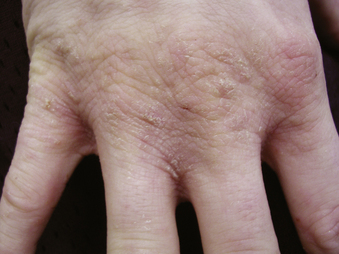
The clinical presentation varies depending on the age of the patient, duration of infestation, and immune status of the patient. Severe and paroxysmal itching is the hallmark, with complaints of itching that is frequently worse than the eruption would suggest. Most children exhibit an eczematous eruption composed of red, excoriated papules and nodules. The classic linear papule or burrow is often difficult to find. Distribution is the most diagnostic finding; the papules are found in the axillae, umbilicus, groin, penis, instep of the foot, and web spaces of the fingers and toes (Fig. 196-1). Infants infested with scabies have diffuse erythema, scaling, and pinpoint papules. Pustules and vesicles are much more common in infants and are found in the axillae and groin and on the palms and soles. The face and scalp usually are spared in adults and older children, but these areas are usually involved in infants. Nodular lesions may occur on the trunk, axillary regions, or genitalia and can represent active infection or prolonged hypersensitivity lesions following resolution of infestation. Chronic lesions may develop a malodorous, scaly crust. Immunocompromised or neurologically impaired persons may develop a severe form of the disease known as Norwegian or crusted scabies, with infestation of 2 million live mites at one time.
Laboratory and Imaging Studies
The diagnosis of scabies can be confirmed by microscopic visualization of the mite, eggs, larvae, or feces in scrapings of papules or burrows examined under oil immersion. Skin biopsy is rarely necessary but may be useful if lesions have become nodular.
The diagnosis of scabies should be considered in any child with severe itching. A thorough search for an infested contact should be undertaken.
Treatment
Curative treatment is achieved by a 12-hour (overnight) application of permethrin 5% cream applied to the entire body, which may be repeated 1 week later if necessary. Gamma benzene hexachloride should be avoided in young children because of a small risk of central nervous system (CNS) toxicity. Parents and all caregivers should be treated simultaneously. Itching persists for 7 to 14 days after the mites have been killed and may continue for up to 6 weeks. Bed linens, towels, pajamas, and clothes worn for the previous 2 days before treatment should be machine-washed in hot water and machine-dried using high heat. Heat is the most effective scabicide. Items that are not washable may be dry-cleaned or placed in a sealed plastic bag for 7 days.
Complications and Prognosis
Secondary bacterial infection may occur but is uncommon. Scabies can be much more severe among immunocompromised persons. In contrast to the pediculoses, scabies is not a vector for infections.
Pruritus may persist for 7 to 14 days after successful therapy because of a prolonged hypersensitivity reaction, which does not indicate treatment failure. Inadequate treatment or reinfestation should be suspected if new lesions develop after treatment.
PEDICULOSES
Etiology
Three species of lice infest humans: Pediculus humanus capitis, the head louse; Pthirus pubis, the pubic louse or crab louse; and Pediculus humanus humanus, (also known as Pediculus humanus corporis), the body louse. Lice are wingless insects 2 to 4 mm in length that cannot fly or jump. Transmission usually occurs by direct contact with the head of another infested individual. Indirect spread through contact with fomites or personal belongings, such as hairbrushes, combs, or caps, is much less frequent.
Pediculosis differs from scabies infestation in that the louse resides on the hair or clothing and intermittently feeds on the host by piercing the skin. The bite causes small urticarial papules and itching. Head lice live close to the skin and may live for 30 days, depositing 100 to 400 eggs as nits on hair shafts, usually within 6 mm of the scalp.
Epidemiology
Head lice are seen most frequently in early school-age children. Head lice infestations are unrelated to hygiene and are not more common among children with long hair or with dirty hair. It is estimated that 6 to 12 million persons in the United States and 1% to 3% of persons in developed countries are infested with head lice each year. In the United States, head lice infestation is rare among African Americans and may be more common in girls, which is attributed to their tendency to play more closely with one another than boys do.
Pubic lice are transmitted by sexual contact. Their presence in children may be a sign of child abuse. Body lice are firm evidence of poor hygiene, such as infrequent washing and clothing changes.
Clinical Manifestations
Itching, if present, is the primary symptom. Pediculosis capitis usually causes pruritus behind the ears or on the nape of the neck or a crawling sensation in the scalp. Pediculosis pubis usually causes mild to severe pruritus in the groin. Eyelash involvement in children may cause crusting and blepharitis. Pediculosis corporis causes pruritus that, because of repeated scratching, may result in lichenification or secondary bacterial infection. Excoriations and crusting, with or without associated regional lymphadenopathy, may be present.
Differential Diagnosis
Infestation with the head louse may be asymptomatic and has little morbidity. The diagnosis can be confirmed by visualizing a live louse. A fine-toothed comb to trap lice is more effective than simply looking at the hair. Wet combing is more time-consuming, but dry combing produces static that may propel the lice away from the comb.
Nits represent the outer casing of the louse ova. Viable nits have an intact operculum (cap) on the nonattached end and a developing louse within the egg. Brown nits located on the proximal hair shaft suggest active infestation. White nits located on the hair shaft 4 cm or farther from the scalp indicate previous infestation. Because nonviable nits can remain stuck in the hair for weeks to months after an infestation has resolved, many children with nits do not have active lice infestation.
Treatment
The treatment of head lice is controversial because of resistance to many established options. Over-the-counter permethrin (1%) and pyrethrin-based products (0.17–0.33%) are the first choices of therapy. Because 20% to 30% of eggs may survive one treatment, a second treatment should be applied in 7 to 10 days. The prevalence of drug resistance has not been determined. Malathion (0.5%) lotion may be used as an alternative for resistant cases.
Everyone in the family should be checked for head lice and treated if live lice are found to reduce the risk of reinfestation. Bed linens, towels, pajamas, and clothes worn for the previous 2 days before treatment should be machine-washed in hot water and machine-dried using high heat. Items that are not washable may be dry-cleaned or placed in a sealed plastic bag for 2 weeks. Brushes and combs should be soaked in dish detergent or rubbing alcohol for 1 hour. Rugs, furniture, mattresses, and car seats should be vacuumed thoroughly.
The finding of active lice infestation indicates their presence for 1 month or more. Manual removal of nits after treatment is not necessary to prevent spread. Children treated for head lice should return to school immediately after completion of the first effective treatment or first wet combing, regardless of the presence of remaining nits. There is no evidence that no nit or nit-free policies reduce transmission of head lice. If required for return to school, nit removal is best achieved by wetting the hair and combing with a fine-toothed metal comb.
Complications
Excoriations can become secondarily infected with skin bacteria, usually Staphylococcus and Streptococcus. The body louse functions as a vector for potentially serious infectious diseases, including epidemic typhus, caused by Rickettsia prowazekii; louse-borne relapsing fever, caused by Borrelia recurrentis; and trench fever, caused by Bartonella quintana. These louse-borne infections are rare in the United States. In contrast to body lice, head lice and pubic lice are not associated with transmission of other infections.
Prognosis and Prevention
Pediculicide treatment along with appropriate disinfestation measures of fomites is highly effective. Reinfestation from untreated contacts or fomites, especially for pediculosis corporis, is more likely than primary treatment failure.
Families, school nurses, and other health care professionals need to be educated about the mode of transmission and precise diagnosis before treatment of contacts is instituted. There is no evidence that group screening is effective.
Callen J., Chamlin S., Eichenfield L.E., et al. A systematic review of the safety of topical therapies for atopic dermatitis. Br J Dermatol. 2007;156:203-221.
Eichenfield L.E., Esterly N.B., Frieden I.J. Textbook of Neonatal Dermatology, 2nd ed. Philadelphia: WB Saunders, 2008.
Oranje A., Harper J., Prose N. Textbook of Pediatric Dermatology, 22nd ed. Hoboken, NJ: John Wiley, 2006.
Paller A.S., Mancini A.J. Hurwitz Clinical Pediatric Dermatology, 3rd ed. Philadelphia: WB Saunders, 2006.
Palmer C.M., Lyon V.B. Stepwise approach to topical therapy for atopic dermatitis. Clin Pediatr. 2008;47:423-434.
Sundine M.J., Wirth G.A. Hemangiomas: An overview. Clin Pediatr. 2007;46:206-221.