 CHAPTER 201 Foot
In newborns and non–weight bearing infants, the difference between posturing and deformity is important. Posturing is the habitual position the infant holds the foot; passive range of motion is normal. Deformity produces an appearance similar to posturing, but passive motion is restricted. Most pediatric foot disorders are painless. Foot pain is more common in older children (Table 201-1).
TABLE 201-1 Differential Diagnosis of Foot Pain by Age
| Age Group | Diagnostic Considerations |
|---|---|
| 0 to 6 years | |
| 6 to 12 years | |
| 12 to 18 years |
CLUB FOOT (TALIPES EQUINOVARUS)
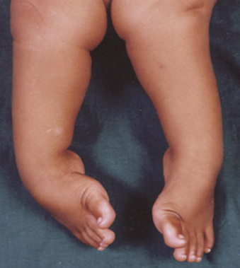
A clubfoot deformity involves the entire leg, not just the foot. It affects 1 in 1000 newborns and is bilateral in one half of the cases. The tarsals in the affected foot are hypoplastic; the talus is most affected. The muscles of the limb are hypoplastic because of the abnormal tarsal interactions, which leads to a generalized limb hypoplasia, mainly affecting and shortening the foot. There is usually atrophy of the calf musculature.
Etiology
Family history is important. Club foot can be congenital, teratologic, or positional. Congenital clubfoot (75% of all cases) is usually an isolated abnormality. Teratologic clubfoot is associated with a neuromuscular disorder, such as myelomeningocele, arthrogryposis, or other syndromes. Positional clubfoot is a normal foot that was held in the deformed position in utero.
Clinical Manifestations
The diagnosis is seldom confused with other disorders (Fig. 201-1). The presence of clubfoot should prompt a careful search for other abnormalities. The infant will have hindfoot equinus and varus, forefoot adduction, and varying degrees of rigidity. All are secondary to the abnormalities of the talonavicular joint. Calf atrophy and foot shortening are more noticeable in older children.
Radiologic Evaluation
In infants, radiographs and advanced imaging are rarely necessary for assessment since their tarsals have incomplete ossification. The navicular ossifies at about 3 years of age for girls and 4 years for boys. As children age, radiographs can be used to follow the tibial calcaneal and lateral talocalcaneal angles and to assess navicular positioning.
Treatment
The goal of treatment is to correct the deformity and preserve mobility. Nonoperative treatment involves the Ponseti method of serial casting. The Ponseti method also relies on a percutaneous tenotomy of the Achilles tendon to help correct the equinus deformity.
About 20% of patients will require an anterior tibialis tendon transfer in early childhood. Rarely, more aggressive surgical procedures may need to be done. Complications of untreated clubfoot include severe disability. Complications of treated clubfoot include recurrence and stiffness.
METATARSUS ADDUCTUS
Metatarsus adductus is the most common foot disorder in infants. It is characterized by a convexity of the lateral foot (Fig. 201-2) and is caused by in utero positioning. It is bilateral in half of the cases. Occurring equally in boys and girls, it is more common in first-born children due to the smaller primigravid uterus. Two percent of infants with metatarsus adductus have developmental dysplasia of the hip.
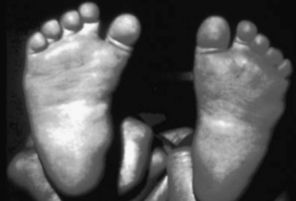
FIGURE 201-2 Clinical picture of metatarsus adductus with a normal foot on opposite side.
(From Kliegman RM, Behrman, RE, Jenson HB, et al: Nelson’s Textbook of Pediatrics, 18th ed. Philadelphia, WB Saunders, 2007, p 2777.)
Clinical Manifestations
The forefoot is adducted and sometimes supinated, but the midfoot and hindfoot are normal. The lateral border of the foot is convex, while the medial border is concave. Ankle dorsiflexion and plantar flexion are normal. With the midfoot and hindfoot stabilized, the deformity can be pushed beyond a neutral position (into abduction). Older children may present with an in-toeing gait.
Treatment
True metatarsus adductus resolves spontaneously over 90% of the time without treatment, so reassurance is all that is needed. Metatarsus adductus that does not improve within 2 years needs evaluation by a pediatric orthopedist. Persistent cases may benefit from serial casting or bracing, and potentially surgery. Many feel that it is acceptable to live with the deformity because it is not associated with a disability.
It is important to differentiate between metatarsus adductus, metatarsus varus, and skewfoot. Metatarsus varus looks like metatarsus adductus, but it is an uncommon rigid deformity that will need serial casting. Skewfoot is an uncommon deformity that is characterized by hindfoot plantar flexion, midfoot abduction, and forefoot adduction, giving the foot a Z or serpentine appearance. This needs to be managed very carefully with serial casting and surgery to help reduce the risk of disability in adulthood.
CALCANEOVALGUS FOOT
The calcaneovalgus foot is another common foot disorder in newborns that is secondary to in utero positioning. It is characterized by a hyperdorsiflexed foot with forefoot abduction and heel valgus. It is usually unilateral. The appearance may be quite severe dorsiflexion, but it is not a rigid deformity like congenital vertical talus. Simulated weight-bearing radiographs may be necessary for questionable diagnoses. The calcaneovalgus foot will appear normal or have minimal hindfoot valgus.
This disorder requires no treatment beyond reassurance. Parents can be taught passive stretching exercises for their infant’s foot. Most affected infants realign by 2 years.
HYPERMOBILE PES PLANUS (FLEXIBLE FLATFOOT)
Hypermobile or pronated feet are seen in 15% of adults. The child with flatfeet is usually asymptomatic and has no activity limitations. Newborn and toddler flatfoot is the result of ligamentous laxity and fat in the medial longitudinal arch. This is called developmental flatfoot and usually improves by 6 years of age. In older children, flatfoot is typically the result of generalized ligamentous laxity, and there is often a positive family history. Hypermobile flatfoot can be thought of as a normal variant.
Clinical Manifestations
In the non-weight bearing position, the older child with a flexible flatfoot will have a medial longitudinal arch. When weight bearing, the foot pronates (arch collapse) with varying degrees of hindfoot valgus. Subtalar motion (essentially all ankle motion except plantar and dorsiflexion) is normal. Any loss of subtalar motion may indicate a rigid flatfoot, which can be related to tarsal coalition, neuromuscular disorders (cerebral palsy), and heel cord contractures. Radiographs of hypermobile flatfeet are usually not indicated.
Treatment
Hypermobile pes planus cannot be diagnosed until after 6 years of age; prior to that, it is developmental pes planus. Reassurance that this is a normal variant is very important. Patients who are symptomatic with activity may require education on proper, supportive footwear, orthotics/arch supports, and heel cord stretching.
TARSAL COALITION
Patients with tarsal coalition will usually present with a rigid flatfoot (loss of inversion and eversion at the subtalar joint). Coalition is produced by a congenital fusion or failure of segmentation of two or more tarsal bones. The attachment may be fibrous, cartilaginous, or osseus. Tarsal coalition can be unilateral or bilateral and will often become symptomatic in early adolescence. The most common forms of tarsal coalition are calcaneonavicular and talocalcaneal.
Clinical Manifestations
The patient will usually present with hindfoot pain, which may radiate laterally due to peroneal muscle spasm. Symptoms are exacerbated by sports, and young athletes can present with frequent ankle sprains. There is a familial component. Pes planus is usually present in both weight bearing and non–weight bearing positions. There is usually a loss of subtalar motion, and passive attempts at joint motion may produce pain.
Radiologic Evaluation
Anteroposterior, lateral, and oblique radiographs should be obtained, but they may not always clearly identify the disorder. The oblique view often identifies the calcaneonavicular coalition. Computed tomography (CT) is the gold standard for diagnosis of tarsal coalition. Even patients with obvious calcaneonavicular coalition on plain radiographs should have a CT scan to rule out a second coalition.
Treatment
Coalitions that are asymptomatic (the majority) do not need treatment. Nonoperative treatment for patients with pain consists of cast immobilization for a few weeks and foot orthotics. The symptoms will often return, necessitating surgery. Surgical excision of the coalition and soft tissue interposition to prevent reossification can be very effective.
CAVUS FOOT
Cavus foot is characterized by increased height of the medial longitudinal arch (high arch) and frequently hindfoot varus. It can be classified as physiologic or neuromuscular. Most patients with physiologic cavus foot are asymptomatic. A thorough neurologic examination on all patients with a cavus foot is important. Patients with painful high arches have a high risk of neurologic (tethered cord) and neuromuscular disease, and there is a strong association with Charcot-Marie-Tooth disease, a familial neuropathy. The underlying disorder should be treated first. Nonoperative treatment using orthotics is usually not helpful. Progressive, symptomatic cavus foot will likely need surgical reconstruction.
IDIOPATHIC AVASCULAR NECROSIS
Kohler disease (tarsal navicular) and Freiberg disease (head of the second metatarsal) are uncommon and due to avascular necrosis. Patients will present with pain at the affected site with activity and weight bearing. Infection, fracture, and neoplasm should be excluded. Treatment consists of immobilization and activity restriction. The majority of the patients will improve upon subsequent revascularization and re-formation of bone.
SEVER DISEASE (CALCANEAL APOPHYSITIS)
Sever disease is common cause of heel pain among active young people. The mean age of presentation for girls is about 9 years of age and for boys about 11 to 12 years. Approximately 60% of cases are bilateral. Sever disease is caused by the forces of the calf musculature through the Achilles tendon at the calcaneal apophysis, causing microfracture. As the child ages and the apophysis begins to close, the pain disappears.
Clinical Manifestations
The common presentation is a young athlete who develops heel pain with activity which decreases with rest. There is rarely swelling, but there may be limping associated with Sever disease. The child will have pain to palpation of the posterior calcaneus and often tight heel cords. Radiographs are rarely indicated, but with persistent pain they should be done to exclude infection or tumor.
TOE DEFORMITIES
Curly toes are the most common deformity of the lesser toes. The fourth and fifth toes are most commonly affected. Curly toes are characterized by flexion at the proximal interphalangeal joint with lateral rotation of the toe. It is caused by contractures of the flexor digitorum brevus and longus tendons. About 25% to 50% of curly toes will spontaneously resolve by 3 to 4 years of age. Persistent deformity may be treated by surgical tenotomy.
Polydactyly (extra toes) are usually found on the initial newborn physical examination. When the extra toe is adjacent to the fifth toe, and attached by only a stalk of soft tissue or skin, simple ligation or amputation is effective. When the deformity involves the great toe or middle toes, or when the extra digit has cartilage or bone, delayed surgical intervention is indicated. Syndactyly (fusion of toes) is more common than polydactyly. It is usually a benign cosmetic problem. Both syndactyly and polydactyly may be associated with malformation syndromes (Table 201-2).
TABLE 201-2 Disorders Associated with Syndactyly and Polydactyly
| Polydactyly | Syndactyly |
|---|---|