CHAPTER 6 Introduction to excitable tissue
Basic anatomy
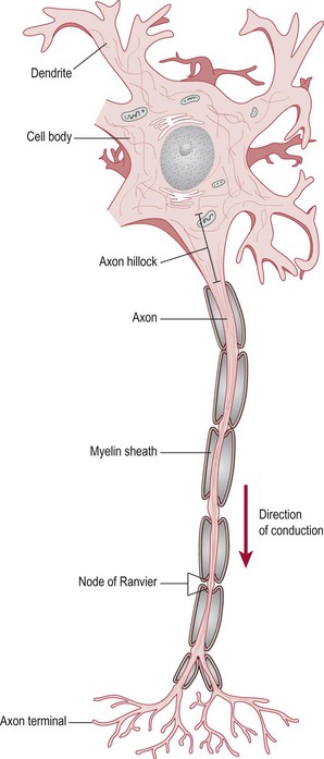
Neurons
Neurons or nerve cells are the main components making up the nervous system and are termed ‘an electrically excitable tissue’ by virtue of their ability to conduct and transmit electrochemical signals throughout the body. There are many different specialized neurons, however a typical neuron (Fig. 6.1) would consist of:
Function of a neuron
Resting potential
The neuronal cell membrane possesses a membrane potential (stored energy). This occurs because of a non-uniform distribution of ions in the intracellular (inside the cell) and extracellular (outside the cell) fluid. This distribution results due to large negative anions in the intracellular fluid and the differential permeability of ions across the membrane, most importantly sodium (Na2+) and potassium (K+). Movement of individual ions occurs according to its electrochemical gradient and continues until a balance is achieved. This balance related to all individual ions results in the inside of the neuron being relatively more negative than the outside of the neuron. This does not mean that the inside is negatively charged, there is just a difference between inside and outside. The typical resting potential of a neuronal cell is −70 millivolts (mV).
Graded potential
During a triggering event (stimulus at a sensory receptor or a signal from another neuron) there is a change in the resting membrane potential at the trigger site, which results in the depolarization of the membrane (i.e. it becomes relatively less negative). In effect, this creates a current flow in the membrane which opens voltage gated ion channels and leads to the passive movement of ions into or out of the cell. This is termed a ‘graded potential’.
The magnitude of the graded potential reflects the magnitude of the triggering event. The current will flow in both directions from the active site but dies out over a short distance. Unlike an action potential, it is possible to produce further graded potentials which can build on the first.
Action potential
When the cell membrane is depolarized to a threshold level (typically −50 mV) at the axon hillock (Fig. 6.1), an ‘action potential’ is initiated. The action potential occurs as a consequence of the high concentration of voltage gated sodium and potassium ion channels at the axon hillock. When depolarized to threshold level, these channels open on mass and initiate conduction along the axon. An action potential once initiated does not fade and therefore the signal may travel over long distances.
Summation
As it is unlikely that a single graded potential will reach threshold level at the axon hillock, further graded potentials will be required. This may occur in two ways:
The fact that an action potential is not initiated immediately following a stimulus is beneficial, as this gives the neuron choice as to whether to respond. If the stimulus is important, the signal will be sent at the appropriate strength producing a stronger graded potential and there is more likelihood of an action potential. In simple terms, if the information is important enough, the signal will be passed on. Summation of a weaker signal gives validity to the signal before the decision is made to transmit it further. In these terms, decision-making is based at an electrochemical level.
Conduction along a neuron
The ability of the signal to travel along the cell membrane is a result of the opening of sodium and potassium ion channels in response to a voltage change. In effect, a current of depolarization moves along the membrane, consecutively opening successive ion channels. Conduction of the signal can occur in two ways:
Synaptic transmission
The junction between two neurons is termed a ‘synapse’ and represents a gap between the two neurons. In order for a signal to be passed from a pre-synaptic neuron to a post-synaptic neuron, the electric signal (the action potential) is transformed into a chemical signal (the neurotransmitter substance), which is released into the synaptic cleft and diffuses across the gap. The neurotransmitter attaches to specific ion receptors on the post-synaptic cell, which results in the opening of the ligand/chemically gated ion channels. Each synapse is highly specific in terms of the ion channels it possesses so that only movement of one particular ion occurs. The direction of ion movement is dictated by the existing chemical gradient. For example, sodium moves into the cell and potassium moves out of the cell.
Ultimately, what is produced in the post-synaptic membrane is a graded potential. Depending upon the ion channels present, the graded potential may result in: