Chapter 8 Appendix

image

Pronation Syndrome

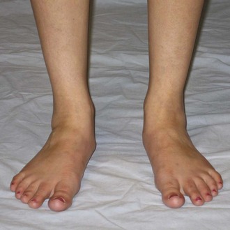
The principal movement impairment in pronation syndrome is pronation at the foot and ankle. Pronation is considered abnormal and an impairment when the amount of pronation during weight-bearing activities is excessive for that individual and/or when there is insufficient movement of the foot in the direction of supination in later stance phase. The pronation impairment can occur in the hindfoot, midfoot, and/or forefoot. A foot with a pronation movement impairment is a flexible foot that compensates for various structural and movement impairments within the foot and ankle, as well as those at the knee and hip.

image

Treatment

Inflammation and Pain Control

Ice
Iontophoresis
Electrical stimulation

Walking and/or Running

The patient is instructed to work on the specific cues that assisted in symptom reduction during the examination or the cues that the physical therapist believes with practice may result in symptom reduction. The following cues are among the possibilities that may assist the patient:

Contract the gastrocnemius muscle by lifting from the heel.
Raise the medial longitudinal arch.
Contract the gluteal muscles (squeeze the buttocks).
Hit with the heel first.

Many of the changes being requested of the patient during walking and running are similar to a strengthening program. As such, the patient should be encouraged to have focused practice time and gradual implementation to avoid injury.

Muscle Performance

Supinators (gastrocnemius, posterior tibialis)
Thera-Band resistance exercise into plantarflexion and plantarflexion inversion
Heel raises
Hopping
Intrinsic muscles of the foot
Towel crunches: Towel placed on the floor, use toes to grab towel and pull it under the foot. Weight can be added to the towel to increase resistance. The arch should lift; do not allow the patient to only use the flexor digitorum longus.
Posterior hip muscles
Sidelying hip lateral rotation progressing to lateral rotation with abduction adding weight as appropriate. Patient may need a pillow between knees for comfort.
Prone hip extension with the knee flexed. Patient should be over a pillow.
Posterior hip muscle strengthening must progress to weight-bearing dynamic activities to prepare the muscles for walking, running, and jumping activities.

After the injured tissue has been protected from excessive stresses and the inflammation has subsided, the involved muscle and tendon should undergo a progressive strengthening program and a progressive return to activity. In general, exercise or activity is permissible if pain remains at 2/10 on a 0 to 10 scale. The strengthening exercise should be completed at a minimum of 70% maximal voluntary contraction for 10 repetitions, 3 sets, 3 to 5 times/week.

Decrease Range of Motion

Short gastrocnemius/soleus muscle/calcaneal (Achilles) tendon.
Wall stretch: The knee is extended for gastrocnemius muscle shortness and flexed for soleus muscle length deficits. The patient should prevent pronation (active patient correction and wearing good footwear). The patient should be instructed to keep the foot facing forward or in line with the femur and tibia. The heel should be kept on the ground during the stretch.
Heel hang stretch: The knee can be extended or flexed as described for the wall stretch. The patient should prevent pronation through active correction and by wearing good footwear.
Long sitting towel-assisted dorsiflexion: The patient should prevent pronation through active correction and by modifying the direction of force through the towel.
Talocrural joint limitation: In addition to the standing stretches, joint limitations can be addressed with mobilization.
Mobilize the talocrural joint using a posterior glide of the talus on the ankle mortise.
Mobilize the talocrural joint using a distraction technique.
Heel lift in the shoe until length changes are apparent.
Night splint to maintain dorsiflexion position.
Short extensor digitorum longus.
Patient plantar flexes the involved foot with the toes flexed. Can be completed in sitting or can be stretched in hands and knees rocking back.
First MTP extension limitation:
Mobilize the MTP joint using an anterior glide of the proximal phalanx on the metatarsal.
Passive ROM (PROM) into first MTP extension (talocrural dorsiflexion with first MTP extension).

Stretching should be held for 30 seconds, 2 to 3 repetitions, completed regularly throughout the day (5 to 8 times/day), and completed 5 to 7 days/week.

Activity

Modify activity level to decrease forces on the foot. May require use of an assistive device if symptoms are severe.
If appropriate for the patient’s goals, progress to dynamic activities such as jumping, hopping, shuttle run, cutting, and so on.
Running progression rules:
Start with a run/walk program gradually progressing to all running.
Start with straight plane jogging and jumping on a smooth flat surface.
Work on distance tolerance first.
Increase speed as tolerated.
Add varied terrain, hills, and cutting (these can include figure 8s).
Finally, add cuts and turns that are unexpected. For example, have the patient run straight forward and call cut right. He or she is expected to change directions immediately. Mix the calls, including cut left, 180 degrees turn, 360 degrees turn, and bend down.

External Tissue Support

Footwear

A last that looks like the foot. The most common last for a pronated foot is a straight or semi-curved last.
Firm heel counter to control hindfoot motion. If pronation is occurring at the hindfoot, the shoe should include rigid material at the medial heel and less rigid material at the lateral heel.
A sole that bends only at the metatarsal heads and rigid from hindfoot to midfoot.
Adequate arch support with medial structures of the footwear generally made of firm, controlling materials.
Appropriate width and depth to accommodate the foot
Cushion indicated as needed for appropriate shock absorption.

Orthoses/Taping

The orthosis should prevent pronation. Start with adding an arch support, then if necessary post at the hindfoot, then the forefoot. If the metatarsal heads are involved, the orthosis may also need to include a metatarsal pad.
For calcaneal tendon involvement, hindfoot eversion must be correct with a medial hindfoot post to maximize tendon alignment.
For tarsal tunnel involvement, hindfoot orthosis additions must be soft to avoid nerve compression.
Arch taping: Used most frequently for involvement of the plantar fascia but is appropriate for any condition that would benefit from additional arch support and prevention of pronation.

Supination Syndrome

The principal movement impairment in supination syndrome is supination at the foot and ankle. Supination is considered abnormal and an impairment when the amount of supination during weight-bearing activities is excessive for that individual or when it occurs from heel strike to midstance in the gait cycle. The supination impairment can occur in the hindfoot, midfoot, and/or forefoot. The foot with a supination impairment is generally a rigid foot with little or no ability to absorb shock and compensate for structural or movement impairments within the foot and ankle, knee, or hip.

image

Treatment

Inflammation and Pain Control

Ice
Iontophoresis
Electrical stimulation

Walking and/or Running

The patient is instructed to work on the specific cues that assist in symptom reduction during the examination or the cues that the physical therapist believes, with practice, may result in symptom reduction. Often, the cues are related to softening the landing, hitting more centrally on the heel, and concentrating on trying to limit lateral loading through the foot.

Muscle Performance

Gastrocnemius/soleus muscle
Thera-Band resistance exercise into plantar flexion
Heel raises
Hopping
Fibularis (peroneus) longus and brevis
Thera-Band resistance exercise into plantar flexion and plantarflexion eversion

After the tissue injury has been protected from excessive stresses and the inflammation has subsided, the involved muscle and tendon should undergo a progressive strengthening program and a progressive return to activity. In general, exercise or activity is permissible if pain remains at 2/10 on a 0 to 10 scale. The strengthening exercise should be completed at a minimum of 70% maximal voluntary contraction for 10 repetitions, 3 sets, 3 to 5 times/week.

Decreased Range of Motion

Short gastrocnemius/soleus muscle length
Wall stretch: The knee is extended for gastrocnemius muscle shortness and flexed for soleus muscle length deficits. The patient should be instructed to keep the foot facing forward or in line with the femur and tibia. The heel should be kept on the ground during the stretch. The patient should prevent foot supination correction and by wearing good footwear. The stretch should be held for 30 seconds, complete 2 to 3 time/session, and done regularly throughout the day (5 to 8 times/day).
Heel hang stretch: The knee can be extended or flexed as described for the wall stretch. The patient should prevent subtalar joint supination through active correction and by wearing good footwear.
Long sitting towel-assisted dorsiflexion. The patient should prevent foot supination through active correction and by modifying the direction of force through the towel.
A night splint to maintain dorsiflexion position is often helpful.
Talocrural joint limitation
Mobilize the talocrural joint using a posterior glide of the talus on the ankle mortise
Mobilize the talocrural joint using a distraction technique
May need to include a heel lift in the shoe until length changes are apparent
Talocrural joint limitation
Decreased length of extensor digitorum longus
Patient plantarflexes the involved foot with the toes flexed. Can be completed in sitting or if extremely short, can be stretched in hands and knees rocking back
Decreased great toe extension
Mobilize the MTP joint using an anterior glide of the proximal phalanx on the metatarsal.
Passive ROM into great toe extension. Patient dorsiflexes the talocrural joint and then extends the great toe.

Stretching should be held for 30 seconds, 2 to 3 repetitions, completed regularly through out the day (5 to 8 times/day), and completed 5 to 7 days/week.

Activity

Modify activity level to decrease forces on the foot. May require use of an assistive device if symptoms are severe.
If appropriate for the patient’s goals, progress to dynamic activities such as jumping, hopping, shuttle run, cutting, and so on.
Running progression rules:
Start with straight plane jogging and jumping on a smooth flat surface.
Work on distance tolerance first.
Increase speed as tolerated.
Add varied terrain, hills, and cutting (these can include figure 8s).
Finally, add cuts and turns that are unexpected. For example, have the patient run straight forward and call cut right. He or she is expected to change directions immediately. Mix the calls, including cut left, 180 degrees turn, 360 degrees turn, or bend down.

Footwear

A last that looks like the foot. The most common last for a supinated foot is a curved last.
Firm heel counter to control hindfoot motion. The hindfoot sole material should NOT include a material density differential that would encourage calcaneal inversion.
Cushioned, conforming insole is a key component in treating the supinated foot.
Appropriate width and depth to accommodate the foot.
Adequate arch support.

External Tissue Support

Accommodative insert with soft materials. May need additional arch support to assist in distributing force through greater weight-bearing surface. If the metatarsal heads are involved, the orthotic may also need to include a metatarsal pad.
Arch taping: Used most frequently for plantar fascia involvement but is appropriate for any condition that would benefit from additional arch support.
Calcaneal (Achilles) tendon taping: Used to reduce stress on the calcaneal tendon.

Insufficient Dorsiflexion Syndrome

The principal movement impairment in insufficient dorsiflexion syndrome is insufficient dorsiflexion. The impairment can occur during midstance to push-off or during swing phase and is not associated with excessive supination or pronation.

image

Treatment

Inflammation and Pain Control

Ice
Iontophoresis
Electrical stimulation

Walking and/or Running

Walking and running cues focus primarily on encouraging active contraction of the gastrocnemius muscle to reduce the reliance on the passive tension of the gastrocnemius/soleus muscle/calcaneal tendon unit. The specific cues are to have the patient actively lift the heel during late stance.

Muscle Performance

Weakness of the anterior tibialis:
Thera-Band resistance exercise into dorsiflexion inversion with toes curled.
Weakness of the intrinsic muscles of the foot.
Towel crunches: Towel placed on the floor, use toes to grab towel and pull it under the foot. Weight can be added to the towel to increase resistance. The arch should lift; do not allow the patient to only use the flexor digitorum longus.

After the tissue injury has been protected from excessive stresses and the inflammation has subsided, the involved muscle and tendon should undergo a progressive strengthening program and a progressive return to activity. In general, exercise or activity is permissible if pain remains at 2/10 on a 0 to 10 scale. The strengthening exercise should be completed at a minimum of 70% maximum voluntary contraction for 10 repetitions, 3 sets, 3 to 5 times/week.

Decreased Dorsiflexion

Short gastrocnemius/soleus muscle/calcaneal tendon
Wall stretch: The knee is extended for gastrocnemius muscle shortness and flexed for soleus muscle length deficits. The patient should be instructed to keep their foot facing forward or in line with the femur and tibia. The heel should be kept on the ground during the stretch. The patient should prevent subtalar joint pronation through active correction and by wearing good footwear. The stretch should be held for 30 seconds, completed 2 to 3 times/session, and done regularly throughout the day (5 to 8 times/day).
Heel hang stretch: The knee can be extended or flexed as described for the wall stretch. The patient should prevent subtalar joint pronation through active correction and by wearing good footwear.
Long sitting towel-assisted dorsiflexion: The patient should prevent subtalar joint pronation through active correction and by modifying the direction of force through the towel.
A night splint to maintain dorsiflexion position is often helpful.
Stretches should be held for 30 seconds, 2 to 3 repetitions, completed regularly throughout the day (5 to 8 times/day), and done 5 to 7 days/week.
Talocrural joint limitation
Mobilize the talocrural joint using a posterior glide of the talus on the ankle mortise.
Mobilize the talocrural joint using a distraction technique.
May need to include a heel lift in the shoe until length changes are apparent.

Activity

Modify activity level to decrease forces on the foot until healed. May require use of an assistive device if symptoms are severe.
If appropriate for the patient’s goals, progress to dynamic activities such as jumping, hopping, shuttle run, cutting etc.
Running progression rules:
Start with straight plane jogging and jumping on a smooth flat surface.
Work on distance tolerance first.
Increase speed as tolerated.
Add varied terrain, hills, and cutting.
Finally, add cuts and turns that are unexpected. For example, have the patient run straightforward and call cut right. He or she is expected to change directions immediately. Mix the calls, including cut left, 180 degrees turn, 360 degrees turn, and bend down.

External Tissue Support

Footwear

Heel-to-toe height of the shoe that accommodates for the lack of dorsiflexion ROM. However, the goal would be to increase dorsiflexion ROM to avoid needing a shoe with a heel.
Appropriate width and depth to accommodate the foot.
A last that looks like the foot. May be curved, straight, or in between (midlast), depending on the shape of the individual’s foot.
Footwear should include the standard shoe components to provide the necessary support and cushion. This includes a firm heel counter, the appropriate amount of arch support, and cushion indicated as needed for shock absorption.
The angle formed by the heel counter and the sole of the shoe at the posterior heel to the vertical line from the floor at the most-posterior portion of the sole of the shoe should be relatively small. A large angle increases the work demand on the anterior tibialis muscle and tendon.
With involvement of the bursa, pain may occur with pressure from the heel of the shoe on the bursa. The patient may need to wear open-heeled shoes temporarily.

Orthoses/Taping

Insert a heel lift into the shoe to relieve stress from decreased talocrural dorsiflexion motion. As the individual completes their home stretching program, the height of the heel lift can be reduced.
Calcaneal tendon taping: Tape the ankle posteriorly to support the tendon, place the foot in plantarflexion, tape from the distal posterior calf to midarch of the foot.

Hypomobility Syndrome

The principal movement impairment in hypomobility syndrome is associated with a limitation in the physiological and accessory motion of the foot and ankle. This may result from degenerative changes in the joint or the effects of prolonged immobilization.

image

Treatment

Inflammation and Pain Control

Ice (may be contraindicated if patient is hypersensitive or with OA or RA)
Heat is often helpful in decreasing feelings of joint stiffness and pain
Compression garment

Walking and/or Running

The presence of OA and/or RA often requires the individual to discontinue weight-bearing, high impact, and high repetition activities (walking or running for fitness). The patient often needs to be guided into lower impact activities, such as stationary bicycling, water aerobics, or use of a StairMaster/elliptical, or activities, such as rowing, that involve aerobic fitness through the upper extremities. Weight loss can also significantly impact pain with weight-bearing activities and should be discussed if appropriate for the patient.
Walking with a limp can result in injury to other areas of the body (knee, hip, back, or uninvolved foot). All efforts to correct the gait pattern should be employed, including work on weight shifting, a gradual increase in weight-bearing tolerance, and addressing the strength and motion impairments contributing to the gait pattern. Use of assistive devices may be temporarily or permanently indicated for patients.

Muscle Performance

Weakness of the plantarflexors (gastrocnemius, posterior tibialis muscles)
Thera-Band resistance exercise into plantar flexion and plantar flexion inversion
Heel raises
Eccentric training
Bilateral and single-leg hopping
Cutting, sprinting, and sport-specific activities
Weakness of the anterior tibialis muscle
Thera-Band resistance exercise into dorsiflexion and/or inversion
Weakness of the fibular (peroneal) muscles
Thera-Band resistance exercise into plantarflexion and/or eversion
Weakness of the intrinsic muscles of the foot
Towel crunches: Towel is placed on the floor, using toes to grab towel, then pull it under the foot. Weight can be added to the towel to increase resistance. The arch should lift; do not allow the patient to only use the flexor digitorum longus.

After the tissue injury has been protected from excessive stresses and the inflammation has subsided, the involved muscle and tendon should undergo a progressive strengthening program and a progressive return to activity. In general, exercise or activity is permissible if pain remains at 2/10 on a 0 to 10 scale. The strengthening exercise should be completed at a minimum of 70% maximum voluntary contraction for 10 repetitions, 3 sets, 3 to 5 times/week.

Decreased Range of Motion

Decreased dorsiflexion: Short gastrocnemius/soleus muscle/calcaneal tendon
Wall stretch: The knee is extended for gastrocnemius muscle shortness and flexed for soleus length deficits. The patient should be instructed to keep the foot facing forward or in line with the femur and tibia. The heel should be kept on the ground during the stretch. The patient should prevent subtalar joint pronation through active correction and by wearing good footwear. The stretch should be held for 30 seconds, completed 2 to 3 times/session, and done regularly throughout the day (5 to 8 times/day).
Heel-hang stretch: The knee can be extended or flexed as described for the wall stretch. The patient should prevent subtalar joint pronation through active patient correction and/or wearing good footwear.
Long sitting towel-assisted dorsiflexion: The patient should prevent subtalar joint pronation through active correction and by modifying the direction of force through the towel.
A night splint to maintain dorsiflexion position is often helpful.
Stretches should be held for 30 seconds, 2 to 3 repetitions, completed regularly throughout the day (5 to 8 times/day), and done 5 to 7 days/week.
Talocrural joint limitation
Mobilize the talocrural joint using a posterior glide of the talus on the ankle mortise.
Mobilize the talocrural joint using a distraction technique.
May need to include a heel lift in the shoe until length changes are apparent.
Decreased plantar flexion: Short dorsiflexor muscles
Hands and knees rocking back.
Sustained active ROM (AROM) and PROM.
Decreased inversion/eversion: Subtalar joint limitation
Mobilize the subtalar joint using lateral and medial glides.
Decreased intertarsal mobility
Mobilize specific intertarsal joints primarily using anterior and posterior glides.
Decreased MTP and interphalangeal flexion and extension
Mobilize MTP and IP joint primarily using anterior (for extension) and posterior (for flexion) glides.
General ROM Activities
Stationary bike.
Baps board or wobble in sitting when weight-bearing tolerance is limited, progressing to standing activities when weight-bearing tolerance increases.

Activity

Progress weight bearing gradually, reducing dependence on and type of assistive device.
Address cardiovascular fitness with use of low impact activities (stationary bike, swimming, rowing, or StairMaster/elliptical training).
If appropriate for the patient’s goals, progress to dynamic activities such as jumping, hopping, shuttle run, cutting, and so on.
Running progression rules:
Start with straight plane jogging and jumping on a smooth flat surface.
Work on distance tolerance first.
Increase speed as tolerated.
Add varied terrain, hills, and cutting (these can include figure 8s).
Finally, add cuts and turns that are unexpected. For example, have the patient run straight forward and call cut right. He or she is expected to change directions immediately. Mix the calls, including cut left, 180 degrees turn, 360 degrees turn, and bend down.

External Tissue Support

Footwear

Appropriate size, width, and depth to accommodate edematous foot.
A last that looks like the foot. May be curved, straight, or in between (midlast), depending on the shape of the individual’s foot.
Footwear should include the standard shoe components to provide the necessary support and cushion. This includes a firm heel counter, the appropriate amount of arch support, and cushion, which is indicated only as needed for shock absorption.
For individuals with OA or RA or those who have had fusion of joints in their foot, a steel shank in the sole of the shoe will make the sole of the shoe rigid and a rocker at the toe break will allow the patient to more easily roll over the foot without needing as much talocrural dorsiflexion or MTP dorsiflexion.

Orthoses

For individuals with OA or RA, a total contact insert made of accommodative material is often indicated. Deformities of the foot should be considered in the design and materials chosen for the orthosis.
A heel lift may be necessary to manage the loss of dorsiflexion ROM.
Temporary orthoses with additional arch support are often indicated to manage foot pain that is often related to the new onset of a pronation impairment that results from the limited foot and ankle mobility.

Scar

Firm and raised
TopiGel sheeting (chemical reaction)
Pressure
Immobile
Gradual application of stress to scar helps the scar remodel in such a way that it allows gliding between structures.
AROM and PROM
Immobile or adhered scar
Massage: Use circular motions and friction, minimum 5 to 6 times/day, 5 minutes each time. Using Dycem or wearing a latex glove on the uninvolved hand may help increase friction.

Hypersensitivity

Desensitization exercises
Progress from light touch to more firm touch.
Progress from soft texture to a rough texture.
Add additional sensation such as vibration and tapping as tolerated.
Emphasize weight-bearing and ROM exercises.
Activity and exercise is better tolerated if heat has been applied (hot pack or exercise in warm whirlpool or pool).

Balance and Proprioception

Stand on the involved leg with eyes open.
Stand on the involved leg with eyes closed.
Stand on the involved leg on foam or uneven surface with eyes open.
Stand on the involved leg on foam or uneven surface with eyes closed.
Stand on one leg and do the following:
Kick and stop a ball.
Throw and catch a ball.
Reach with hand.
Do similar activities on a mini-trampoline or other challenging surface.

Foot and Ankle Impairment

A key criterion for placement into the foot and ankle impairment classification is the need to protect tissue. Usually, the tissue involved is stressed by a surgical procedure or trauma and may cause significant pain at rest and during movement. The patient is unable to tolerate a typical movement system examination. The limitation in movement is not primarily related to a chronic pain condition. Tissue healing and normal movement are expected. These are general guidelines and not intended to stand alone. Consult the physician’s protocol for specific precautions and progressions. The physical therapist must be familiar with the tissues that were affected in the surgical procedure.

Symptoms and History

Patient has history of surgery or acute injury.
Knowledge of specific surgical approach or injury is mandatory.
Patient may report severe pain.

Physiological Factors

Factors that affect the physical stress of tissue and/or thresholds of tissue adaptation and injury1 specific to the foot are as follows.

Tissue Factors

Bone
Fractures of the base of the fifth metatarsal have a high probability of nonunion secondary to the pull of the fibularis brevis. Additionally, stress fractures of the anterior lateral tibial diaphysis, medial malleolus, talus, navicular, and sesamoids are high-risk areas that often fail to heal, re-fracture, and/or need operative intervention.2
Cartilage
Muscle
Common to lose dorsiflexion ROM with immobilization.
Common to experience atrophy of gastrocnemius/soleus muscle with immobilization.
Tendon
Calcaneal tendon rupture: Gastrocnemius/soleus muscle contraction contraindicated; may have dorsiflexion ROM restrictions.
Posterior tibial tendon injury or insufficiency: If severe, may result in significant deformity of the arch (flattening); may require long periods of immobilization or surgery. Contraction of the posterior tibial muscle would be contraindicated after surgery.
Ligament
Lateral ankle sprain: Often accompanied by an avulsion fracture of the lateral malleolus.
Syndesmotic ankle sprain (“high ankle sprain”): Associated with greater discomfort during weight-bearing function and longer healing time and may require immobilization.3
Skin
Edema has the potential to limit mobility and compromise the space occupied by nerves and arteries; often encountered under the extensor retinaculum and the flexor retinaculum; note location and measure extent of edema; assess whether it is brawny or pitting.
Scar: Note location, appearance, mobility, sensitivity, and if incision appears healed (approximately 10 days).
Color: Note location and size of discoloration, including bruising and other important changes in color (red, white, blue, or black).
Temperature: Note location of warmth.
Nerve
Deep fibular (peroneal) nerve can be compromised as it runs beneath the extensor retinaculum.
Tibial nerve can be compressed as it runs beneath the flexor retinaculum.
Interdigital nerve.

Types of Surgeries (Indications)

Stabilization (fracture, avascular necrosis [AVN], osteosarcoma)
ORIF
External fixation
Fusion
Bone graft
Osteotomy (malalignment or osteosarcoma)
Calcaneal
Metatarsal
Phalangeal
Arthroplasty (DJD, arthritis, joint destruction)
Compartment decompression (crush injury, overuse with loss of arterial blood flow)
Debridement (tear, arthritis, or infection)
Capsule
Cartilage
Wound
Repair (tear, graft, or cell injections—open or arthroscopic)
Ligament
Cartilage
Tendon
Soft tissue release (short tissue or spastic muscle)
Gastrocnemius/soleus muscle/calcaneal tendon
Plantar fascia
Excision
Neuromas
Tumors
Bone

Medications

Consider side effects and effects of medications on tissue, exam, and intervention
Nonsteroidal antiinflammatory drugs (NSAIDs)
Muscle relaxants
Analgesics
Steroids

Medical Complications

DVT
Pulmonary embolus
Fibular (peroneal) nerve neurapraxia
Neurovascular compromise
Compartment syndrome
Infection
Nonunion and malunion

Movement and Alignment Factors

Variations
Anthropomorphics
Structural impairments
Scar adhesions
Standing alignment
May demonstrate protective stance or rotational impairments
Underlying Movement Impairment Syndromes
Pronation
Supination
Insufficient dorsiflexion
Hypomobility

Extrinsic Factors

Assistive devices to unload extremity
Orthotic devices or braces

Psychosocial Factors

Response to pain and/or anxiety

Treatment for Foot and Ankle Impairment

Emphasis of treatment is to restore ROM of the ankle and strength of the lower extremity without adding excessive stresses to the injured tissues and within the precautions outlined by the physician. Underlying movement impairments should be addressed during rehabilitation and functional activities to ensure optimal stresses to the healing tissues.

Impairments

Pain

Be sure to clarify the location, quality, and intensity of the pain.

Stage 1

Surgical: Within the first 2 weeks of the postoperative period, some pain will be associated with exercises. Gradually over the next few weeks, pain associated with the exercise should lessen. Pain can be used as a guide to rehabilitation. Sharp, stabbing pain should be avoided. Mild aching is expected after exercises but should be tolerable for the patient. This postexercise discomfort should decrease within 1 to 2 hours of the rehabilitation. A sudden increase in symptoms or symptoms that last longer than 2 hours after exercise may indicate that the rehabilitation program is too aggressive. Coordinating the use of analgesics with exercise sessions is important.

Acute Injury: Despite discomfort, tests may need to be performed to rule out serious injury. Modalities and taping/bracing may be helpful to decrease pain. The patient may also require the use of an assistive device, walker, or crutches in the early phases of healing.

Stage 2 to 3

Surgical/Acute Injury: Pain associated with the specific tissue that was involved in the surgery should be decreased by weeks 4 to 6. As the activity level of the patient is progressed, the patient may report increased pain or discomfort with new activities such as returning to daily activities and fitness. Pain or discomfort location should be monitored closely. Muscle soreness is expected, similar to the response of muscle to overload stimulus (e.g., weight training). General muscle soreness should be allowed to resolve, usually 1 to 2 days before repeating the bout of activity. Pain described as stabbing should always be avoided.

Edema

Stage 1

Surgical/Acute Injury: Edema is quite common in the foot and ankle s/p surgery or injury. The patient should be educated in use of edema-controlling techniques, such as the following:

Ice4
Elevation
Compression: Ace wraps, compression stockings

Patient should be encouraged to keep extremity elevated as much as possible particularly in the early phases (1 to 3 weeks). Application of ice after exercise is recommended. Other methods to control edema in the foot and ankle include electrical stimulation or compression pumps.

Measurement of edema should be taken at each visit. A sudden increase in edema may indicate that the rehabilitation program is too aggressive or the patient possibly has an infection.

Stage 2 to 3

Surgical/Acute Injury: Time until swelling is resolved is variable among patients and surgical procedures. As the patient increases the time spent on their feet in regular daily activities or more weight-bearing exercises, the patient may experience a slight increase in edema. This is to be expected; however, the patient should be encouraged to continue to use techniques stated previously to manage the edema.

Appearance

Stage 1

Surgical: Infections should be suspected if the area around the incision or the involved joint is red, hot, and/or swollen. An increase in drainage from the incision, particularly if it has a foul odor or is no longer a clear color, is also indication of an infection. Red streaks following the lymphatic system can also appear with infection. The physician should be consulted immediately if infection is suspected. It is common to observe bruising after surgery. This should be monitored continuously for any changes; an increase in bruising during the rehabilitation phases may indicate infection. Stitches are typically removed in 7 to 14 days.

Stage 2 to 3

Surgical: Incisions should be well healed. Bruising may still be present for as long as 3 to 4 weeks; however, it should be diminishing. Signs of increased bruising are a red flag and should be immediately referred to the physician.

ROM

Stage 1

Surgical/Acute Injury: To prevent contracture, ROM exercises should begin as soon as possible as allowed by the precautions. In the early phases of rehabilitation, the patient should perform ROM exercises at least three times per day and all exercises should be performed within pain tolerance. All uninvolved lower extremity joints should be exercised to prevent the development of restricted ROM at those joints. The typical exercise progression begins with gentle PROM, assisted AROM, or AROM. The choice between PROM, assisted AROM, and AROM is based in part on the tissue injured or repaired. If resistance is allowed, proprioceptive neuromuscular facilitation (PNF) techniques, such as contract-relax or hold-relax, can assist in achieving greater ROM. During Stage 1, resistance should be very gentle and can be progressed to a submaximum level as the patient tolerates. When performing ROM exercises of the ankle in the patient with a fracture, attention to hand placement during the exercises can minimize the stresses placed on the healing fracture site. Decreasing pain and edema and improving ROM are typical signs that it is safe to progress the exercises. Refer to specific protocols for guidelines regarding progression of the exercises.

The patient may have ROM precautions per the physician. A common example is tendon transfer with no ROM of the ankle.

Mobilizations to the following specific joints may be indicated (see the next section):

Talocrural joint
Midtarsal joints
Tarsometatarsal joints
Metatarsophalangeal and interphalangeal joints

Stage 2 to 3

Surgical/Acute Injury: Precautions are typically lifted by the time the patient reaches this stage. ROM should be approaching normal. Exercises may need to be progressed using passive force. Patient should be instructed that a stretching discomfort is expected; however, sharp pain should be avoided. Mobilizations may be indicated in later stages of rehabilitation to improve ROM. Consult with the physician before initiating joint mobilization after surgery of the knee.

Decreased Dorsiflexion

Short gastrocnemius/soleus muscle/calcaneal tendon
Wall stretch: The knee is extended for gastrocnemius muscle shortness and flexed for soleus muscle length deficits. The patient should be instructed to keep their foot facing forward or in line with the femur and tibia. The heel should be kept on the ground during the stretch. The patient should prevent subtalar joint pronation through active correction and by wearing good footwear. The stretch should be held for 30 seconds, completed 2 to 3 times/session, and done regularly throughout the day (5 to 8 times/day).
Heel hang stretch: The knee can be extended or flexed as described for the wall stretch. The patient should prevent subtalar joint pronation through active correction and by wearing good footwear.
Long sitting towel-assisted dorsiflexion: The patient should prevent subtalar joint pronation through active correction and by modifying the direction of force through the towel.
A night splint to maintain dorsiflexion position is often helpful.
Stretches should be held for 30 seconds, 2 to 3 repetitions, completed regularly throughout the day (5 to 8 times/day), and done 5 to 7 days/week.
Talocrural joint limitation
Mobilize the talocrural joint using a posterior glide of the talus on the ankle mortise.
Mobilize the talocrural joint using a distraction technique.
May need to include a heel lift in the shoe until length changes are apparent.

Decreased Plantar Flexion

Short dorsiflexor muscles
Hands and knees rocking back
Sustained AROM/PROM
Talocrural joint limitation
Mobilize the talocrural joint using an anterior glide of the talus on the ankle mortise.
Mobilize the talocrural joint using a distraction technique.

Decreased Inversion/Eversion

Subtalar joint limitation
Mobilize the subtalar joint using lateral and medial glides.

Decreased Intertarsal Mobility

Mobilize specific intertarsal joints, primarily using anterior and posterior glides.

Decreased Metatarsophalangeal and Interphalangeal Flexion and Extension

Mobilize MTP and interphalangeal joints, primarily using anterior (for extension) and posterior (for flexion) glides.

General Range-of-Motion Activities

Stationary bike.
Baps board or wobble in sitting when weight-bearing tolerance is limited, progressing to standing activities when weight-bearing tolerance increases.

Muscle Performance

Stage 1

Surgical/Acute Injury: Strengthening often begins after the initial phase of healing (4 weeks). The emphasis should be placed on proper movement patterns in preparation for strengthening activities. After 4 weeks, strengthening may be gradually incorporated. Progression to resistive exercise is based on the patient’s ability to perform ROM with a good movement pattern and without increase in pain. Weights, Thera-Band, or isokinetic equipment may be used. Specific exercise protocols provided by physicians and physical therapists should be evaluated to ensure that all exercises are appropriate for the individual’s situation. Gastrocnemius/soleus muscles are most commonly affected with surgery or injury to the ankle; however, others may be involved. Electrical stimulation or biofeedback may be used to improve strengthening (see the “Medications, Modalities, and Additional Interventions” section). The patient may have strengthening precautions per the physician.

Stage 2 to 3

Surgical/Acute Injury: After the tissue injury has been protected from excessive stresses and the inflammation has subsided, the involved muscle and tendon should undergo a progressive strengthening program and a progressive return to activity. At this stage, precautions are typically lifted. Strength activities can be progressed as tolerated by the patient. In general, exercise or activity is permissible if pain remains at 2/10 on a 0 to 10 scale. The strengthening exercise should be completed at a minimum of 70% maximum voluntary contraction for 10 repetitions, 3 sets, 3 to 5 times/week.

Plantarflexors (gastrocnemius, posterior tibialis muscles)
Thera-Band resistance exercise into plantarflexion and plantarflexion inversion.
Heel raises: This exercise can be started with both feet on the ground with greater weight on the uninvolved side. As strength improves, the patient is instructed to increase weight on the involved extremity with the goal of completing the heel raise with all of the individual’s weight on the involved side (single heel raise). The exercise should be performed with good control during the concentric and eccentric portions of the exercise.
Bilateral and single-leg hops.
Anterior tibialis muscle
Thera-Band resistance exercise into dorsiflexion inversion: Have the patient curl the toes down if the extensor digitorum and extensor hallucis are dominant.
Fibular (peroneal) muscles
Thera-Band resistance exercise into plantarflexion eversion.
Intrinsic muscles of the foot
Towel crunches: Towel placed on the floor, use the toes to grab the towel, pulling it under the foot. Weight can be added to the towel to increase resistance. The arch should lift; do not allow the patient to only use the flexor digitorum longus.

Proprioception and Balance5-12

Stage 1

Surgical/Acute Injury: Activities to improve proprioception of the lower extremity should be incorporated as soon as possible. Begin early in treatment, using activities such as weight shifting, progressive increases in weight-bearing function on the involved lower extremity, and then eventually unilateral stance. As the patient improves, the eyes should be closed to increase the challenge for the lower extremity. As the patient can take full weight on the involved lower extremity, activities are progressed to use of a balance board and closed chained activities such as wall sits and lunges.

Stage 2 to 3

Surgical/Acute Injury: In this stage, precautions are typically lifted. Activities should be progressed to prepare patient to return to daily activities, fitness routines, and work or sporting activities. As the patient progresses, proprioception can be challenged by asking the patient to stand on unstable surfaces (pillows, trampoline, or BOSU ball), perturbations can be applied through having the patient catch a ball being thrown to him or her while standing on one leg. Sliding board activities have been shown to be beneficial to patients after surgery.13 The prescription regarding frequency and duration of proprioceptive exercise training remains unclear, but research in this area supports a measurable and sustainable change in balance measures with a maximum of 10 weeks of training, 3 to 5 days/week, for 10 to 15 minutes.6-12

Cardiovascular and Muscular Endurance

Stage 1

Surgical/Acute Injury: Stationary bicycle riding can be started early in the rehabilitation if the patient’s weight-bearing precautions allow. If weight-bearing precautions prohibit riding with the involved extremity, unilateral cycling can be performed with the uninvolved extremity. The involved extremity is supported on a stationary surface, while the patient pedals with the uninvolved extremity. The individual should start with low resistance stationary cycling and as strength improves, resistance should be increased. Water walking and swimming are good substitutes for full weight-bearing activities. For swimming, if kicking against the resistance is contraindicated, the patient may participate in swim drills that mainly challenge the upper extremities for conditioning.

Stage 2 to 3

Surgical/Acute Injury: The patient may be progressed to activities such as water walking, to walking on a treadmill, to an elliptical machine, to a Nordic ski machine, to a StairMaster, and then running and hopping when appropriate. The patient should be given specific instructions in gradual progression of these activities. See the “Work, School, and Higher-Level Activities” section for progression of running.

Patient Education

Stage 1 to 3

Surgical/Acute Injury: Educate the patient in specific medical precautions when indicated.

Instruct the patient in proper method to don and doff brace if indicated.
Educate the patient in timeline to return to activity, often driven by physician’s guidelines.
Educate the patient in maintaining precautions during various functional activities (e.g., ambulation, stairs, and transfers).
Educate the patient in appropriate wound care and monitoring.

Scar and Sensitivity

Stage 1 to 3

Surgical: The gradual application of stress to scars, incisions, or port holes helps the scar remodel. Exercise, massage, compression, silicone gel sheets, and vibration are used in the management of scar. A hypersensitive scar requires desensitization. A dry incision that has been closed and reopens as the result of the stresses applied with scar massage indicates that the scar massage is too aggressive. Refer to specific guidelines for management of scar for more treatment suggestions. Scars may require desensitization exercises as follows:

Progress from light touch to more firm touch.
Progress from soft texture to a rough texture.
Add additional sensation such as vibration and tapping as tolerated.
Emphasize weight-bearing and ROM exercises.
Activity and exercise is better tolerated if heat has been applied (hot pack or exercise in warm whirlpool or pool).

Changes in Status

Stage 1 to 3

Surgical/Acute Injury: Consider carefully reports of increased pain or edema or significant change in ROM, especially in combination. The patient should be questioned regarding precipitating events (e.g., time of onset, activity, and so on). If the integrity of the surgery is in doubt, contact the physician promptly. If the patient has fever and erythema spreading from the incision, the physician should be contacted because of the possibility of infection.

Functional Mobility

Basic Mobility

Stage 1

Surgical/Acute Injury: The patient should be instructed in mobility while following medical precautions.

Sit to Stand: The patient should be instructed in the proper use of assistive device if a device is indicated and performance maintains prescribed weight-bearing precautions.

Ambulation: The patient may have weight-bearing precautions. The patient should be instructed in the proper use of an assistive device and proper gait pattern. Emphasis should be placed on normalizing the patient’s gait pattern. If the patient is given partial or toe-touch weight-bearing restrictions, the patient should be instructed in using a heel-to-toe pattern while restricting the amount of weight that is accepted by the lower extremity. The patient should not place his weight on the ball of his foot only.

Stairs: The patient should be instructed in the proper stair ambulation with use of an assistive device (if indicated). In the early phases of healing (s/p surgery or acute injury), the patient should be instructed to use a step-to cadence, lead with the involved lower extremity when descending stairs, and lead with the uninvolved lower extremity when ascending stairs.

Stage 2 and 3

Surgical/Acute Injury:

All Mobility: As weight-bearing precautions are lifted, the patient should be instructed to gradually reduce the level or type of assistive device required. Progression away from the device depends on the ability of the patient to achieve a normal gait pattern. If the patient demonstrates a significant gait deviation secondary to pain or weakness, the patient should continue to use the device. This may prevent the adaptation of movement impairment and development of pain problems in the future. A progression may be as follows: walker to crutches to one crutch to cane to no assistive device.

Stairs: As the patient progresses through the healing stages and can accept more weight on the involved leg, he or she should be instructed in normal stair ambulation for ascending and descending.

Work, School, and Higher-Level Activities

Stage 1

Surgical/Acute Injury: The patient may be off work or school in the immediate postoperative period or after acute injury. When the patient is cleared to return to work/school, he or she should be instructed in gradual resumption of activities. Emphasis should also be placed on edema control, particularly elevation and compression.

Stage 2 and 3

Surgical/Acute Injury: The patient should be prepared to return to their previous activities. Suggestions for improving proprioception and balance are provided in a previous section. In preparation for the return to sports, sport-specific activities should be added. The initial phases of these activities will include straight plane activities at a slow pace and then gradually increase the level of difficulty. The following sections are examples of activity progression:

Agility Exercises: Emphasis is placed on proper form.

Hopping timed: Within each level, begin with short bouts of hopping and longer rests between (15 seconds on, 30 seconds off), then increase on time and decrease off time (30 seconds on, 15 seconds off).
Hopping bilateral lower extremities with support of the upper extremities to decrease the amount of stress through the knee.
Hopping bilateral lower extremities without support.
Hopping bilateral lower extremities in different directions: Side-to-side, back and forth, box, V, and zigzag hopping.
Progress to same activities with unilateral lower extremities.
Jumping from short surface (2 inches). Emphasis should be placed on landing on both feet evenly, knees flexed, and with neutral knees over toes (avoid excessive valgus or femoral medial rotation).The patient should also think about landing softly to help absorb the landing.5,14
Jump forward, backward, and off to each side.
Progress by increasing the height of the surface.
Jumping up on to surface: Begin with shorter surface and increase height when appropriate.
Other plyometrics: Ladder drills.

Running: Early in the phases of running, the emphasis is placed on achieving an ideal gait pattern; speed or distance should not be emphasized. Assess gait pattern and instruct as appropriate. Cues are often needed to achieve a heel-toe gait pattern. The patient should run on even and soft surfaces initially. It is expected that the patient may experience some generalized discomfort or swelling with the initiation of running. If this generalized pain and swelling persists longer than 48 hours, then the running must be decreased. If the patient describes a stabbing pain or a pain that is consistent with tissue injury, running should be stopped and the patient reevaluated.

Patient should be able to walk 30 minutes without an increase in pain or swelling to begin.
Run 1: Walk 4 minutes, run 1 minute, repeat 4 times for 20 minutes.
Rest day.
Run 2: Walk 3 minutes, run 2 minutes, repeat 4 times for 20 minutes.
Rest day.
Run 3: Walk 2 minutes, run 3 minutes, repeat 4 times for 20 minutes.
Continue to progress running appropriately. This example will not be appropriate for all patients and must be adjusted as needed.

Once the patient can run 1 mile without increasing pain or swelling, begin with other running drills such as the following:

Figure 8 running, beginning with a large “8,” then decreasing the size of the “8” gradually.
Zigzag running with soft cuts, hard cuts, cut and spin: Care should be taken to evaluate how the patient chooses to cut. Often (particularly in noncontact injuries), you will note that the patient has adopted in inefficient cutting pattern such as planting the left foot when trying to cut to the left.

Drills: Once the patient can complete cutting drills without pain or swelling and demonstrates good control of the lower extremity, variations can be added such as the following:

Drills with sport-specific equipment (e.g., basketball, hockey stick, soccer ball)
Partner drills

Special notes: If plyometrics and strengthening are to be performed during the same visit, plyometrics should be performed before the strengthening activities.5

Functional testing: Consider functional tests before the patient’s return to sport. There are many functional tests available. The validity of these tests is controversial; however, each test can offer some insight to how the patient may perform in their specific sport. It is recommended that a battery of tests be used to assess the aspects of balance, coordination, agility, and strength. Common test items for the ankle/foot include the following:

Single-leg hop for distance15
Triple-leg hop for distance15
6-Meter hop for time15,16
Crossover hop for distance15
6-Meter shuttle run17,18
Vertical jump19,20
Lateral step21

Sleeping

Stage 1 to 3

Surgical/Acute Injury: Sleeping is often disrupted in the immediate postoperative period or after acute injury. The lower extremity should be slightly elevated (foot higher than the knee and knee higher than the hip) to minimize edema.

Support

Stage 1

Surgical: A brace may be used to protect the surgical site, depending on the procedure or type of fracture. A brace should fit comfortably. The patient should be educated in the timeline for wearing the brace. Refer to specific protocol or consult with physician if the wearing time is not clear.

Stage 2 to 3

Surgical: The recommendations concerning the need for bracing long term are varied. Communication among the team (patient, physician, and physical therapist) is necessary. Functional bracing is recommended if the patient wishes to return to high level sporting activities and demonstrates either laxity in the joint and/or performs poorly on functional tests.

Medications, Modalities, and Additional Interventions

Medications

Surgical: During the acute stage, physical therapy treatments should be timed with analgesics, typically 30 minutes after administration of oral medication. If medication is given intravenously, therapy often can occur immediately after administration. Communication with nurses and physicians is critical to provide optimal pain relief for the patient.

Acute Injury: The patient’s medications should be reviewed to ensure that the patient is taking the medications appropriately.

Modalities: Thermal

Surgical/Acute Injury: Instruct the patient in proper home use of thermal modalities to decrease pain. Ice has been shown to be beneficial, particularly in the immediate postoperative phases.22

Electrical Stimulation

Stage 1 to 3

Surgical/Acute Injury: Electrical stimulation can be used for three purposes: Pain relief, edema control, and strengthening. Interferential current has been shown to be helpful in decreasing pain and edema.23-25 Sensory level transcutaneous electrical stimulation (TENS) can assist in decreasing pain.

Currently, there is no definitive answer for the use of electrical stimulation for gastrocnemius/soleus muscle strengthening. It was once believed that electrical stimulation did not provide a distinct advantage over high-intensity exercise training.26,27 However, more recent studies support the use of stimulation to improve motor recruitment and strength.27-30 When strengthening the gastrocnemius/soleus muscles, portable units may not provide adequate stimulation and wall units are preferred; however, recent advances have produced more efficient portable units.

Be sure to check for contraindications. Avoid areas where metal is in close approximation of the skin.

Aquatic Therapy

Surgical/Acute Injury: Aquatic therapy to decrease weight bearing during ambulation may be helpful in the rehabilitation of patients after fracture or surgical procedures. Often, this medium is not available but should be considered if the patient’s progress is slowed secondary to pain or the patient has difficulty maintaining weight-bearing precautions. Incisions must be healed before aquatic therapy is initiated.

Discharge Planning: Equipment

Stage 1

Surgical: Equipment that may be needed depends on the patient’s abilities, precautions, and home environment.

Assistive device: Walker, crutches, or cane
Reacher
Tub bench and hand-held shower

Discharge Planning: Therapy

Assess the need for physical therapy after discharge from an acute stay at a skilled nursing or rehabilitation facility, or if the patient has been discharged from a home health program or outpatient physical therapy.

After the acute phase of recovery, the patient should be reassessed to determine whether a movement impairment diagnosis exists. The patient should be given documentation for consistency of care. Documentation should include the following:

Physician protocol, including precautions and progression of activities
Progress of patient during physical therapist’s care
Expected outcomes

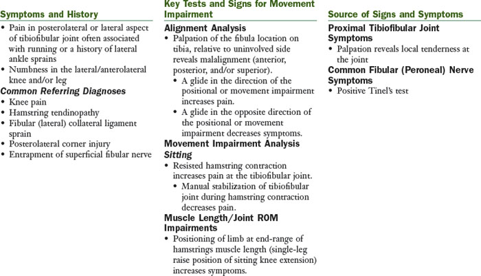
Proximal Tibiofibular Glide Syndrome

The principal movement impairment in proximal tibiofibular glide syndrome is posterior or superior motion of the fibula on the tibia during active hamstring contraction (especially during running). The principal positional impairment is the fibula located anterior, posterior, superior, or inferior to the normal position on the tibia after trauma, particularly an ankle sprain.

image

Treatment

Inflammation and Pain Control

Ice
Iontophoresis
Electrical stimulation

Walking and/or Running

Walking and running cues focus on using proximal hip extensors and lateral rotator (gluteus maximus, gluteus medius, and intrinsic hip lateral rotators) to assist with controlling hip motion and decreasing use of lateral hamstrings.

Muscle Performance

Gluteus maximus
Prone hip extension with the knee flexed.
Positioning: Patient’s that have short hip flexors will require a pillow under the pelvis. Patient must be able to control the tibial positioning during prone knee flexion to begin this exercise.
Lunges, squats.
Intrinsic hip lateral rotators and posterior gluteus medius (if indicated)
Prone hip lateral rotation isometrics (prone foot pushes)
Prone hip abduction
Sidelying hip abduction with lateral rotation (level 1, 2, or 3)

Monitor that patient feels the contraction in the “seat” region; the therapist must palpate to be sure that the patient is recruiting the correct muscles. Common cues for improve performance of the hip lateral rotators include the following:

Positioning: The pelvis may be rotated posteriorly too far. Ask the patient to roll the pelvis anteriorly.
Positioning: Place a pillow between the knees.
Spin the thigh around an axis longitudinally through the femur.
Weight shifting with gluteal squeeze on the stance lower extremity; progress to standing on one leg with correct alignment; progress to resisted activities of the opposite leg while standing on the affected leg.
Lunges: Resisted; using Thera-Band around proximal thigh, the therapist pulls in the direction of medial rotation and adduction.

After the tissue injury has been protected from excessive stresses and the inflammation has subsided, the involved muscle and tendon should undergo a progressive strengthening program and a progressive return to activity. In general, exercise or activity is permissible if pain remains at 2/10 on a 0 to 10 scale. The strengthening exercise should be completed at a minimum of 70% maximum voluntary contraction for 10 repetitions, 3 sets, 3 to 5 times/week.

Decreased Dorsiflexion

Short gastrocnemius/soleus muscle/calcaneal tendon
Wall stretch: The knee is extended for gastrocnemius muscle shortness and flexed for soleus muscle length deficits. The patient should be instructed to keep their foot facing forward or in line with the femur and tibia. The heel should be kept on the ground during the stretch. The patient should prevent subtalar joint pronation through active patient correction and by wearing good footwear. The stretch should be held for 30 seconds, completed 2 to 3 times/session, and done regularly throughout the day (5 to 8 times/day).
Heel hang stretch: The knee can be extended or flexed as described for the wall stretch. The patient should prevent subtalar joint pronation through active correction and by wearing good footwear.
Long sitting towel-assisted dorsiflexion. The patient should prevent subtalar joint pronation through active correction and by modifying the direction of force through the towel.
A night splint to maintain dorsiflexion position is often helpful.
Stretches should be held for 30 seconds, 2 to 3 repetitions, completed regularly throughout the day (5 to 8 times/day), and done 5 to 7 days/week.
Talocrural joint limitation
Mobilize the talocrural joint using a posterior glide of the talus on the ankle mortise.
Mobilize the talocrural joint using a distraction technique.
May need to include a heel lift in the shoe until length changes are apparent.

Decreased Hamstring Muscle Length

Hamstrings
Active sitting knee extension with dorsiflexion with hip in neutral rotation.

Positional Fault

Mobilization/manipulation of the fibula on the tibia as indicated by evaluation findings.
The choice of which grade of movement to choose depends on how much shortening has occurred to the associated joint structures. Chronic conditions typically have shortening of the tissues surrounding the joint. Thus chronic conditions usually respond to prolonged stretching (creep) of the tissues gained with grade IV oscillations, primarily ending with grade III oscillations to ease the joint gently out of the grade IV stretching that was just performed.
Acute conditions are not likely to have shortening of surrounding tissues and often are too painful to be mobilized back and forth. Rather, a high-velocity movement is much less painful and corrects the fault.

External Tissue Support

Orthoses/Taping

Potential to develop a taping strategy to attempt to immobilize or limit motion between the proximal tibia and fibula

References

1 Mueller MJ, Maluf KS. Tissue adaptations to physical stress: a proposed “Physical Stress Theory” to guide physical therapist practice, education and research. Phys Ther. 2002;82(4):383-403.

2 Kaeding CC, Yu JR, Wright R, et al. Management and return to play of stress fractures. Clin J Sport Med. 2005;15(6):442-447.

3 Boytim MJ, Fischer DA, Neumann L. Syndesmotic ankle sprains. Am J Sports Med. 1991;19(3):294-298.

4 Lessard L, Scudds R, Amendola A, et al. The efficacy of cryotherapy following arthroscopic knee surgery. J Orthop Sports Phys Ther. 1997;26(1):14-22.

5 Hewett TE, Paterno MV, Myer GD. Strategies for enhancing proprioception and neuromuscular control of the knee. Clin Orthop. 2002;1(402):76-94.

6 Tropp H, Askling C, Gillquist J. Prevention of ankle sprains. Am J Sports Med. 1985;13(4):259-262.

7 Bernier JN, Perrin DH. Effect of coordination training on proprioception of the functionally unstable ankle. J Orthop Sports Phys Ther. 1998;27(4):264-275.

8 Wester JU, Jespersen SM, Nielsen KD, et al. Wobble board training after partial sprains of the lateral ligaments of the ankle: a prospective randomized study. J Orthop Sports Phys Ther. 1996;23(5):332-336.

9 Rozzi SL, Lephart SM, Sterner R, et al. Balance training for persons with functionally unstable ankles. J Orthop Sports Phys Ther. 1999;29(8):478-486.

10 Eils E, Rosenbaum D. A multi-station proprioceptive exercise program in patients with ankle instability. Med Sci Sports Exerc. 2001;33(12):1991-1998.

11 Osborne MD, Chou LS, Laskowski ER, et al. The effect of ankle disk training on muscle reaction time in subjects with a history of ankle sprain. Am J Sports Med. 2001;29(5):627-632.

12 Matsusaka N, Yokoyama S, Tsurusaki T, et al. Effect of ankle disk training combined with tactile stimulation to the leg and foot on functional instability of the ankle. Am J Sports Med. 2001;29(1):25-30.

13 Blanpied P, Carroll R, Douglas T, et al. Effectiveness of lateral slide exercise in an anterior cruciate ligament reconstruction rehabilitation home exercise program. Phys Ther. 2000;30(10):609-611.

14 Hewett TE, Lindenfeld TN, Riccobene JV, et al. The effect of neuromuscular training on the incidence of knee injury in female athletes. A prospective study. Am J Sports Med. 1999;27(6):699-706.

15 Ross MD, Langford B, Whelan PJ. Test-retest reliability of 4 single-leg horizontal hop tests. J Strength Cond Res. 2002;16(4):617-622.

16 Bolgla LA, Keskula DR. Reliability of lower extremity functional performance tests. J Orthop Sports Phys Ther. 1997;26(3):138-142.

17 Demeritt KM, Shultz SJ, Docherty CL, et al. Chronic ankle instability does not affect lower extremity functional performance. J Athl Train. 2002;37(4):507-511.

18 Nadler SF, Malanga GA, Feinberg JH, et al. Functional performance deficits in athletes with previous lower extremity injury. Clin J Sport Med. 2002;12(2):73-78.

19 Petschnig R, Baron R, Albrecht M. The relationship between isokinetic quadriceps strength test and hop tests for distance and one-legged vertical jump test following anterior cruciate ligament reconstruction. J Orthop Sports Phys Ther. 1998;28(1):23-31.

20 Blackburn JR, Morrissey MC. The relationship between open and closed kinetic chain strength of the lower limb and jumping performance. J Orthop Sports Phys Ther. 1998;27(6):430-435.

21 Ross M. Test-retest reliability of the lateral step-up test in young adult healthy subjects. J Orthop Sports Phys Ther. 1997;25(2):128-132.

22 Lessard L, Scudds R, Amendola A, et al. The efficacy of cryotherapy following arthroscopic knee surgery. J Orthop Sports Phys Ther. 1997;26(1):14-22.

23 Christie AD, Willoughby GL. The effect of interferential therapy on swelling following open reduction and internal fixation of ankle fractures. Physiother Theory Pract. 1990;6:3-7.

24 Johnson MI, Wilson H. The analgesic effects of different swing patterns of interferential currents on cold-induced pain. Physiotherapy. 1997;83:461-467.

25 Young SL, Woodbury MG, Fryday-Field K. Efficacy of interferential current stimulation alone for pain reduction in patients with osteoarthritis of the knee: a randomized placebo control clinical trial. Phys Ther. 1991;71(Suppl):252.

26 Lieber RL, Silva PD, Daniel DM. Equal effectiveness of electrical and volitional strength training for quadriceps femoris muscles after anterior cruciate ligament surgery. J Orthop Res. 1996;14(1):131-138.

27 Van Swearingen J. Electrical stimulation for improving muscle performance. In: Nelson RM, Hayes KW, Currier DP, editors. Clinical electrotherapy. Stamford, CT: Appleton & Lange, 1999.

28 Delitto A, Rose SJ, Lehman RC, et al. Electrical stimulation versus voluntary exercise in strengthening the thigh musculature after anterior cruciate ligament surgery. Phys Ther. 1988;68:660-663.

29 Stevens JE, Mizner RL, Snyder-Mackler L. Neuromuscular electrical stimulation for quadriceps muscle strengthening after bilateral total knee arthroplasty: a case series. J Orthop Sports Phys Ther. 2004;34(1):21-29.

30 Fitzgerald GK, Piva SR, Irrgang JJ. A modified neuromuscular electrical stimulation protocol for quadriceps strength training following anterior cruciate ligament reconstruction. J Orthop Sports Phys Ther. 2003;33(9):492-501.