26 Urticaria, angio-oedema and anaphylaxis
Urticaria, angio-oedema and anaphylaxis may occur as a result of a variety of different stimuli, predominantly immunological but also nonimmunological, that induce mast cell (and basophil) degranulation. The predominant immunological mechanism is a type I hypersensitivity (HS) reaction whereby antigen activates mast cells (and basophils) by binding to immunoglobulin E (IgE) molecules on surface receptors. Previous exposure to the antigen in question (sensitization) results in an exaggerated immune response and excessive production of IgE which is then available for binding to mast cells and mediating an abnormally intense response to repeat antigen exposure.
Type I HS is ‘immediate’ as a reaction occurs within seconds or minutes of antigen exposure. This may then be followed by an additional late phase reaction 6–12 hours later due to the release of mediators from activated eosinophils (and neutrophils). Nonimmunological mast cell activation does not require prior sensitization and occurs either directly or more commonly through complement activation.
Clinical signs result from the sudden and excessive release of inflammatory mediators from mast cells (and perhaps basophils) and subsequently eosinophils. The severity and location of the response depend on the number and location of the cells involved that in turn depend on the degree of sensitization of the animal (immunological), the amount of antigen involved and the route of exposure. Mast cell degranulation and mediator release can be localized or systemic in more severe cases.
Localized Reactions
Localized reactions are most common, with the site dependent on the route of antigen exposure. For example:
Urticaria and Angio-oedema
Urticaria refers to local superficial (dermis) lesions grossly visible as discrete erythematous wheals. Lesions usually develop within 30 minutes to a few hours and are variably pruritic. Lesions usually disappear within 24–48 hours (but may be more chronic). Differential diagnoses for urticaria include:
In angio-oedema, subcutaneous tissues and deep blood vessels become affected by the inflammatory process, resulting in oedema and consequent diffuse swelling that is usually not pruritic. Angio-oedema typically affects the face (especially around the eyes, mouth and ears) although other areas, in particular the extremities, may be involved (Figure 26.1).
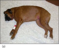
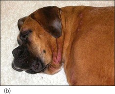
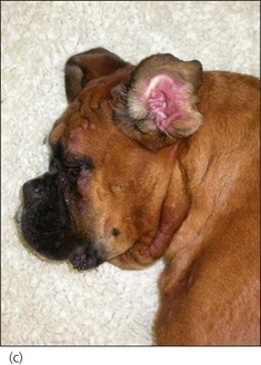
Figure 26.1 (a–c) Photographs of a Boxer dog showing cutaneous erythema, urticaria and angioedema especially affecting the head.
(Photographs courtesy of Filippo de Bellis)
Angio-oedema of the laryngeal or pharyngeal mucosa may cause potentially severe upper respiratory tract obstruction. Differential diagnoses for angio-oedema include:
Treatment of urticaria and angioedema is described in the case example.
Systemic Reactions – Anaphylaxis/Anaphylactic Shock
A severe sudden systemic release of inflammatory mediators from activated mast cells (and perhaps basophils) is the underlying pathogenesis of acute anaphylaxis/anaphylactic shock. The rate of mediator release exceeds the animal’s ability to compensate for their effects. The majority of mast cell mediators have vasoactive properties and systemic vascular effects are therefore most prominent.
Maldistributive shock occurs and in addition a marked increase in capillary permeability causes significant loss of fluid from the intravascular to the extravascular compartment, resulting in hypovolaemic shock (see Ch. 2). The cardiovascular picture during anaphylaxis (hypoperfusion that is refractory to appropriate fluid therapy) is also a hallmark of maldistributive shock secondary to systemic inflammatory response syndrome (SIRS) or sepsis (SIRS due to infection, most commonly bacterial). Thorough evaluation to exclude sepsis may be indicated in some cases prior to making a presumptive diagnosis of anaphylaxis.
Treatment of anaphylaxis
The aims of therapy in anaphylaxis are to correct systemic hypoperfusion and to attempt to address some of the underlying pathogenic mechanisms. If appropriate, suspected inciting causes must be discontinued and reexposure prevented. Aggressive individually tailored intravenous fluid therapy and adrenaline (epinephrine) are the mainstay of treatment, and regular monitoring of cardiovascular and other major systems is vital. Recommended doses of adrenaline (epinephrine) are:
Adrenaline (epinephrine) should be administered immediately once the diagnosis is made and administration can be repeated after 20–30 min (or sooner if necessary). Appropriate fluid therapy and adrenaline (epinephrine) administration should bring about clinical improvement within minutes.
Corticosteroids may also be administered in the treatment of anaphylaxis (e.g. methylprednisolone sodium succinate 1–2 mg/kg i.v. over 15–20 min; dexamethasone 0.25–1 mg/kg i.v. bolus). However, they should not be used in place of adrenaline (epinephrine) as they are likely to be much less effective in the immediate life-threatening phase. Corticosteroids may be helpful, however, in controlling on-going mediator release following initial stabilization. Additional therapy may include the use of bronchodilators (e.g. terbutaline, aminophylline), furosemide and antihistamines.
Clinical Tip
Causes of urticaria, angioedema and anaphylaxis are listed in Box 26.1. There are a large number of potential causes (not all have been reported in dogs/cats) but in a significant proportion of cases the responsible antigen cannot be identified; fortunately this does not impact on short-term emergency management.
Case example 1
Presenting Signs and Case History
A 2-year-old male neutered Boxer dog presented for treatment of a suspected allergic reaction. The dog had been out in the garden with his owners and shortly after returning indoors, he was noted to have progressive skin changes. The dog’s eyes had also puffed up en route to the practice and he appeared to be irritated by his skin. The owners reported a similar milder self-limiting episode the previous year but there was no other significant history.
Major body system examination
On presentation the dog was alert and responsive but appeared mildly agitated. Cardiovascular examination was unremarkable but the dog was panting. Abdominal palpation was unremarkable and rectal temperature was mildly elevated at 39.3°C. Facial oedema was present along with diffuse cutaneous erythema and urticaria of the inguinal region, ventral abdominal and thoracic walls. The lesions were variably pruritic.
Case management
Although the dog’s respiration was normal at presentation, given his breed, there was some concern over the potential for him to develop upper respiratory tract obstruction. Bearing in mind the potential for a late phase hypersensitivity reaction 6–12 hr following the initial signs as well, it was decided to hospitalize the dog for treatment and observation. As the dog appeared agitated and pruritic, both chlorpheniramine (8 mg total dose i.m.) and dexamethasone (0.25 mg/kg i.m.) were administered parenterally.
The dog was discharged after 12 hr of hospitalization at which time urticaria and angioedema were still present but no longer progressing, and the dog appeared stable and comfortable. He was discharged on oral prednisolone (1 mg/kg q 24 hr for 48 hr) and chlorpheniramine (8 mg total dose p.o. q 12 hr for 48 hr). It was discussed with the owners that while avoidance and elimination of aetiological antigens are ideal, in this case – as in many others – it was not possible to identify specifically such an antigen.