Chapter 41 Hybrid scanners
Chapter contents
41.1 Aim
The aim of this chapter is to introduce the reader to the basic principles and development of hybrid scanners.
41.2 Background
In medical imaging, we wish to produce images which will enable us to diagnose, treat (if possible) and monitor a disease process. Some images will produce good anatomical images (e.g. computed tomography (CT) and magnetic resonance imaging (MRI)) of pathology but will give little information about the physiological process occurring around the pathology. Other imaging devices will produce good images of the physiological processes involved in pathology (e.g. radionuclide imaging (RNI) and positron emission tomography (PET)) but give little information about associated changes in the anatomy around the pathology. Hybrid scanners attempt to collect both anatomical and physiological data and to merge this information to give a composite image, and for this reason they are sometimes referred to as multimodal imaging (MMI). Such has been the success of hybrid scanners that no manufacturer now produces a ‘stand-alone’ PET scanner.
41.3 Historical development
Historically there have been two approaches to producing the composite images. These will now be discussed.
41.3.1 The software approach
The software approach took images produced by two different scanners and then merged these images using appropriate software. To achieve effective fusion of the images, accurate spatial and temporal alignment is crucial. This is difficult to achieve when the images have been produced on two separate scanners and so this approach is susceptible to differences in patient position and noise artefacts because of the patient being in different phases of the respiratory cycle. In recent years, this has been superseded by the hardware approach.
41.3.2 The hardware approach
The hardware approach utilizes a hybrid system where two (or more) scanners are housed in a single device. The principal advantage of this system is that the imaging modalities collect data sequentially while the patient is in the same position on the couch thus limiting noise artefacts caused by inaccurate alignment. These systems will now be discussed in more detail.
41.4 The SPECT/CT hybrid scanner
This system consists of a patient couch and two gamma cameras ring mounted in front of an adapted CT scanner. The system has been around in various forms since 1999 but has seen significant improvement over the years. Figure 41.1 shows the layout of such a scanner.
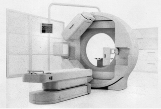
The gamma cameras can rotate around the patient and so can collect single photon emission computed tomography (SPECT) scan data. This produces an axial SPECT scan. The CT scanner part will produce an axial CT image of the same area. The two images can be displayed separately and a merged image can also be displayed. The ability to collect anatomical and physiological information simultaneously has great merit in oncology and also has potential in cardiac studies.
41.5 The PET/CT hybrid scanner
This is again an example of a hybrid scanner which will collect functional data (from the PET scan) and anatomical data (from the CT scan). The first of these entered clinical use in 2001 and at least 2000 have been installed worldwide. Figure 41.2 shows such a scanner.
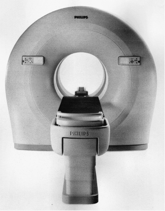
There is a patient couch and a gantry which contains the PET scanner (normally at the front) and a CT scanner. Data from both scanners can be displayed separately or merged to produce a composite image of the anatomy and physiological detail of a lesion. The main use of this type of scanner is in cardiac studies and in oncology where it gives useful data on the anatomy and activity of tumours.
41.6 The PET/MRI hybrid scanner
Images from a PET/MRI hybrid scanner were first published in 2007. The logic of moving from a PET/CT hybrid to a PET/MRI hybrid is that MRI can often provide better anatomical detail of soft tissue structures than CT. MRI can also deliver specific information about molecular cell structure. One of the problems in designing such a hybrid scanner is that the photomultiplier tubes used in conventional PET scanners are very sensitive to magnetic fields. These photomultipliers are replaced in the hybrid scanner by silicon avalanche photodiode detectors. Again the unit consists of a PET scanner and a CT scanner in the same housing. The images from this type of scanner are still being evaluated but are producing encouraging results in neurological studies, where tumours are revealed earlier and in more detail than with other scanners, and in oncology.
41.7 Other hybrid scanner combinations and current research
Efforts to develop an ultrasound/MRI hybrid are currently ongoing although no results have been published at the time of writing.
Another area of current research in hybrid imaging is the development of a multimodal contrast agent. These could be used to enhance specific areas in both scans and could also be used as markers to produce better control points for image alignment. At present only very limited clinical studies have been published.
In this chapter, you should have learnt the following:
• The historical development of hybrid scanners (see Sect. 41.3).
• The general operation of the SPECT/CT hybrid scanner (see Sect. 41.4).
• The general operation of the PET/CT hybrid scanner (see Sect. 41.5).
• The general operation of the PET/MRI scanner (see Sect. 41.6).
• Areas of current research in the area of hybrid scanners (see Sect. 41.7).